Introduction
Innovative models of human development and disease, together with animal models, are critical to further understand genetic disorders, tissue regeneration, and disease onset and progression.[Reference Miller and Spence1–Reference Drost and Clevers7] Human-derived organoids have emerged as a model system that recapitulates some aspects of human organ function in a dish. Human organoid models can be derived from either primary human tissue stem cells or from human pluripotent stem cells (hPSCs), which include human embryonic stem cells (hESCs) or induced pluripotent stem cells (iPSCs). Since there are ethical concerns pertaining to the source of hESCs, primary tissues and iPSCs have been used as an alternative to alleviate these concerns. In contrast to traditional two-dimensional monolayer cell culture, which are often made of a single transformed cell type, organoids possess multiple cell types along with structural and cellular organization, and can possess multiple tissue layers such as an epithelium and mesenchyme.[Reference Dye, Miller and Spence4, Reference Dedhia, Bertaux-Skeirik, Zavros and Spence8–Reference Rookmaaker, Schutgens, Verhaar and Clevers12] Organoids can be seen as bridging the gap between cell lines, and organotypic explant cultures (such as whole-organ or slice cultures), which have the cellular and structural complexity of the native tissue, but which are typically short-lived in culture.[Reference Shamir and Ewald13]
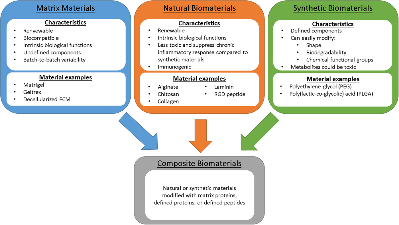
Organoid cultures are appealing due to their ability to survive for long periods of time in vitro, for their cellular heterogeneity, and for their architecture and cellular organization that recapitulate some aspects of the native tissue. The three-dimensional growth environment is critical for this structural and cellular organization by mimicking features of the physical and chemical environment of native tissue. While complex cell-derived extracellular matrices (ECM), such as Matrigel, have been the most commonly used scaffolds for organoid growth, new biomaterial systems (Fig. 1) are being developed as a means to molecularly dissect or to promote the formation of these complex tissue structures. Biomaterial composites and scaffolds offer advantages over cell-derived matrix mixtures by providing a fully defined system that enables mechanistic studies to gain detailed insights into how properties of the physical environment influence tissue growth and homeostasis. This review will cover the most recent applications of biomaterials with established organoid models of the digestive and respiratory system along with future applications and opportunities of biomaterials to enhance organoid models.
Materials for organoid, maintenance, differentiation, and transplantation. Material characteristics and examples of matrix materials, natural, synthetic, and composite biomaterials used in organoid applications.

Intestinal organoids
The intestinal organoid models derived from primary human tissue and hPSCs have been one of the pioneering organoid models in the field.[Reference Clevers5, Reference Sato, Stange, Ferrante, Vries, van Es, Van den Brink, Van Houdt, Pronk, Van Gorp, Siersema and Clevers14–Reference Watson, Mahe, Múnera, Howell, Sundaram, Poling, Schweitzer, Vallance, Mayhew, Sun, Grabowski, Finkbeiner, Spence, Shroyer, Wells and Helmrath22] Intestinal organoids have been used to model epithelial barrier function, interactions with microbes, infectious diseases and cancer.[Reference Finkbeiner, Zeng, Utama, Atmar, Shroyer and Estes23–Reference Mizutani, Tsukamoto and Clevers32]
Intestinal organoids derived from human samples were first grown from isolated small intestinal or colonic crypts, which are localized at the base of the intestinal and colonic epithelium, respectively. In the crypt, an adult stem cell population can be found with the capacity to replenish all intestinal epithelial cells, including absorptive and secretory cells (goblet cells, enterocytes/colonocyte, enteroendocrine cells and Paneth cells in the small intestine).[Reference Sato, Stange, Ferrante, Vries, van Es, Van den Brink, Van Houdt, Pronk, Van Gorp, Siersema and Clevers14–Reference Jabaji, Brinkley, Khalil, Sears, Lei, Lewis, Stelzner, Martin and Dunn17, Reference Jung, Sato, Merlos-Suárez, Barriga, Iglesias, Rossell, Auer, Gallardo, Blasco, Sancho, Clevers and Batlle33] Once placed in three-dimensional culture, whole crypts or individual intestinal stem cells form cystic or budded organoids that contain the differentiated intestinal epithelial cell types while maintaining the stem cell population. In addition to healthy crypts, patient tissue with specific mutations and cancer tissue has been used to derive organoid models in order to conduct drug screens.[Reference Sato, Stange, Ferrante, Vries, van Es, Van den Brink, Van Houdt, Pronk, Van Gorp, Siersema and Clevers14, Reference Cristobal, van den Toorn, van de Wetering, Clevers, Heck and Mohammed27–Reference Fumagalli, Drost, Suijkerbuijk, van Boxtel, de Ligt, Offerhaus, Begthel, Beerling, Tan, Sansom, Cuppen, Clevers and van Rheenen30, Reference Dekkers, Berkers, Kruisselbrink, Vonk, de Jonge, Janssens, Bronsveld, van de Graaf, Nieuwenhuis, Houwen, Vleggaar, Escher, de Rijke, Majoor, Heijerman, de Winter-de Groot, Clevers, van der Ent and Beekman34–Reference van de Wetering, Francies, Francis, Bounova, Iorio, Pronk, van Houdt, Van Gorp, Taylor-Weiner, Kester, McLaren-Douglas, Blokker, Jaksani, Bartfeld, Volckman, van Sluis, Li, Seepo, Sekhar Pedamallu, Cibulskis, Carter, McKenna, Lawrence, Lichtenstein, Stewart, Koster, Versteeg, van Oudenaarden, Saez-Rodriguez, Vries, Getz, Wessels, Stratton, McDermott, Meyerson, Garnett and Clevers36] Both the healthy and diseased organoids were maintained in Matrigel,[Reference Sato, Stange, Ferrante, Vries, van Es, Van den Brink, Van Houdt, Pronk, Van Gorp, Siersema and Clevers14–Reference Mahe, Sundaram, Watson, Shroyer and Helmrath16, Reference Jung, Sato, Merlos-Suárez, Barriga, Iglesias, Rossell, Auer, Gallardo, Blasco, Sancho, Clevers and Batlle33] yet was also shown that intestinal organoids derived from primary crypts can be maintained in ECM protein, Collagen I.[Reference Jabaji, Brinkley, Khalil, Sears, Lei, Lewis, Stelzner, Martin and Dunn17]
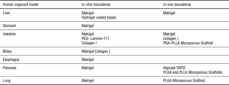
Matrigel is the most common ECM scaffold used for the culture and transplantation of organoids including intestinal organoids (Table I). Matrigel is derived from mouse tumor cells that secrete basement membrane and consists of ECM proteins (enriched for laminin) and numerous undefined growth factors.[Reference Czerwinski and Spence37] The main limitations of Matrigel is that it contains undefined materials, differs from batch-to-batch, and may have tumorgenic affects, and thus cannot be easily translated into the clinic for cell replacement therapies or small tissue transplantations. Additionally, the physical and chemical properties of Matrigel cannot be easily modified.[Reference Hughes, Postovit and Lajoie38] For these reasons, there has been an effort to define the matrix and materials suitable for intestinal organoids. For example, organoids can be grown in purified Collagen I [Reference Jabaji, Brinkley, Khalil, Sears, Lei, Lewis, Stelzner, Martin and Dunn17]; Natural materials, such as purified collagen or alginate, or synthetic materials (e.g., PEG) have more well-defined components, along with the potential to modified the physical and chemical properties (Table I).
Biomaterials used for organoid models both in vitro and in vivo.

All the human organoid models of the digestive and respiratory systems have used Matrigel to provide a three-dimensional environment. In vivo organoid transplants have used more of a variety of biomaterials such as scaffolds in order to generate the specific shapes of the organ such as tube shape for lung airways or intestine.
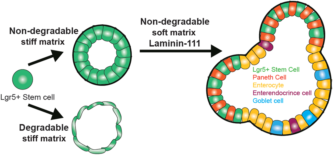
More recently, synthetic hydrogels have been developed for intestinal organoid cultures in order to define the both the physical and adhesive properties of the scaffold and without the need for Matrigel[Reference Gjorevski, Sachs, Manfrin, Giger, Bragina, Ordóñez-Morán, Clevers and Lutolf18] (Fig. 1). Polyethylene glycol (PEG) hydrogels were utilized to initiate and maintain organoid cultures due to their physical and chemical properties, which can be easily manipulated. For example, the rate and mechanism of degradation, stiffness, and adhesive properties can all be carefully controlled in PEG hydrogels. When crypts or individual intestinal stem cells were placed in unmodified, soft PEG (~300 Pa) hydrogels that mimicked the approximate stiffness as Matrigel, organoids did not form and remained as single cells, suggesting that the adhesive properties in Matrigel were crucial for maintaining organoid cultures. After screening a series of full-length ECM proteins, and adhesive peptides, RGD (Arg–Gly–Asp) was found to be sufficient to replace fibronectin, providing environment suitable for epithelial growth within the hydrogel. Interestingly, scaffold stiffness was found to correlate tightly with the maintenance of stem cells, or with differentiation. A stiff (1.3 kPa) RGD conjugated PEG hydrogel maintained the intestinal adult stem cell population and formed organoids, but did not possess differentiated intestinal cell types. This led the investigators to use a soft (190 Pa) PEG in combination with RGD and full-length Laminin-111. The combination of soft PEG, RGD, and Laminin-111 led to the formation of intestinal epithelial organoids with all four epithelial cell types along with the adult stem cells[Reference Gjorevski, Sachs, Manfrin, Giger, Bragina, Ordóñez-Morán, Clevers and Lutolf18] (Fig. 2). This landmark study was the first to define the physical and chemical properties of the extracellular environment that was required to maintain or differentiate human epithelial intestinal organoids derived from primary tissue (Fig. 2). While the PEG system provided a defined physical and adhesive environment, the PEG hydrogels do not have the same adhesive and structural properties as native extracellular matrix proteins and thus the correlation with normal development is unclear.
Changes in biomaterial properties causes different cell types and structures to form with intestinal epithelial organoids. This figure summarizes the findings of Gjorevski et al. where the biomaterial properties are defined for an organoid culture system, the intestinal organoid. Organoids were derived from the intestinal adult stem cell population, Lgr5+ cells. The stiff (1.3 kPa) non-degradable matrix was essential to maintain the adult stem cells, while the degradable biomaterial caused the cells to become thin and could not be sustained. To differentiate the adult stem cells, a softer (190 Pa) PEG hydrogel along with Laminin 111 was used to allow the organoids to grow and differentiate while maintaining the stem cell population (green cells).[Reference Gjorevski, Sachs, Manfrin, Giger, Bragina, Ordóñez-Morán, Clevers and Lutolf18] The goal will be to define the biomaterial properties for each organoid system in order to replace the non-defined Matrigel matrix.

Intestinal organoids derived from hPSCs have only been successfully grown and maintained in Matrigel to date.[Reference Spence, Mayhew, Rankin, Kuhar, Vallance, Tolle, Hoskins, Kalinichenko, Wells, Zorn, Shroyer and Wells19, Reference Finkbeiner and Spence39] hPSCs-derived intestinal organoids are derived using a step-wise differentiation process known as “directed differentiation”, which is thought to mimic the key developmental events in vitro. For more details, differentiation of hPSC-derived organoids has been reviewed by others,[Reference Aurora and Spence2, Reference Dedhia, Bertaux-Skeirik, Zavros and Spence8, Reference Wells and Spence20, Reference Finkbeiner and Spence39] but the process leads to the differentiation of small polarized spheroids consisting of an inner epithelial layer and surrounded by a mesenchymal layer.[Reference Spence, Mayhew, Rankin, Kuhar, Vallance, Tolle, Hoskins, Kalinichenko, Wells, Zorn, Shroyer and Wells19] Spheroids are placed in a Matrigel droplet and overlaid with intestinal growth media, causing the spheroids to grow several orders of magnitude at which point they are called intestinal organoids. hPSC-derived intestinal organoids are immature (fetal) in nature, represent the small intestine, possess many epithelial cell types, and are surrounded by mesenchyme.[Reference Finkbeiner, Freeman, Wieck, El-Nachef, Altheim, Tsai, Huang, Dyal, White, Grikscheit, Teitelbaum and Spence21, Reference Watson, Mahe, Múnera, Howell, Sundaram, Poling, Schweitzer, Vallance, Mayhew, Sun, Grabowski, Finkbeiner, Spence, Shroyer, Wells and Helmrath22, Reference Finkbeiner, Hill, Altheim, Dedhia, Taylor, Tsai, Chin, Mahe, Watson, Freeman, Nattiv, Thomson, Klein, Shroyer, Helmrath, Teitelbaum, Dempsey and Spence40, Reference Tsai, Nattiv, Dedhia, Nagy, Chin, Thomson, Klein and Spence41] Further investigation will be needed to define the matrix suitable for the hPSC-derived organoids that supports the derived intestinal epithelium along with the surrounding mesenchyme, unlike the primary intestinal-derived organoids, which consists of epithelium only.[Reference Sato and Clevers15, Reference Spence, Mayhew, Rankin, Kuhar, Vallance, Tolle, Hoskins, Kalinichenko, Wells, Zorn, Shroyer and Wells19]
The ultimate goal of a human intestinal organoid model is to form a long tubular structure lined with villi that contain the proper intestinal cells and cellular organization.[Reference Sato, Stange, Ferrante, Vries, van Es, Van den Brink, Van Houdt, Pronk, Van Gorp, Siersema and Clevers14–Reference Watson, Mahe, Múnera, Howell, Sundaram, Poling, Schweitzer, Vallance, Mayhew, Sun, Grabowski, Finkbeiner, Spence, Shroyer, Wells and Helmrath22] Previous reports have shown that hPSC intestinal organoids further mature in an in vivo environment such as a mouse kidney capsule. The intestinal organoids gain villi structures and integrate with the host vasculature.[Reference Finkbeiner, Freeman, Wieck, El-Nachef, Altheim, Tsai, Huang, Dyal, White, Grikscheit, Teitelbaum and Spence21, Reference Watson, Mahe, Múnera, Howell, Sundaram, Poling, Schweitzer, Vallance, Mayhew, Sun, Grabowski, Finkbeiner, Spence, Shroyer, Wells and Helmrath22] To establish a tube-like structure, a microporous tube composed of poly(glycolic acid) (PLGA) and poly-L-lactic acid (PLLA) was seeded with intestinal organoids along the outside of the tube and placed into the omentum of immunocompromised mice. The organoids matured and grew, however still lacked the tube structure since the organoids could not infiltrate into the lumen of the tubular scaffold.[Reference Finkbeiner, Freeman, Wieck, El-Nachef, Altheim, Tsai, Huang, Dyal, White, Grikscheit, Teitelbaum and Spence21] Collectively, many improvements remain for the intestinal organoid models, yet great strides have been made by defining a matrix to culture primary tissue-derived organoids, formation of villus structures in an in vivo environment, and modeling infectious disease.
Pancreatic organoids
Islet organoids
The prevalence of type 1 diabetes, an autoimmune disease that leads to the destruction of insulin-producing islets, has focused extensive research on the derivation of insulin-producing cells.[Reference Mojibian, Glavas and Kieffer42–Reference Loo, Lau, Jasmen, Lim and Teo45] Pancreatic islets consist of multiple cell types, including alpha, beta, delta, and pancreatic polypeptide (pp) cells. The beta cells within the islets secrete insulin in response to elevated levels of blood glucose.[Reference Cryer46, Reference Pambianco, Costacou, Ellis, Becker, Klein and Orchard47] Allogeneic islets have been transplanted into the portal vein, which leads to their entrapment within the sinusoids of the liver. Allogeneic islet transplantation combined with systemic immunosuppression has led to insulin independence [Reference Barton, Rickels, Alejandro, Hering, Wease, Naziruddin, Oberholzer, Odorico, Garfinkel, Levy, Pattou, Berney, Secchi, Messinger, Senior, Maffi, Posselt, Stock, Kaufman, Luo, Kandeel, Cagliero, Turgeon, Witkowski, Naji, O'Connell, Greenbaum, Kudva, Brayman, Aull, Larsen, Kay, Fernandez, Vantyghem, Bellin and Shapiro48, Reference Desai and Shea49]; however, engraftment is inefficient and long-term graft survival and function is challenged by a combination of the hepatic environment and immunosuppression.[Reference Robertson50–Reference Srinivasan, Huang, Amiel and Heaton52] Ongoing studies are attempting to enhance engraftment post-transplantation, identify alternative extrahepatic sites for optimal engraftment and survival, and identify alternative cell sources, motivated by the shortage of donor islets.
Culture conditions have been identified for the differentiation of hPSCs to immature beta-like cells, which can undergo the final steps of maturation in vivo.[Reference Pagliuca, Millman, Gürtler, Segel, Van Dervort, Ryu, Peterson, Greiner and Melton53–Reference Schulz, Young, Agulnick, Babin, Baetge, Bang, Bhoumik, Cepa, Cesario, Haakmeester, Kadoya, Kelly, Kerr, Martinson, McLean, Moorman, Payne, Richardson, Ross, Sherrer, Song, Wilson, Brandon, Green, Kroon, Kelly, D'Amour and Robins59] The hPSC-derived beta-like cells have been cultured in suspension causing cell aggregates to form three-dimensional clusters, which has demonstrated enhanced differentiation of beta-like cells compared with monolayer differentiation approaches.[Reference Pagliuca, Millman, Gürtler, Segel, Van Dervort, Ryu, Peterson, Greiner and Melton53, Reference Rezania, Bruin, Arora, Rubin, Batushansky, Asadi, O'Dwyer, Quiskamp, Mojibian, Albrecht, Yang, Johnson and Kieffer55–Reference Kim, Kim, Ko, Oh, Lim, Sohn, Shin, Kim and Han60] The derived beta-like suspension cultures are not considered islet organoids because the clusters do not contain all the cell types of an islet, lack islet cellular organization and do not possess the cellular ratios of the native islets.[Reference Vegas, Veiseh, Gürtler, Millman, Pagliuca, Bader, Doloff, Li, Chen, Olejnik, Tam, Jhunjhunwala, Langan, Aresta-Dasilva, Gandham, McGarrigle, Bochenek, Hollister-Lock, Oberholzer, Greiner, Weir, Melton, Langer and Anderson57–Reference Bosco, Armanet, Morel, Niclauss, Sgroi, Muller, Giovannoni, Parnaud and Berney61]
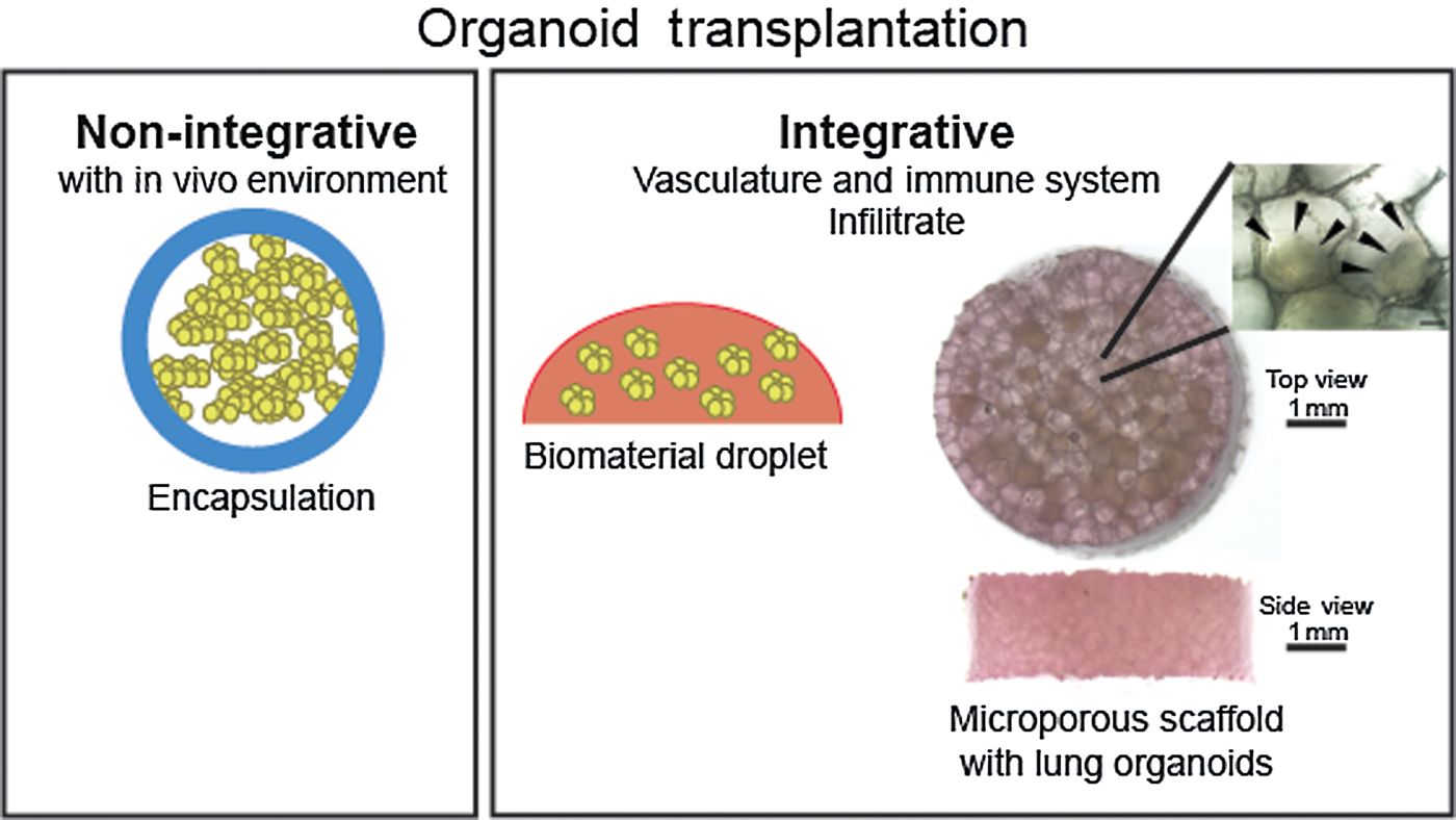
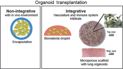
A number of materials have been applied to the transplantation of islets or hPSC-derived beta-like cells, which can be broadly placed in two categories: encapsulation for immune-isolation or porous scaffolds for integration with the host (Fig. 3).[Reference Desai and Shea49, Reference Vegas, Veiseh, Gürtler, Millman, Pagliuca, Bader, Doloff, Li, Chen, Olejnik, Tam, Jhunjhunwala, Langan, Aresta-Dasilva, Gandham, McGarrigle, Bochenek, Hollister-Lock, Oberholzer, Greiner, Weir, Melton, Langer and Anderson57, Reference Gibly, Zhang, Graham, Hering, Kaufman, Lowe and Shea62–Reference Kheradmand, Wang, Gibly, Zhang, Holland, Tasch, Graham, Kaufman, Miller, Shea and Luo66] A number of encapsulation materials have been investigated,[Reference Desai and Shea49] and a recent report involved the transplantation of hPSC-derived beta-like cell clusters encapsulated with non-degradable alginate triazole–thiomorpholine dioxide (TMTD). The encapsulated beta-like cells established normoglycemia in diabetic mice.[Reference Vegas, Veiseh, Gürtler, Millman, Pagliuca, Bader, Doloff, Li, Chen, Olejnik, Tam, Jhunjhunwala, Langan, Aresta-Dasilva, Gandham, McGarrigle, Bochenek, Hollister-Lock, Oberholzer, Greiner, Weir, Melton, Langer and Anderson57] The modified alginate was developed to reduce the foreign body response, and the encapsulation can prevent direct access of the adaptive cell response to the entrapped islets (Fig. 3).[Reference Desai and Shea49, Reference Vegas, Veiseh, Gürtler, Millman, Pagliuca, Bader, Doloff, Li, Chen, Olejnik, Tam, Jhunjhunwala, Langan, Aresta-Dasilva, Gandham, McGarrigle, Bochenek, Hollister-Lock, Oberholzer, Greiner, Weir, Melton, Langer and Anderson57] An alternative to encapsulation involves the use of microporous scaffolds that leads to integration with the host tissue. This allows the transplanted tissue to connect with the host vasculature providing direct access to nutrients for survival, and also the ability to readily sense blood glucose and for the distribution of secreted insulin specifically for islet transplants (Fig. 3). Microporous scaffolds have been widely employed for islet transplantation,[Reference Gibly, Zhang, Graham, Hering, Kaufman, Lowe and Shea62–Reference Hlavaty, Gibly, Zhang, Rives, Graham, Lowe, Luo and Shea64, Reference Gibly, Zhang, Lowe and Shea67–Reference Tuch, Gao and Lees70] primarily in rodent models, though studies have begun transplantation into non-human primates.[Reference Berman, Molano, Fotino, Ulissi, Gimeno, Mendez, Kenyon, Kenyon, Andrews, Ricordi and Pileggi71, Reference Berman, O'Neil, Coffey, Chaffanjon, Kenyon, Ruiz, Pileggi, Ricordi and Kenyon72] Reports are beginning to emerge with the transplantation of hPSC-derived immature beta cells on microporous PLGA and PLLA scaffolds, but thus far have only reported some beta cell maturation in vivo and a short rescue of normoglycemia in diabetic mice.[Reference Tuch, Gao and Lees70, Reference Weizman, Michael, Wiesel-Motiuk, Rezania and Levenberg73, Reference Mao, Chen, Bai, Song and Wang74]
The incorporation of biomaterials with organoid transplantation. Non-integrative approaches include encapsulation where the material surrounds one or more organoids and does not let the organoids interact directly with the host tissue. Integrative approaches involve degradable biomaterials such as an ECM protein droplet or microporous scaffolds where the organoids can interact directly with the host tissues. The microporous scaffolds shown are seeded with lung organoids. The inset view shows the organoids occupying individually pores. The scale bar in the inset represents 100 µm. The inset image was taken from Dye et al.[Reference Dye, Dedhia, Miller, Nagy, White, Shea and Spence129].

Acinar and ductal organoids
In addition to endocrine cells, the pancreatic epithelium also consists of exocrine cell types, which include the ductal and acinar cells.[Reference Shih, Wang and Sander75] Pancreatic organoids have been derived from primary human pancreatic ductal cells and hPSCs by using Matrigel to provide a three-dimensional environment.[Reference Broutier, Andersson-Rolf, Hindley, Boj, Clevers, Koo and Huch76–Reference Hohwieler, Illing, Hermann, Mayer, Stockmann, Perkhofer, Eiseler, Antony, Müller, Renz, Kuo, Lin, Sendler, Breunig, Kleiderman, Lechel, Zenker, Leichsenring, Rosendahl, Zenke, Sainz, Mayerle, Costa, Seufferlein, Kormann, Wagner, Liebau and Kleger78] Pancreatic organoids derived from the primary ductal cells only contain ductal cells with no acinar or endocrine cells. Even when non-ductal cells were mixed with the ductal cell population, the ductal cell population out-competed the non-ductal cells within the organoid cultures.[Reference Broutier, Andersson-Rolf, Hindley, Boj, Clevers, Koo and Huch76] Similarly, pancreatic organoids derived from hPSCs contained acinar and ductal cells along with ductal structures but contained no endocrine cells. After orthotropic transplantation of the pancreatic organoids, ductal and acinar cells were observed with a small population of endocrine cells, resembling both transcriptionally and structurally the fetal pancreas.[Reference Hohwieler, Illing, Hermann, Mayer, Stockmann, Perkhofer, Eiseler, Antony, Müller, Renz, Kuo, Lin, Sendler, Breunig, Kleiderman, Lechel, Zenker, Leichsenring, Rosendahl, Zenke, Sainz, Mayerle, Costa, Seufferlein, Kormann, Wagner, Liebau and Kleger78] The challenge thus far is to derive a pancreatic organoid that possesses exocrine and endocrine cell populations. This is particularly challenging due to the nature of the exocrine compartment, which produces a significant number of digestive enzymes and proteases.[Reference Shih, Wang and Sander75] If exocrine–ductal–endocrine organoids are established in the future, they will need to incorporate tubular and islet-like structures, with appropriate secretory function and the proper flow through network in order to protect the tissues from secreted enzymes. Instead of Matrigel, biomaterials may be used to establish these barriers between exocrine and endocrine functions, while also promoting tissue shape into islets and ductal tubes.
Liver organoids
As widely reported by the literature, deriving models of the liver or simply culturing liver cells, called hepatocytes, has been a major focus to generate improved models of drug metabolism and liver disease.[Reference Broutier, Andersson-Rolf, Hindley, Boj, Clevers, Koo and Huch76, Reference Ramachandran, Schirmer, Münst, Heinz, Ghafoory, Wölfl, Simon-Keller, Marx, Øie, Ebert, Walles, Braspenning and Breitkopf-Heinlein79–Reference Park, Chen, Ordovas and Verfaillie85] Hepatocyte cultures from primary tissue or from hPSCs provide models to study hepatocyte function, including production of serum proteins, detoxification of metabolites, and metabolism.[Reference Broutier, Andersson-Rolf, Hindley, Boj, Clevers, Koo and Huch76, Reference Ramachandran, Schirmer, Münst, Heinz, Ghafoory, Wölfl, Simon-Keller, Marx, Øie, Ebert, Walles, Braspenning and Breitkopf-Heinlein79–Reference Duncan, Dorrell and Grompe87] Specifically hepatocytes derived from hPSCs have been used for drug testing and discovery.[Reference Cai, Zhao, Liu, Ye, Song, Qin, Meng, Chen, Zhou, Song, Guo, Ding and Deng82–Reference Touboul, Hannan, Corbineau, Martinez, Martinet, Branchereau, Mainot, Strick-Marchand, Pedersen, Di Santo, Weber and Vallier84, Reference Davidson, Ware and Khetani88] When hepatocytes fail to function, the onset of liver disease occurs and in extreme cases requires organ transplantation.[Reference Si-Tayeb, Lemaigre and Duncan86, Reference Davidson, Ware and Khetani88, Reference Gissen and Arias89] Cultured hepatocyte cells could be used as a source to restore normal liver function in patients with liver disease.[Reference Park, Chen, Ordovas and Verfaillie85, Reference Davidson, Ware and Khetani88] Although two-dimensional cultures of hepatocytes have been used for drug discovery and cell transplantation, these systems lack some hepatocyte function, cellular and structural organization, and interactions with the surrounding non-hepatocyte tissues such as the ducts, stroma, and vasculature. Moreover, hepatocyte function varies, and depends if the hepatocyte is near a central vein or bile duct.[Reference Gissen and Arias89, Reference Si-Tayeb, Noto, Nagaoka, Li, Battle, Duris, North, Dalton and Duncan90] Therefore, the presence of vasculature or ducts is essential to fully recapitulate the function of the hepatocytes. Current liver organoid models have incorporated the vasculature by either co-culturing with vasculature cells and/or transplantation using the host vasculature.[Reference Broutier, Andersson-Rolf, Hindley, Boj, Clevers, Koo and Huch76, Reference Huch, Gehart, van Boxtel, Hamer, Blokzijl, Verstegen, Ellis, van Wenum, Fuchs, de Ligt, van de Wetering, Sasaki, Boers, Kemperman, de Jonge, Ijzermans, Nieuwenhuis, Hoekstra, Strom, Vries, van der Laan, Cuppen and Clevers80, Reference Takebe, Sekine, Enomura, Koike, Kimura, Ogaeri, Zhang, Ueno, Zheng, Koike, Aoyama, Adachi and Taniguchi81] Yet, the current liver organoid models still lack the cellular complexity, which are required to fully recapitulate the native in vivo organ.
Liver organoids derived from primary ductal tissue were able to maintain and propagate hepatocytes in vitro,[Reference Broutier, Andersson-Rolf, Hindley, Boj, Clevers, Koo and Huch76, Reference Ramachandran, Schirmer, Münst, Heinz, Ghafoory, Wölfl, Simon-Keller, Marx, Øie, Ebert, Walles, Braspenning and Breitkopf-Heinlein79, Reference Huch, Gehart, van Boxtel, Hamer, Blokzijl, Verstegen, Ellis, van Wenum, Fuchs, de Ligt, van de Wetering, Sasaki, Boers, Kemperman, de Jonge, Ijzermans, Nieuwenhuis, Hoekstra, Strom, Vries, van der Laan, Cuppen and Clevers80] which is in contrast to two-dimensional primary liver cultures where hepatocytes cannot be propagated.[Reference Mitaka91] Using donor tissue, isolated bile ducts were cultured as a bi-potent progenitor that can become bile duct cells called cholangiocytes or hepatocytes. The progenitor population was cultured within a Matrigel droplet and overlaid with hepatocyte-specific media, which caused the progenitor cells to form hepatocyte organoids. Although the hepatocyte organoids expressed the relevant liver markers, the organoid lacked a vasculature system, which is essential for liver function.[Reference Broutier, Andersson-Rolf, Hindley, Boj, Clevers, Koo and Huch76, Reference Huch, Gehart, van Boxtel, Hamer, Blokzijl, Verstegen, Ellis, van Wenum, Fuchs, de Ligt, van de Wetering, Sasaki, Boers, Kemperman, de Jonge, Ijzermans, Nieuwenhuis, Hoekstra, Strom, Vries, van der Laan, Cuppen and Clevers80] To incorporate the vasculature, the differentiated hepatocyte organoids were transplanted by injecting intrasplenically into mice. The transplanted liver organoids were able to secrete human albumin and human α1-antitrypsin (A1AT) into the host's blood.[Reference Huch, Gehart, van Boxtel, Hamer, Blokzijl, Verstegen, Ellis, van Wenum, Fuchs, de Ligt, van de Wetering, Sasaki, Boers, Kemperman, de Jonge, Ijzermans, Nieuwenhuis, Hoekstra, Strom, Vries, van der Laan, Cuppen and Clevers80] Liver organoids derived from the A1AT-deficient patients modeled the disease by having reduction of human A1AT in the host's blood along with aggregation of A1AT in the endoplasmic reticulum within the liver organoid cells.[Reference Huch, Gehart, van Boxtel, Hamer, Blokzijl, Verstegen, Ellis, van Wenum, Fuchs, de Ligt, van de Wetering, Sasaki, Boers, Kemperman, de Jonge, Ijzermans, Nieuwenhuis, Hoekstra, Strom, Vries, van der Laan, Cuppen and Clevers80, Reference Stoller and Aboussouan92] Similar approaches were applied to the adult liver cell line, UPcyte.[Reference Ramachandran, Schirmer, Münst, Heinz, Ghafoory, Wölfl, Simon-Keller, Marx, Øie, Ebert, Walles, Braspenning and Breitkopf-Heinlein79] Overall, transplanted liver organoids derived from patient hepatocytes do not need a biomaterial in order to engraft. However, in vitro grown organoids are expanded in Matrigel. For the in vitro studies, a defined matrix would be beneficial in order to control and standardize the system for drug screening using healthy and diseased liver organoid models.
Similarly to transplanted patient hepatocyte-derived organoids, hPSC-derived liver organoids incorporated the vasculature by co-culturing with commercially available human umbilical cord endothelial vasculature cells (HUVECs) along with human mesenchymal stem cells (MSCs) in order to form the three-dimensional liver buds. The hPSC-derived liver organoids incorporated an endothelial network on Matrigel coated plates. Other matrices were tested, including 1.5% agarose, Collagen I, Laminin, and a combination of Laminin and Entactin; yet Matrigel was the only matrix that maintained healthy liver organoid cultures. The attempted matrix proteins and materials alone did not provide the proper environment to maintain liver organoids compared with Matrigel. Thus, either the specific physical properties and/or the chemical properties of Matrigel maintained the liver organoids.
The hPSC-derived liver organoids have been transplanted to integrate with a functional vasculature system and ultimately mature the liver organoids. The functionality of the hPSC-derived liver organoids was tested through drug metabolism, using specific drugs such as ketoprofen and debrisoquine that are metabolized differently between mice and humans. The organoids had a similar metabolite profile compared with human adult liver. In addition, the liver organoids were transplanted underneath the kidney capsule of a humanized liver mouse model called TK-NOG, and were able to rescue a drug-induced lethal liver model similar to the transplanted primary hepatocytes. The transplanted liver organoids behaved similar to human adult liver and can be used to study metabolite profiles and liver diseases.[Reference Takebe, Sekine, Enomura, Koike, Kimura, Ogaeri, Zhang, Ueno, Zheng, Koike, Aoyama, Adachi and Taniguchi81]
Overall, liver organoids derived from primary tissue or hPSCs use Matrigel as the three-dimensional environment for in vitro cultures. Another approach to initiate three-dimensional cultures is applying synthetic beads in suspension culture. This approach results in random clusters of cells, which lack the cellular organization of organoid models, but has resulted in improved scaling up methods for hPSC-derived hepatocytes.[Reference Park, Chen, Ordovas and Verfaillie85] For hPSC-derived hepatocytes, cells were cultured on dextran crosslinked microbeads that were coated with gelatin. The beads allowed for efficient differentiation to hepatocyte lineage along with increasing the number of cells produced in a single differentiation.[Reference Park, Chen, Ordovas and Verfaillie85] Suspension bead cultures have also been used for a liver metastatic cancer model. Liver cancer cell line called human hepatoma cells (HepG2m) were co-cultured with human colon carcinoma cells (HT-116) in a 10:1 ratio on PEG hydrogel-coated beads in a rotating culture vessel. The hydrogel was hyaluronic acid crosslinked to thiol functionalized gelatin by a disulfide bond to allow the cells to adhere to the beads. Although both microbead models served as a three-dimensional hepatocyte culture and liver metastases site, the vasculature was not incorporated.[Reference Park, Chen, Ordovas and Verfaillie85, Reference Skardal, Devarasetty, Rodman, Atala and Soker93] Both suspension culture models could be better served by incorporating the vasculature and moving the clusters to a three-dimensional environment of defined biomaterials to initiate and/or improve cellular organization and tissue structure.
Overall liver organoid models derived from primary tissue or hPSCs still do not have a defined matrix or biomaterials to be propagated and maintained in culture. Although synthetic beads have been applied to form liver cell clusters of primary tissue, these clusters lack cellular organization and structure of the native organ. Instead of using Matrigel for culturing liver organoids derived from patient hepatocytes or hPSCs, a defined set of biomaterials may allow the cultures to be standardized in order to use for clinical applications such as drug testing and/or to easily manipulate the matrix to incorporate other proteins such as growth factors to enhance the liver epithelial structures. In addition to improvements for the matrix, liver organoids still need to incorporate all cell types of the liver, including hepatocytes, Kupffer cells, and stellate cells.
Gastric organoids
Gastric organoids provide a physiological relevant environment to study stomach infections caused by bacteria or virus. Organoids derived from primary tissue and hPSC have been microinjected with Helicobacter pylori bacteria within the epithelial lumen of the organoid. The bacteria is contained within the lumen similar to the space within the stomach and the relationship between the stomach epithelium and the bacteria can be elucidated.[Reference McCracken, Catá, Crawford, Sinagoga, Schumacher, Rockich, Tsai, Mayhew, Spence, Zavros and Wells94, Reference Bartfeld, Bayram, van de Wetering, Huch, Begthel, Kujala, Vries, Peters and Clevers95] In addition to stomach infections, stomach cancer has been modeled by deriving organoids from stomach tumors and comparing these to organoids derived from healthy tissue. The cancerous gastric organoids provided an environment similar to the structure of the stomach where cancer drugs can be tested compared with health gastric organoids.[Reference Bartfeld, Bayram, van de Wetering, Huch, Begthel, Kujala, Vries, Peters and Clevers95]
Gastric organoids derived from primary stomach tissue begin as isolated glands or single gland cells from the stomach. Both isolated glands and single cells were able to give rise to gastric organoids that could either stay as cell types within the gastric gland or change lineages and become cell types of the gastric pit by manipulating Wnt signaling.[Reference Bartfeld, Bayram, van de Wetering, Huch, Begthel, Kujala, Vries, Peters and Clevers95] These organoids contain only stomach epithelium and may require different matrix properties than gastric organoids derived from hPSCs, which possess both mesenchyme and epithelium.[Reference McCracken, Catá, Crawford, Sinagoga, Schumacher, Rockich, Tsai, Mayhew, Spence, Zavros and Wells94]
Although gastric organoids have successfully been grown in Matrigel, the three-dimensional matrix has not been defined.[Reference McCracken, Catá, Crawford, Sinagoga, Schumacher, Rockich, Tsai, Mayhew, Spence, Zavros and Wells94, Reference Bartfeld, Bayram, van de Wetering, Huch, Begthel, Kujala, Vries, Peters and Clevers95] Developing a defined matrix that recapitulates the native environment of the stomach would reveal the necessary chemical and physical properties for gastric development and maintenance of cell types, cellular organization and gland structures. The stomach has three separate muscle layers along with a submucosa and mucosa that surround the epithelium.[Reference McCracken and Wells96] Thus, to better recapitulate the surrounding gastric environment, a stiffer matrix may need to be employed to maintain and/or further mature the gastric organoids.
Esophageal organoids
Deriving an esophageal organoid will provide an esophageal epithelium that represents the shape and cellular organization specific to the human esophagus and can serve as a model for Barrett's esophagus, where the esophagus gains an intestinal phenotype that is associated with cancer.[Reference Sato, Stange, Ferrante, Vries, van Es, Van den Brink, Van Houdt, Pronk, Van Gorp, Siersema and Clevers14, Reference Odze97, Reference Lisovsky and Srivastava98] Currently, esophageal organoids have only been established from primary esophageal tissue from mouse and human, but not from hPSCs.[Reference Sato, Stange, Ferrante, Vries, van Es, Van den Brink, Van Houdt, Pronk, Van Gorp, Siersema and Clevers14, Reference DeWard, Cramer and Lagasse99] Healthy mice esophageal organoids have been successfully cultured within a Matrigel droplet.[Reference DeWard, Cramer and Lagasse99] However, only diseased human Barrett's esophageal organoids has been successfully cultured.[Reference Sato, Stange, Ferrante, Vries, van Es, Van den Brink, Van Houdt, Pronk, Van Gorp, Siersema and Clevers14] Human esophagus can be cultured on an air–liquid interface or on the matrix of decellularized esophageal tissue.[Reference Mou, Vinarsky, Tata, Brazauskas, Choi, Crooke, Zhang, Solomon, Turner, Bihler, Harrington, Lapey, Channick, Keyes, Freund, Artandi, Mense, Rowe, Engelhardt, Hsu and Rajagopal100, Reference Green, Huang, Khan, Battaglia, Corfe, MacNeil and Bury101] Yet, these cultures lack the supporting tissue around the esophagus along with a tubular structure that an esophageal organoid could incorporate. The proper matrix and media composition will need to be determined in order to generate esophageal organoids from primary tissue and hPSCs.
Biliary gland organoids
The biliary gland functions to control the bile being transported from the liver to the intestine, which includes controlling drug secretion. Deriving biliary organoids will allow for an in vitro human model of the drug secretion system and biliary gland diseases.[Reference Ogawa, Ogawa, Bear, Ahmadi, Chin, Li, Grompe, Keller, Kamath and Ghanekar102–Reference Sampaziotis, Cardoso de Brito, Madrigal, Bertero, Saeb-Parsy, Soares, Schrumpf, Melum, Karlsen, Bradley, Gelson, Davies, Baker, Kaser, Alexander, Hannan and Vallier105] For new and current drugs, it is crucial to understand the process of elimination through the biliary gland, which could help facilitate drug development and design,[Reference Hosey, Broccatelli and Benet106, Reference Sharifi and Ghafourian107] and the biliary organoids could provide a system to model this process. In addition to drug secretion, biliary gland organoids derived from iPSC lines can model genetic diseases such as cystic fibrosis (CF) where the cystic fibrosis transmembrane conductance regulator (CFTR) is impaired and disrupts fluid secretion within the glands.[Reference Colombo, Battezzati, Strazzabosco and Podda108] Biliary gland organoids derived from CF iPSC lines did not swell when induced representing impaired fluid secretion compared with control iPSC-derived biliary organoids.[Reference Ogawa, Ogawa, Bear, Ahmadi, Chin, Li, Grompe, Keller, Kamath and Ghanekar102]
Biliary organoids consist of biliary epithelial cells called cholangiocytes.[Reference Si-Tayeb, Lemaigre and Duncan86, Reference Cardinale, Wang, Carpino, Mendel, Alpini, Gaudio, Reid and Alvaro109] These cells have been derived from hPSC and liver progenitor cell lines.[Reference Ogawa, Ogawa, Bear, Ahmadi, Chin, Li, Grompe, Keller, Kamath and Ghanekar102–Reference Sampaziotis, Cardoso de Brito, Madrigal, Bertero, Saeb-Parsy, Soares, Schrumpf, Melum, Karlsen, Bradley, Gelson, Davies, Baker, Kaser, Alexander, Hannan and Vallier105, Reference Tanimizu, Miyajima and Mostov110] The liver and biliary gland are derived from the same progenitor population during development, called hepatoblasts.[Reference Si-Tayeb, Lemaigre and Duncan86, Reference Cardinale, Wang, Carpino, Mendel, Alpini, Gaudio, Reid and Alvaro109, Reference Gordillo, Evans and Gouon-Evans111] This population has been derived from hPSCs and has the capability of becoming either hepatocytes or cholangiocytes depending on the growth factors added to the culture. The hPSC-derived cholangiocytes can be cultured in a three-dimensional environment that consists of Matrigel and Collagen type I.[Reference Ogawa, Ogawa, Bear, Ahmadi, Chin, Li, Grompe, Keller, Kamath and Ghanekar102–Reference Sampaziotis, Cardoso de Brito, Madrigal, Bertero, Saeb-Parsy, Soares, Schrumpf, Melum, Karlsen, Bradley, Gelson, Davies, Baker, Kaser, Alexander, Hannan and Vallier105] In the three-dimensional environment, the hPSC-derived cholangiocytes had proper cellular organization forming an apical–basal polarity with barrier function, similar to native cholangiocytes.[Reference Ogawa, Ogawa, Bear, Ahmadi, Chin, Li, Grompe, Keller, Kamath and Ghanekar102–Reference Sampaziotis, Cardoso de Brito, Madrigal, Bertero, Saeb-Parsy, Soares, Schrumpf, Melum, Karlsen, Bradley, Gelson, Davies, Baker, Kaser, Alexander, Hannan and Vallier105, Reference Tanimizu, Miyajima and Mostov110] When hPSC-derived cholangiocytes were co-cultured with Op9 stromal cell to stimulate Notch signaling, ductal, and tubular structures formed similar to native bile ducts.[Reference Ogawa, Ogawa, Bear, Ahmadi, Chin, Li, Grompe, Keller, Kamath and Ghanekar102] Here, chimeric aggregates of Op9 stromal cells and hPSC-derived cholangiocytes were cultured in the Matrigel and Collagen I environment.[Reference Ogawa, Ogawa, Bear, Ahmadi, Chin, Li, Grompe, Keller, Kamath and Ghanekar102] Cholangiocytes derived without stromal cells did form the apical–basal polarity, but resembled a cyst-like structure rather than the native ductal structures.[Reference Zhao, Chen, Cai, Guo, Song, Che, Liu, Wu, Ding and Deng103–Reference Sampaziotis, Cardoso de Brito, Madrigal, Bertero, Saeb-Parsy, Soares, Schrumpf, Melum, Karlsen, Bradley, Gelson, Davies, Baker, Kaser, Alexander, Hannan and Vallier105]
To model the secretion system, the cell barrier with the MDR1 transporter was tested as a function of the biliary gland.[Reference Ogawa, Ogawa, Bear, Ahmadi, Chin, Li, Grompe, Keller, Kamath and Ghanekar102–Reference Sampaziotis, Cardoso de Brito, Madrigal, Bertero, Saeb-Parsy, Soares, Schrumpf, Melum, Karlsen, Bradley, Gelson, Davies, Baker, Kaser, Alexander, Hannan and Vallier105, Reference Tanimizu, Miyajima and Mostov110] Thus, an intact cell barrier is crucial to the biliary gland function. Since the combination of Matrigel and Collagen have been used to maintain the biliary organoids, defining the matrix parameters necessary to establish the biliary organoid barrier could facilitate the development of synthetic systems that provide a consistent and reproducible screening system. This culture system could provide insight into the barrier function of biliary glands and overall secretory system.
Lung organoids
The complex architecture of the lung can be categorized into two basic structures, the airways and alveoli. The airways form a tree-like network for air to move in and out of the body, while the alveoli are where the gas is exchanged with the blood.[Reference Dye, Miller and Spence4]
Airway Organoids
The airway network consists of a pseudo-stratified epithelium that is lined with an adult stem cell population, called basal cells.[Reference Rock, Onaitis, Rawlins, Lu, Clark, Xue, Randell and Hogan112–Reference Pardo-Saganta, Law, Tata, Villoria, Saez, Mou, Zhao and Rajagopal117] Common airway disorders include chronic obstructive pulmonary disease and asthma, for which airway organoids could provide useful models.[Reference Dye, Miller and Spence4] Most recent studies have shown organoids that represent early stages of lung development can be injected with respiratory syncytial virus and the lung epithelium sloughs off, which is a known consequence of this virus.[Reference Chen, Huang, de Carvalho, Ho, Islam, Volpi, Notarangelo, Ciancanelli, Casanova, Bhattacharya, Liang, Palermo, Porotto, Moscona and Snoeck118] The airway organoid model, derived from primary tissue, involves isolating the basal cell population from the primary tissue and culturing these cells in a three-dimensional environment of Matrigel. Here, the basal cells form cyst structures that contain the basal cells along with other mature cell types of the airway.[Reference Rock, Onaitis, Rawlins, Lu, Clark, Xue, Randell and Hogan112, Reference Butler, Hynds, Gowers, Lee, Brown, Crowley, Teixeira, Smith, Urbani, Hamilton, Thakrar, Booth, Birchall, De Coppi, Giangreco, O'Callaghan and Janes119–Reference Barkauskas, Chung, Fioret, Gao, Katsura and Hogan125] Recently, a cell surface marker specific to NKX2.1+ lung progenitors was discovered and used to sort out NKX2.1+ lung progenitors from hPSC-derived lung cultures using flow cytometry.[Reference Konishi, Gotoh, Tateishi, Yamamoto, Korogi, Nagasaki, Matsumoto, Muro, Hirai, Ito, Tsukita and Mishima126, Reference Gotoh, Ito, Nagasaki, Yamamoto, Konishi, Korogi, Matsumoto, Muro, Hirai, Funato, Mae, Toyoda, Sato-Otsubo, Ogawa, Osafune and Mishima127] The single cells were placed in a Matrigel droplet overlaid with airway-specific media, which resulted in airway cysts that contain basal cells and a few other mature epithelial airway cells.[Reference Konishi, Gotoh, Tateishi, Yamamoto, Korogi, Nagasaki, Matsumoto, Muro, Hirai, Ito, Tsukita and Mishima126] Both the hPSC and primary basal cell airway cysts contain a single layer of epithelial cells that contained basal cells and other mature airway cells.[Reference Rock, Onaitis, Rawlins, Lu, Clark, Xue, Randell and Hogan112, Reference Butler, Hynds, Gowers, Lee, Brown, Crowley, Teixeira, Smith, Urbani, Hamilton, Thakrar, Booth, Birchall, De Coppi, Giangreco, O'Callaghan and Janes119–Reference Gao, Bali, Randell and Hogan124, Reference Konishi, Gotoh, Tateishi, Yamamoto, Korogi, Nagasaki, Matsumoto, Muro, Hirai, Ito, Tsukita and Mishima126]
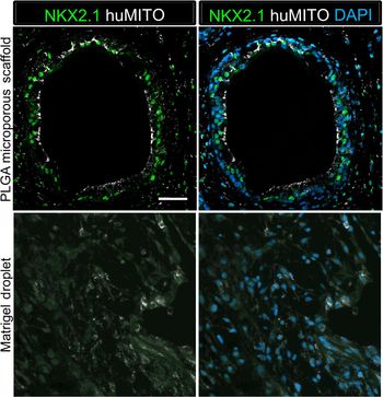
Airway models have also been derived from a cluster of hPSC-derived lung progenitors and mesenchymal cells referred to as lung spheroids.[Reference Dye, Hill, Ferguson, Tsai, Nagy, Dyal, Wells, Mayhew, Nattiv, Klein, White, Deutsch and Spence128, Reference Dye, Dedhia, Miller, Nagy, White, Shea and Spence129] These lung spheroids were derived similar to hPSC-derived intestinal organoids that relied on growth factor stimulated self-assembly to induce spheroid formation from the endodermal monolayer.[Reference Spence, Mayhew, Rankin, Kuhar, Vallance, Tolle, Hoskins, Kalinichenko, Wells, Zorn, Shroyer and Wells19, Reference Dye, Hill, Ferguson, Tsai, Nagy, Dyal, Wells, Mayhew, Nattiv, Klein, White, Deutsch and Spence128, Reference Dye, Dedhia, Miller, Nagy, White, Shea and Spence129] Lung spheroids were collected and placed into a Matrigel droplet and after several months in culture, gave rise to organoids that possessed airway cell types, including basal cells and the alveolar progenitors, along with pertinent mesenchymal populations. To further mature the tissue, the organoids were transplanted within a Matrigel droplet into an immunocompromised mouse. Multiple transplant sites were attempted with the organoids including the kidney capsule, omentum, and epididymal fat pad, but only the transplantation of the organoids onto a microporous scaffold was able to promote maturation of the organoids (Fig. 4).[Reference Dye, Dedhia, Miller, Nagy, White, Shea and Spence129] The scaffolds were fabricated from polylactide D,L co-glycolide (PLGA), a biodegradable polymer, into a cylindrical shape, 5 mm diameter and 2 mm height, with pore sizes ranging from 250 to 425 µm in diameter. These scaffolds have been used previously for islet transplantation, with islets filing the pores of the scaffold while allow for vasculature to infiltrate and integrate with the islets.[Reference Gibly, Zhang, Graham, Hering, Kaufman, Lowe and Shea62–Reference Kheradmand, Wang, Gibly, Zhang, Holland, Tasch, Graham, Kaufman, Miller, Shea and Luo66, Reference Dye, Dedhia, Miller, Nagy, White, Shea and Spence129] Lung spheroids similarly fit within the pores of the scaffold and were vascularized from the host (Fig. 4). Transplantation of the lung spheroids into the epididymal fat pad of an immunocompromised mouse resulted in the development of airway-like structures indistinguishable from adult airways in structure, cellular organization, and cellular composition, with no traces of the scaffold remaining. The microporous PLGA scaffold allowed vascularization and engraftment with the host, while also providing a stiff three-dimensional matrix to support the lung organoids, which subsequently degraded allowing the native organoid extracellular matrix to support the transplant.[Reference Dye, Dedhia, Miller, Nagy, White, Shea and Spence129] In separate work, different methodologies generated lung organoids from hPSCs that had a branched phenotype. The organoids were transplanted within a Matrigel droplet under the kidney capsule of immunocompromised mice. The retrieved tissue contained airway-specific markers lining the luminal branched structures, but lacked the cellular organization and cellular ratios of the native human lung airway.[Reference Chen, Huang, de Carvalho, Ho, Islam, Volpi, Notarangelo, Ciancanelli, Casanova, Bhattacharya, Liang, Palermo, Porotto, Moscona and Snoeck118]
Lung organoids engraft and form NKX2.1+ airway structures with PLGA scaffolds. Lung organoids were transplanted either on a PLGA scaffold coated with Matrigel (top panel) or a Matrigel droplet (bottom panel) into the epididymal fat pad of immunocompromised mice. The tissue was retrieved after 8 weeks. The lung organoids on the scaffold formed airway-like structures with lumens that expressed lung marker NKX2.1 (green) and human mitochondria marker (huMito, white) indicating the tissue was from the lung organoid (top panel). The lung organoids in the Matrigel droplet did not form airway-like structures, did not express lung marker NKX2.1 (green), but still expressed huMito (white) indicating that the organoid tissue remained but the lung identity was lost (bottom panel). Scale bar represents 50 μm.

Both the primary tissue-derived organoids and the hPSC-derived branched lung organoids contained mature cell types, but overall, these tissues lacked a proper pseudostratified epithelium.[Reference Rock, Onaitis, Rawlins, Lu, Clark, Xue, Randell and Hogan112, Reference Butler, Hynds, Gowers, Lee, Brown, Crowley, Teixeira, Smith, Urbani, Hamilton, Thakrar, Booth, Birchall, De Coppi, Giangreco, O'Callaghan and Janes119–Reference Gao, Bali, Randell and Hogan124] On the other hand, scaffold-transplanted lung organoids derived from hPSCs formed a tube that was organized into a pseudostratified epithelium similar to the adult airway. However, this tissue organization could not be generated in vitro.[Reference Dye, Dedhia, Miller, Nagy, White, Shea and Spence129] In addition, the scaffold was coated in Matrigel in order for the initial organoids to adhere to the scaffold. A fully defined matrix either from natural or synthetic materials would determine the necessary properties, including stiffness, degradability, and ECM proteins that allow for airway development. The matrix and stiffness that resembles the native environment of the airways, including the thick rings of cartilage and smooth muscle may be necessary to derive airway structures in either in vitro or in vivo and replace the non-tissue-specific Matrigel.[Reference Dye, Miller and Spence4]
Alveolar organoids
An alveolar organoid could serve as a model for severe diseases such as idiopathic fibrosis and lung cancer.[Reference Dye, Miller and Spence4, Reference Barkauskas, Chung, Fioret, Gao, Katsura and Hogan125] Alveolar organoid models, also called alveolospheres, have also been derived from primary tissue and hPSCs.[Reference Dye, Miller and Spence4, Reference Barkauskas, Chung, Fioret, Gao, Katsura and Hogan125, Reference Gotoh, Ito, Nagasaki, Yamamoto, Konishi, Korogi, Matsumoto, Muro, Hirai, Funato, Mae, Toyoda, Sato-Otsubo, Ogawa, Osafune and Mishima127, Reference Barkauskas, Cronce, Rackley, Bowie, Keene, Stripp, Randell, Noble and Hogan130, Reference Lee, Bhang, Beede, Huang, Stripp, Bloch, Wagers, Tseng, Ryeom and Kim131] Previous reports have established that one of the main alveolar epithelial cell types, type II alveolar epithelial cells (AECII), can act as a self-renewing progenitor, and can also give rise to type I alveolar epithelial cells (AECI) under injury circumstances or in culture.[Reference Barkauskas, Cronce, Rackley, Bowie, Keene, Stripp, Randell, Noble and Hogan130] When AECIIs were isolated from the primary tissue, they required additional support of a fetal lung fibroblast cell line, in order to form alveolar organoids. The organoids formed thin cysts similar to the squamous epithelium of alveoli consisting of both AECI and AECII cells within a Matrigel droplet.[Reference Barkauskas, Cronce, Rackley, Bowie, Keene, Stripp, Randell, Noble and Hogan130]
To generate hPSC-derived alveolospheres, lung epithelial progenitors obtained through directed differentiation were purified using flow cytometry and cultured with a human fetal lung fibroblast cell line, and given specific biochemical cues through specialized media. This approach resulted in the formation of thin cysts within a Matrigel droplet, similar to alveolospheres from primary cells.[Reference Gotoh, Ito, Nagasaki, Yamamoto, Konishi, Korogi, Matsumoto, Muro, Hirai, Funato, Mae, Toyoda, Sato-Otsubo, Ogawa, Osafune and Mishima127] Another lung organoid models derived from hPSCs that started as lung spheroids resulted in cells that expressed markers of an alveolar progenitor, called a bipotent progenitor, as well as AECIs and AECIIs.[Reference Dye, Hill, Ferguson, Tsai, Nagy, Dyal, Wells, Mayhew, Nattiv, Klein, White, Deutsch and Spence128] Even after transplanting the lung organoids on a microporous PLGA scaffold, the alveolar bi-potent progenitor population was not present and no alveolar structures or alveolar cells were observed.[Reference Dye, Dedhia, Miller, Nagy, White, Shea and Spence129] More recently, in a separate work the transplanted hPSC-derived branched lung organoids contained mature alveolar markers of AEI and AEII, but lacked the structure and shape of native airway and alveoli.[Reference Chen, Huang, de Carvalho, Ho, Islam, Volpi, Notarangelo, Ciancanelli, Casanova, Bhattacharya, Liang, Palermo, Porotto, Moscona and Snoeck118]
The alveolar structure is unique and does not form spontaneously in vitro. Biomaterial scaffolds provide an opportunity to direct the organization of cells into sac structures either in vitro or in vivo. Decellularized adult lung matrix has been used to provide cells with an alveolar-shaped scaffold matrix. However, seeding of the decellularized matrix using primary human alveolar cells or hPSC-derived alveolar progenitors, has proved challenging, with very few cells migrating into the decellularized matrix of the alveoli.[Reference Dye, Hill, Ferguson, Tsai, Nagy, Dyal, Wells, Mayhew, Nattiv, Klein, White, Deutsch and Spence128, Reference Booth, Hadley, Cornett, Dreffs, Matthes, Tsui, Weiss, Horowitz, Fiore, Barker, Moore, Martinez, Niklason and White132] This lack of cells in the alveolar matrix may result from the spacing and the small surface area between AECI and AECII cell adhesion. A biomaterial scaffold could be used to start with concentrating the AECI and AECII cells in one area increasing the cell adhesion, and then can be stretched to establish the thin squamous epithelium of the alveoli. The expansion and decompression of alveolar sacs with each breath supports the use of an elastic biomaterial with properties similar to the native environment.[Reference Dye, Miller and Spence4]
Future perspectives
Defining the specific biomaterials to culture respiratory and digestive organoid models could allow these organoids to be used for translational applications such as tissue transplants or disease studies. In addition, defining the biomaterials that support specific tissue types within a three-dimensional environment can enhance organoid maturation, growth, and/or structure. To enhance established organoid models, the native environment needs to be replicated such as through the stiffness, flexibility, matrix proteins, and growth factors. The majority of organoids from digestive and respiratory systems have been grown in Matrigel, which comprises undefined factors and derived from a mouse tumor[Reference Czerwinski and Spence37] (Table I). The physical and chemical parameters of Matrigel cannot be easily changed compared with defined biomaterials, including polymers and hydrogels. For instance, hydrogels can be cross-linked with matrix proteins or growth factors that are specific to the tissue along with changing the stiffness and degradability of the gel. In addition, microporous scaffold comprised polymers can be coated with matrix proteins.
Biomaterials have also provided enhanced engraftment via scaffolds when organoids are transplanted in an in vivo environment.[Reference Finkbeiner, Freeman, Wieck, El-Nachef, Altheim, Tsai, Huang, Dyal, White, Grikscheit, Teitelbaum and Spence21, Reference Tuch, Gao and Lees70, Reference Weizman, Michael, Wiesel-Motiuk, Rezania and Levenberg73, Reference Dye, Dedhia, Miller, Nagy, White, Shea and Spence129] Biomaterials more specifically scaffolds can also be employed to promote tissue structure. For instance, many epithelial-derived organoids contain tube-like structures such as the lung airway and intestine. The next steps may be to derive a tube-shaped scaffold that could be employed in order to promote a tube-shaped epithelium to form. A major difference in moving from in vitro to in vivo is the immune response, which may influence the growth and maturation of the organoids. The hPSC-derived organoids mimic normal developmental during in vitro growth; however, the role of the immune system during organ development is not entirely clear, and it is important to keep in mind that tissue transplanted into adult animals may be influenced by the host immune system. Transplantation is often associated with a surgical injury and a subsequent inflammatory response that may influence progenitor cell differentiation, regardless of whether the cells are encapsulated and therefore immune-isolated within a material[Reference Desai and Shea49, Reference Vegas, Veiseh, Gürtler, Millman, Pagliuca, Bader, Doloff, Li, Chen, Olejnik, Tam, Jhunjhunwala, Langan, Aresta-Dasilva, Gandham, McGarrigle, Bochenek, Hollister-Lock, Oberholzer, Greiner, Weir, Melton, Langer and Anderson57] (Fig. 3). In this context, biomaterials may be used as platforms to deliver factors specific to reduce the immune response and to minimize the role of the injury and inflammatory response on organoid engraftment.[Reference Liu, Zhang, Zhang, Hlavaty, Ricci, Leonard, Shea and Gower133, Reference Margul, Park, Boehler, Smith, Johnson, McCreedy, He, Ataliwala, Kukushliev, Liang, Sohrabi, Goodman, Walthers, Shea and Seidlits134]
Collectively, biomaterial systems have the potential to recapitulate the physical and chemical properties of the native organ in order to maintain and mature organoid models of the respiratory and digestive systems.[Reference Gjorevski, Sachs, Manfrin, Giger, Bragina, Ordóñez-Morán, Clevers and Lutolf18, Reference Finkbeiner, Freeman, Wieck, El-Nachef, Altheim, Tsai, Huang, Dyal, White, Grikscheit, Teitelbaum and Spence21, Reference Dye, Dedhia, Miller, Nagy, White, Shea and Spence129] Creating an environment with the necessary properties for tissue maintenance and maturation will allow for further manipulation to enhance organoid growth and maturation in vitro, or may promote specific structures and engraftment in vivo.