Introduction
Sinonasal tumours are a rare phenomenon, accounting for only 3 per cent of head and neck malignancies and 1 per cent of all malignancies.Reference Haerle, Gullane, Witterick, Zweifel and Gentili1 Sinonasal tumours pose a diagnostic challenge to clinicians. Firstly, they have an indolent presentation, with vague, non-specific symptoms such as nasal congestion and facial pain. As a result, sinonasal tumours often go undetected until secondary symptoms develop from invasion into adjacent structures, such as proptosis or diplopia from extension into the orbit,Reference Mahalingappa and Khalil2 unilateral epiphora from extension into the nasolacrimal system, or lateral facial mass from extension into the infratemporal fossa, or intra-cranial extension. The second diagnostic challenge is characterising the tumour. Computed tomography (CT) and magnetic resonance imaging (MRI) can help to separate benign lesions from malignancy, but confirmation of the tumour type requires histological examination.
The wide variety of sinonasal tumour types means that there can be differences in terms of: tumour behaviour, the likelihood of recurrence, the extent of margin resection required and the response to chemoradiotherapy. Therefore, it is important that sinonasal tumours are discussed at a multidisciplinary team (MDT) meeting before a management plan is devised.
This paper reports the diagnostic journey of a 25-year-old man with a sinonasal tumour.
Case report
A 25-year-old man presented with nasal congestion for 1 year, which was worse on the left side, associated with a reduced sense of smell and occasional post-nasal drip. He reported no rhinorrhoea, epistaxis nor facial pain, and had not undergone any previous surgical procedures. His medical history was otherwise unremarkable and a trial of intranasal corticosteroid had not improved his symptoms. He had no allergies, did not smoke, consumed a moderate amount of alcohol and had a studio-based job.
Anterior rhinoscopy revealed a large, unilateral, left-sided nasal polyp filling the left nares. This was confirmed on flexible nasendoscopy with a small septal spur to the right, and with a normal nasopharyngeal appearance.
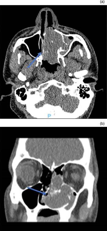
An urgent CT scan of the sinuses demonstrated an expansile left-sided sinonasal mass centred around the left inferior turbinate, with remodelling of the adjacent bones, indicating chronicity, and contiguous opacification reaching the ethmoid roof (Figure 1).
Axial (a) and coronal (b) computed tomography scans, showing a 6.4 cm anteroposterior mass filling the nasal cavity, which appears centred around the left inferior turbinate (arrows). P = posterior

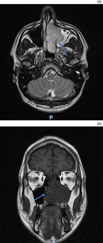
An MRI scan showed the mass extending into the left maxillary sinus and ethmoid air cells, with no intra-orbital or intra-cranial extension demonstrated (Figure 2). The MRI scan was unable to characterise the tissue. However, it showed a high T2-weighted signal in the nasolacrimal sac and mandible. An orthopantomogram of the mandible revealed a dentigerous cyst, which was asymptomatic and did not require treatment.
Axial T2-weighted (a) and coronal T1-weighted (b) magnetic resonance imaging scans, showing a left-sided nasal cavity mass extending into the maxillary sinus and ethmoid cells (arrows). The tumour has contact with the orbital floor and left lamina papyracea without breach of the periorbita; it extends into the left posterior choana and sphenopalatine foramen, and anteriorly into the vestibule. P = posterior; I = inferior

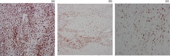
A biopsy was performed under local anaesthesia. Initial histological findings suggested a grade 2 chondrosarcoma (Figure 3). Therefore, following a head and neck MDT and locoregional discussion in the sarcoma MDT meeting, the patient was offered resection by endoscopic sinus surgery.
Immunohistochemistry analysis showed that the tumour expressed (a) glial fibrillary acidic protein (GFAP; ×??), (b) S100 (×??) and (c) pan cytokeratin (AE1/AE3; ×??), confirming that it is a low-grade myoepithelioma.

Surgery was performed without complication and with seemingly good margins of normal macroscopic mucosal appearance. However, both the posterior and anterior margins were microscopically positive for grade 2 chondrosarcoma. His case was discussed at the regional sarcoma MDT meeting for review of his histological findings and further management planning.
Amidst careful discussion about further treatment options, additional pathology was performed on the specimen. Sections showed sinonasal mucosa infiltrated by a cellular tumour with vague multi-lobular architecture, composed of stellate and spindle-shaped cells set within a diffuse myxochondroid matrix. There were scattered enlarged atypical cells with multilobulated nuclei, but a very low proliferative index (Ki67 antigen: 1 per cent). Tumour necrosis was not a feature, but the tumour permeated through the host sinonasal bone. Immunohistochemistry showed that the tumour was strongly positive for glial fibrillary acidic protein (GFAP) and cytokeratin AE1/AE3, and weakly positive for S100 protein and epithelial membrane antigen (EMA).
The differential diagnoses included both chondromyxoid fibroma and myoepithelioma with diffuse chondroid differentiation. On balance, it was agreed that the lesion was difficult to classify, but it was more in keeping with a low-grade myoepithelioma. Separately, the tumour was thought to have a high risk of local recurrence because of the observed infiltrative pattern. In view of a more benign revised diagnosis, a post-operative MRI was performed to exclude residual macroscopic disease. Frequent local surveillance with follow up by the local skull base MDT was planned in favour of further surgical resection or adjuvant therapy.
Discussion
A high level of clinical suspicion for malignancy should be maintained for patients presenting with a unilateral polyp. The first step is to perform a thorough history and examination, including full rhinological and endoscopic examinations of the rest of the upper aerodigestive tract, as well as a neck examination. The history and examination findings of our patient were in keeping with a sinonasal benign inverted papilloma. Benign papilloma is the commonest cause of unilateral polyps, especially in the younger age group. Benign sinonasal lesions are related to allergens, air pollution, and human papillomavirus types 6 and 11, whilst malignant lesions tend to be associated with tobacco, alcohol and occupational exposure to heavy metal particles. Nevertheless, it is impossible to separate benign from malignant lesions by history and clinical examination alone.Reference Lund, Clarke, Swift, McGarry, Kerawala and Carnell3 Furthermore, benign inverted papilloma carries a 9 per cent risk of malignant transformation and so requires further investigation.Reference Nygren, Kiss, von Buchwald and Bilde4
Following history-taking and clinical examination, the next investigation recommended in the UK National Multidisciplinary Guidelines for Head and Neck Cancer is CT or MRI.Reference Lund, Clarke, Swift, McGarry, Kerawala and Carnell3,Reference Dragonetti, Gera, Sciuto, Scotti, Bigoni and Barbaro5 The CT scan of the sinuses showed an expansile left-sided sinonasal mass in the left inferior turbinate that was associated with bone remodelling, but there was no infiltration of adjacent structures. This would be in keeping with a more benign diagnosis, but is not characteristic of a specific condition. There are few reports of CT or MRI findings for myoepithelioma given the rarity of disease. One study reported that a non-enhanced CT scan of myoepithelioma typically shows a well-circumscribed lesion with homogeneous attenuation.Reference Yue, Feng, Ning, Han and YaHong6 Magnetic resonance imaging findings demonstrate high intensity in T2-weighted and low intensity in T1-weighted images.Reference Hiwatashi, Matsumoto, Kamoi, Yamashita and Nakashima7,Reference Monzen, Fukushima and Fukuhara8 The MRI scans of the sinus and neck demonstrated a sinonasal tumour extending into the left maxillary sinus and ethmoid cells, with an area of high T2-weighted signal in the mandible and nasolacrimal sac. However, neither the CT nor MRI findings are specific to myoepithelioma, and they are also in keeping with benign cartilaginous tumours.
A biopsy was then taken for histopathological analysis, to help characterise the nature of the lesion. Microscopic analysis showed a chondroid matrix with atypical nuclei, which was initially thought to be a grade 2 chondrosarcoma. Interestingly, the tumour was also positive for chondrosarcoma in the posterior and anterior margins. Chondrosarcomas are most often found in the long bones, pelvis and ribs. They constitute 15 per cent of all primary malignant bone tumours, but are extremely rare in the sinonasal region. Previous cases have recognised that cartilaginous tumours are difficult to differentiate prior to surgery. Often chondrosarcoma is misdiagnosed as chondromyxoid fibroma or chordoma.Reference Lee, Jung, Yoon, Lee, Joo and Lim9
Chondromyxoid fibroma is also a rare tumour; it constitutes 0.5 per cent of all primary lesions, and occurs in the head and neck region in less than 5 per cent of cases. Similar to chondrosarcoma, chondromyxoid fibroma is most typically found in the metaphysis of long bones, in particular the femur and tibia.Reference Rahimi, Beabout, Ivins and Dahlin10
Immunochemistry analysis of chondromyxoid fibroma is usually positive for vimentin, GFAP, SOX9 and S100. However, the tumour in the case reported here was only positive for GFAP, EMA, S100 and AE1/AE3, which is more commonly seen in myoepithelial tumours.Reference Veras, Santamaria and Luna11 Myoepithelioma is a rare benign lesion that accounts for 1 per cent of salivary gland neoplasias, and most commonly affects the parotid. Myoepithelial cells are found in both acini and intercalated ducts, and play an important role in glandular excretion.
• Diagnosis of sinonasal tumours is often unclear, and requires detailed clinical history, examination and investigation
• Sinonasal tumour diagnosis hinges more on histopathology than imaging, because it can distinguish between various conditions labelled as sinonasal tumours
• Differentiation between sinonasal tumours is pivotal for management planning
• The multidisciplinary team is vital for evaluating tumour types and formulating a post-operative plan
• If there is uncertainty regarding the diagnosis, a second specialist opinion is advised
A recent review of English-language literature, published in 2019, revealed only 12 previous cases of sinonasal myoepithelioma,Reference Kang, Im, Song and Jung12 and there have been no subsequent publications of case reports in the literature since this review. Patients present with chronic nasal obstruction without secondary symptoms and with a painless mass on examination. Diagnosis requires immunohistochemistry analysis; tumours are typically positive for S100, calponin, GFAP, vimentin, EMA and carcinoembryonic antigen (CEA).Reference Sayed, Kazi, Jagade, Palav, Shinde and Pawar13 The mainstay treatment is surgical resection, performed either as an open or endoscopic procedure.Reference Cunha, Camurugy, Cavalcante Ribeiro, Costa, Azevedo and Vinhaes14 The risk of recurrence is extremely uncommon in most cases. However, cases with positive excision margins have been reported to have disease recurrence on follow up. Therefore, wide healthy margins on excision are necessary to avoid recurrence where possible, and close follow up is advised.Reference Jafarian, Omidi, Roshan, Montazer and Joushan15
A study focusing on the clinicopathological profile of sinonasal masses found variations between clinical, radiological and pathological findings in 3.63 per cent of cases.Reference Bist, Varshney, Baunthiyal, Bhagat and Kusum16 In our case, the patient presented with a unilateral nasal polyp that clinically behaved in a similar manner to a benign inverted papilloma. Initially, histology suggested a grade 2 chondrosarcoma, but, on further specialist review, a chondromyxoid fibroma or myoepithelioma was thought to be more likely. On balance, after further immunohistochemical analysis, myoepithelioma with diffuse chondroid differentiation was the preferred diagnosis. This uncertainty in diagnosis can lead to mismanagement in terms of patient care. Knowledge of the tumour type is important for calculating the risk of recurrence, the extent of resection and the plans for post-operative management. For example, adjuvant proton radiotherapy may play a role in treating small volume recurrent or residual chondrosarcomas in high-risk areas such as the brainstem or cranial nerves, whereas myoepitheliomas are both radio- and chemo-insensitive.Reference Lund, Clarke, Swift, McGarry, Kerawala and Carnell3,Reference Bist, Varshney, Baunthiyal, Bhagat and Kusum16
Conclusion
The diagnosis of sinonasal tumours is often unclear and requires a detailed clinical history, examination and investigation. Diagnosis depends on histopathology, which can distinguish the range of conditions labelled as a sinonasal tumour. The MDT plays a vital role in evaluating tumour types and formulating a post-operative plan. If there is uncertainty in the diagnosis, a second specialist opinion is advised.
Competing interests
None declared