A 51-year-old woman presented to a tertiary hospital with shortness of breath, cough, and recurrent palpitations. She reported fatigue and poor exercise tolerance for the last 2 years. The patient was aware that she had a congenital cardiac condition but was unaware of the details. A review of her chart revealed that she had only recently been diagnosed with a ventricular septal defect, bicuspid aortic valve, and Eisenmenger syndrome after presenting with similar symptoms to another institution. She was venesected for secondary polycythaemia. Her past medical history included type II diabetes mellitus, hypertension, and hypothyroidism.
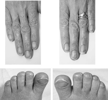
On examination, she was hypertensive (140/70 in the right arm) with no upper limb–lower limb blood pressure differential. The femoral artery pulses were easily palpable. There was obvious clubbing and cyanosis affecting the feet but not the hands. Oxygen saturations in room air in the upper limbs were 87–93%, and 70–80% in the feet. No murmurs were audible. There was moderate hepatomegaly.
Transthoracic echocardiogram demonstrated dilatation of the inferior vena cava and hepatic veins, right atrial and right ventricular dilatation, marked right ventricular hypertrophy, and reduced right ventricular systolic function. There was mild to moderate tricuspid regurgitation, and right ventricular systolic pressure was estimated at 125 mmHg with septal flattening in systole and diastole consistent with right ventricular pressure overload. There was marked dilatation of the main and branch pulmonary arteries. The left ventricular wall thickness, size, and systolic function was normal. A large perimembranous ventricular septal defect was present, although the septal leaflet of the tricuspid valve appeared to abut the right ventricular border, leading to functional closure of the defect, with no shunting – neither right-to-left or left-to-right – appreciated by colour flow Doppler. A bicuspid aortic valve was demonstrated, without stenosis or regurgitation. There was continuous right to left shunting across a large patent ductus arteriosus at low velocity consistent with severe pulmonary arterial hypertension.
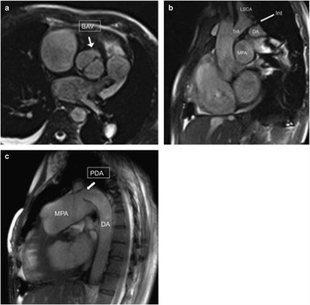
A cardiac magnetic resonance imaging was performed. This demonstrated left aortic arch branching pattern with interruption of the aortic arch beyond the left subclavian artery (type A) with antegrade flow in the distal thoracic aorta via a large patent ductus arteriosus.
The patient was treated with supportive treatment in the form of diuretics and systemic vasodilators given her systemic hypertension and biventricular dysfunction. Despite the theoretical risk of increasing right-to-left shunting with the use of vasodilators, this was not observed, and there was a general improvement in her clinical status. In addition, serial venesections were discontinued. She had documented paroxysmal supraventricular tachycardia (typical AV nodal re-entry tachycardia), which was successfully treated by radiofrequency catheter ablation. The patient did not consent to invasive haemodynamic testing and this was not pursued because of safety concerns. The patient has since returned to full-time work but remains in New York Heart Association functional class III.
Discussion
Interruption of the aorta is a rare congenital cardiac anomaly.Reference Silva, Nunes, Ferreira, Pedro, Uva and Macalhaes 1 It is usually diagnosed shortly after birth, and is associated with other congenital cardiac defects in over 90% of cases – most commonly patent ductus arteriosus and ventricular septal defect.Reference Silva, Nunes, Ferreira, Pedro, Uva and Macalhaes 1 This patient exhibited a type A interruption, distal to the left subclavian artery.Reference Dillman, Yarram, D’Amico and Hernandez 2 The differential clubbing and cyanosis in the feet but not the hands occurs where there is flow of deoxygenated blood to the lower half of the body via the patent ductus arteriosus associated with interrupted aorta in the presence of the Eisenmenger syndrome. Although a right-to-left (or left-to-right) shunt across the ventricular septal defect could not be demonstrated on colour flow Doppler, some right-to-left shunting was suspected on the basis of the upper limb oxygen saturations being intermittently low (87–93%) (Fig 1).
Clubbing and cyanosis was present in both feet but not in the hands.

This case highlights the importance of good clinical examination as it can direct and focus the cardiac imaging. It further highlights the importance of cardiologists and imaging specialists with experience and understanding of complex congenital heart disease, which may rarely present later in adulthood (Fig 2).
( a , b , c ): Magnetic resonance images the BAV is evident in ( a ) in short axis. In ( b ), the TrA is relatively hypoplastic. There is a short segment Int of the aorta, immediately distal to the LSCA. In ( c ), the MPA is grossly dilated. The DA emanates from the main pulmonary artery via a large PDA. BAV=bicuspid aortic valve; DA=descending aorta; Int=interruption; LSCA=left subclavian artery; MPA=main pulmonary artery; PDA=patent ductus arteriosus; TrA=transverse aortic arch.

Acknowledgements
The authors acknowledge and thank Dr Lawrence Dembo and Dr Conor Murray in their help in coordination and the performance of the diagnostic imaging.
Financial Support
This research received no specific grant from any funding agency, commercial or not-for-profit sectors.
Conflicts of Interest
None.