




No CrossRef data available.
Article contents
Proteolytic expression in Blastocrithidia culicis: influence of the endosymbiont and similarities with virulence factors of pathogenic trypanosomatids
Published online by Cambridge University Press: 02 November 2004
Abstract
Blastocrithidia culicis is an insect trypanosomatid that presents bacterial endosymbionts. The cell-associated and secreted proteinases of the endosymbiont-bearing and aposymbiotic strains were compared through the incorporation of proteinaceous substrates into sodium dodecyl sulfate-polyacrylamide gel electrophoresis (SDS-PAGE). Few qualitative changes could be detected in the proteolytic zymograms in the 2 strains studied when gelatin, casein, haemoglobin or bovine serum albumin (BSA) were tested. However, the level of proteolytic activities was significantly higher in the aposymbiotic strain. Some of the B. culicis proteins reacted in Western blots with antibodies raised against gp63, a zinc-metalloproteinase, and cruzipain, a cysteinyl-proteinase, which are virulence factors of the human pathogenic trypanosomatids, Leishmania spp. and Trypanosoma cruzi, respectively. The anti-cross-reacting determinant (CRD) antibody recognized 2 polypeptides (50 and 58 kDa) in the spent culture media and in the supernatant from glycosylphosphatidylinositol-phospholipase C (GPI-PLC)-treated cells, suggesting that these proteins are GPI-anchored to the plasma membrane. In addition, the anti-gp63 reacted with the 50 kDa protein. The identification of protein homologues in trypanosomatids with distinct life-cycles may help to determine the importance of proteinases in trypanosomatids.
Keywords
Information
- Type
- Research Article
- Information
- Copyright
- 2005 Cambridge University Press
INTRODUCTION
Proteinases occur ubiquitously in biological systems and have functions that range from exhaustive digestion of proteins for nutritive purposes to exquisite control of protein function by the hydrolysis of a highly specific peptide bond in a protein substrate. Because of the diverse functions of proteinases in the parasites' life-cycles and parasitic infections, they have increasingly become a focus for drug and vaccine development (Sajid & McKerrow, 2002).
The Trypanosomatidae family comprises protozoa pathogenic to humans, animals, plants and insects (Wallace, 1966). The major proteolytic activity in Trypanosoma cruzi, the causative agent of Chagas' disease, is a lysosomal cysteine-proteinase (cruzipain, cruzain or gp57/51) that is present in all life-cycle stages of this parasite. This enzyme appears to be involved in the infection of mammalian macrophages by the trypomastigote form of the parasite as well as having a role in the parasite nutrition and differentiation (Cazzulo, Stoka & Turk, 2001). In Leishmania spp., the causative agents of leishmaniasis, the major surface proteinase (MSP, gp63 or leishmanolysin) is a metalloproteinase mainly anchored to the plasma membrane of promastigotes by a glycosylphosphatidylinositol (GPI) anchor. The function of gp63 is not totally clear. This proteinase has been implicated in the parasite attachment and entry into macrophages, in the resistance to complement-mediated lysis and in the survival within the phagolysosome of macrophages (Yao, Donelson & Wilson, 2003). However, proteolytic activities with similar biochemical properties have also been detected in lower trypanosomatids belonging to the Crithidia (Etges, 1992; Melo et al. 2001; d'Avila-Levy et al. 2003a), Herpetomonas (Etges, 1992; Schneider & Glaser, 1993; Santos et al. 1999, 2002) and Leptomonas (Jaffe & Dwyer, 2003) genera, which suggests that this metalloproteinase might fulfill a nutritional role for these trypanosomatids in the vector midgut, since insect colonization is the only life-cycle stage common to the monoxenous and heteroxenous flagellate trypanosomatids.
Trypanosomatids are an interesting model for studying cellular interactions since this family comprises species which are intracellular parasites of various hosts and also species that harbour intracellular bacterial symbionts. The study of intracellular symbiosis is relevant to the understanding of many biological problems including the origin of eukaryotic cells and the colonization and survival of parasites within host cells. In trypanosomatids, the possibility of elimination of the endosymbiont by the usage of antibiotics (cure) has increased the interest in the study of endosymbiont-harbouring species, since several bacterium–protozoa interactions can be analysed by the comparison of cured and wild strains (de Souza & Motta, 1999).
The endosymbiont induces several morphological changes in the trypanosomatids and markedly influences the composition of surface membrane carbohydrates and surface anionic groups, the heme synthesis pathway, the urea cycle, the enzyme threonine deaminase, the nutritional requirements (de Souza & Motta, 1999) and the secretion of proteinases (d'Avila-Levy et al. 2001, 2003a). Blastocrithidia culicis is an insect trypanosomatid, found in the midgut of different culicidae species, which harbours an endosymbiotic bacterium in its cytoplasm, and presents the amastigote, promastigote and epimastigote forms (Wallace, 1966). Additionally, B. culicis was able to differentiate into trypomastigotes in vitro, which are similar to those observed during T. cruzi metacyclogenesis (Sousa, 1994), a fact that emphasizes the importance of biochemical studies in this species.
The aim of the present study is to characterize cell-associated and secreted proteinases of the wild and cured strains of B. culicis using different proteinaceous substrates, in order to verify whether the amino acids supplied by the endosymbiont influences the proteolytic pattern, since one of the functions of this enzymatic class is the turnover of proteins for nutritive purposes. In addition, we have assessed, by antibody cross-reactivity, the relationship of these enzymes to the well-described virulence factors from Leishmania spp. and T. cruzi, named gp63 and cruzipain, respectively.
MATERIALS AND METHODS
Chemicals
Media constituents, reagents used in electrophoresis, buffer components, nitrocellulose membrane and reagents for chemiluminescence detection were purchased from Amersham Life Science (Little Chalfont, England). The proteolytic inhibitors (trans-epoxysuccinyl-L-leucylamido-(4-guanidino) butane [E-64], phenyl-methyl sulfonyl-fluoride [PMSF], pepstatin A and 1,10-phenanthroline), the proteinaceous substrates (gelatin, haemoglobin, casein and bovine serum albumin [BSA]), the secondary antibody, polyethyleneglycol 4000 and dithiothreitol (DTT) were obtained from Sigma Chemical Co. (St Louis, MO, USA).
Parasites and cultivation
The trypanosomatid Blastocrithidia culicis and a bacteria-free strain of this protozoan were kindly provided by Dr Maria Cristina M. Motta (Instituto de Biofísica Carlos Chagas Filho, UFRJ, Brazil). These microorganisms were grown at 28 °C in 2 different media[ratio ]Roitman's complex medium (RCM) (Roitman, Roitman & Azevedo, 1972) and brain–heart infusion supplemented with 2% haemoglobin (BHI). The flagellated Trypanosoma cruzi, Dm28c strain (kindly provided by Dr Thais Souto-Padrón, Instituto de Microbiologia Prof. Paulo de Góes, UFRJ, Brazil) was cultured in liver infusion trypticase (LIT) medium, for 72 h at 26 °C to obtain epimastigote forms. Leishmania amazonensis Josefa strain (MHOM/BR/75Josefa) was grown for 4 days at 26 °C in BHI medium, to obtain promastigote forms. All these media were supplemented with 10% (v/v) heat-inactivated fetal bovine serum.
Parasites and spent culture media extracts
B. culicis (1×10⁸ cells) were harvested at the exponential phase by centrifugation at 1500 g for 15 min at 4 °C and washed 3 times with cold PBS (150 mM NaCl, 20 mM phosphate buffer, pH 7·2). Cells were then resuspended in 30 μl of PBS and lysed at 4 °C by the addition of 70 μl of sodium dodecyl sulfate-polyacrylamide gel electrophoresis (SDS-PAGE) sample buffer (250 mM Tris, pH 6·8, 8% SDS, 40% glycerol and 4% bromophenol blue) (Branquinha et al. 1996). Samples intended for spectrophotometric analysis were resuspended in 100 μl of PBS and lysed at 4 °C by the addition of 1% SDS (w/v). The cells were broken in a vortex by alternating 1 min shaking and 2 min cooling intervals. The spent culture media from 5×10⁸ cells were centrifuged, passed over a 0·22 μm filtration unit (Millipore), and concentrated 20-fold by dialysis (cut off 9000 Da) against polyethyleneglycol 4000 overnight at 4 °C. When samples were intended for substrate-gel electrophoresis analysis, the SDS-PAGE sample buffer was added in a 7[ratio ]3 (extract[ratio ]buffer – v[ratio ]v) proportion.
Growth evaluation and cellular viability
Growth was estimated by determining the cell number in a Neubauer chamber. Cellular viability was assessed by mobility, trypan blue cell dye exclusion and the absence of malate dehydrogenase, an intracellular enzyme, in the spent culture medium supernatants (d'Avila-Levy et al. 2003b).
Parasite treatment with GPI-PLC
Parasites (1×10⁸ cells) were fixed for 30 min in 0·5% paraformaldehyde, washed exhaustively in PBS and then incubated with 0·5 U/μl of glycosylphosphatidylinositol-phospholipase C (GPI-PLC) in a final reaction mixture (200 μl) containing 100 mM Tris-acetate, pH 7·4, supplemented with 0·1% Triton X-100 for 16 h at 37 °C. Control cells were subjected to the same experimental conditions, except for the presence of GPI-PLC. The supernatants from the reaction mixtures were collected by centrifugation (10 min at 1500 g), filtered in a 0·22-μm membrane (Millipore) and mixed with SDS-PAGE sample buffer (Santos et al. 2002).
Substrate-gel electrophoresis
The ability of the proteinases to degrade different proteinaceous substrates was evaluated by 10% SDS-PAGE containing 0·1% gelatin, casein, haemoglobin or BSA (Melo et al. 2002). The gels were loaded with the equivalent to 5×10⁶ cells of parasite extracts, or the equivalent to 3·75×106 cells of spent culture media extracts per slot, and following electrophoresis at a constant voltage of 200 V at 4 °C, they were soaked for 1 h at 25 °C in 2·5% Triton X-100. The gels were then incubated for 48 h at 37 °C in 50 mM sodium phosphate buffer, pH 5·5, in the presence or in the absence of 2 mM DTT, 10 μM E-64, 1 mM PMSF, 1 μM pepstatin A or 10 mM 1,10-phenanthroline. The gels were stained for 1 h with 0·2% Coomassie brilliant blue R-250 in methanol-acetic acid-water (50[ratio ]10[ratio ]40) and destained in the same solvent. The molecular weight of the proteinases was calculated by comparison with the mobility of low molecular weight standards.
Quantitative proteolytic assay
Proteinase activity was measured spectrophotometrically using the substrate gelatin according to Jones et al. (1998). Briefly, the parasites and the spent culture medium extracts (equivalent to 2×10⁷ cells) were mixed with 50 mM sodium phosphate buffer, pH 5·5, up to a final volume of 400 μl, and then 600 μl of the substrate solution (1% (w/v) gelatin in distilled water) were added. After incubation at 37 °C for 60 min, a 300 μl aliquot was removed from the reaction mixture and added to 400 μl of cold isopropanol. The supernatant was collected by centrifugation (16000 g for 10 min at 4 °C) and the absorbance was measured at 280 nm. One unit of enzyme activity was defined as the amount of enzyme that caused an increase of 0·01 in absorbance unit under standard assay conditions.
Protein immunoblotting
Total protein extracts were separated in SDS-PAGE and the polypeptides were electrophoretically transferred (100 V/300 mA for 2 h at 4 °C) to nitrocellulose membranes. The blots were blocked with 10% non-fat dried milk in PBS containing 0·05% Tween 20 for 1 h at room temperature. The membranes were incubated for 2 h with the following primary antibodies at 1[ratio ]500, 1[ratio ]250 and 1[ratio ]400 dilutions, respectively: anti-gp63 (provided by Dr Kwang-Poo Chang, University of Health Sciences/Chicago Medical School, USA), anti-cruzipain (provided by Dr Thais Souto-Padrón, UFRJ, Brazil), and anti-cross-reacting determinant (CRD) (provided by Dr Michael A. J. Ferguson and Dr Maria Lúcia Guther, University of Dundee, UK). The secondary antibody used was horseradish peroxidase-conjugated goat anti-rabbit Fc followed by immunodetection by chemiluminescence (ECL reagent).
Statistical analysis
Statistical analysis of data was performed using Student's t-test version EPI-INFO 6.04 (Database and Statistics Program for Public Health) computer software. P values of 0·05 or less were considered statistically significant.
RESULTS
The extracellular proteinases detected in this survey are considered to be genuine secretions based on (a) the detection in the culture supernatant of enzymes with molecular weights and enzymatic class not present in the cells; (b) the detection of parasites unable to exclude trypan blue dye only in the stationary growth phase, and (c) the absence of malate dehydrogenase activity, an intracellular enzyme, in the culture supernatant.
The spent culture media analysed displayed only metalloproteinases in the symbiote-containing and symbiote-free B. culicis, based on inhibition by 1,10-phenanthroline (Figs 1 and 2, lanes Phe), lack of stimulation by the thiol-reducing agent, DTT, and lack of inhibition by E-64, pepstatin A and PMSF, which are inhibitors of cysteine, aspartic and serine proteinases, respectively (data not shown).

Fig. 1. Detection of extracellular proteolytic activities in Roitman's complex medium (RCM) in the wild (w) and cured (c) strains of Blastocrithidia culicis using gelatin (1), and either casein, haemoglobin or BSA as substrates incorporated into SDS-PAGE. For simplicity, only the results containing casein (2) are shown, since the others are similar. The gel strips were incubated for 48 h at pH 5·5 supplemented with 2 mM DTT. Phe, representative gel strip showing the inhibition by 10 mM 1,10-phenanthroline in the wild strain using casein incorporated into SDS-PAGE. Numbers on the left indicate relative molecular weight markers (in kDa).

Fig. 2. Detection of extracellular proteolytic activities in brain heart infusion medium (BHI) in the wild (w) and cured (c) strains of Blastocrithidia culicis using gelatin (1), and either casein, haemoglobin or BSA as substrates incorporated into SDS-PAGE. For simplicity, only the results containing casein (2) are shown, since the others are similar. The gel strips were incubated for 48 h at pH 5·5 supplemented with 2 mM DTT. Phe, representative gel strip showing the inhibition by 10 mM 1,10-phenanthroline in the cured strain using gelatin incorporated into SDS-PAGE. Numbers on the left indicate relative molecular weight markers (in kDa).
Both strains presented the same extracellular gelatinolytic profile in RCM comprising 4 bands migrating at 110, 70, 58 and 50 kDa (Fig. 1, lane 1). Besides the gelatinolytic activity, these enzymes were also able to degrade casein, haemoglobin and BSA (Fig. 1, lane 2). Interestingly, a distinct proteinase (35 kDa) was detected only in the wild strain when casein, haemoglobin or BSA were used as substrates (Fig. 1, lane 2, w).
When both strains were cultured in BHI, a completely distinct pattern of released enzymes was observed, composed of proteinases with molecular weights of 66 and 56 kDa (Fig. 2). These enzymes were able to degrade the 4 substrates tested (Fig. 2, lanes 1 and 2). A 20 kDa enzyme capable of degrading solely gelatin was detected only in the symbiote-free B. culicis (Fig. 2, lane 1, c).
The analysis of the cell-associated proteinases from the cured and wild strains of B. culicis in the BHI and RCM media, using the 4 different substrates, is presented in Fig. 3A. The profile obtained in BHI and RCM is identical, in contrast to the results found in the spent culture media. The cellular gelatinolytic profile of both strains in the two media comprised 6 bands ranging from 35 to 76 kDa (Fig. 3A, lane 1). The 76, 64, 55, 50 and 40 kDa enzymes were also able to hydrolyse casein (Fig. 3A, lane 2). The substrates haemoglobin and BSA were degraded by the 55 kDa proteinase and more weakly by the 76 and 40 kDa proteolytic enzymes (Fig. 3A, lanes 3 and 4). The inhibitory pattern revealed that the bands migrating at 64, 40 and 35 kDa were completely inhibited by E-64, which strongly suggests that they belong to the cysteine proteinase class, while the 76, 55 and 50 kDa are metalloproteinases, since they were restrained by 1,10-phenanthroline (Fig. 3B). The proteolytic inhibitors PMSF and pepstatin A had no significant effect on enzymatic activities (data not shown).

Fig. 3. (A) Detection of cellular proteolytic activities in the wild (w) and cured (c) strains of Blastocrithidia culicis using gelatin (1), casein (2), haemoglobin (3) and BSA (4) as substrates incorporated into SDS-PAGE. Only the proteolytic profile obtained in the BHI culture medium is shown, as it is similar to the one detected in RCM. Gel strips were incubated for 48 h at pH 5·5 supplemented with 2 mM DTT. (B) Gel strips (containing gelatin incorporated) showing the inhibitory pattern by 10 mM 1,10-phenanthroline (Phe) and 10 μM E-64 (E-64). Numbers on the left indicate relative molecular weight markers (in kDa).
The comparison of the cellular proteinases of the wild and bacteria-free strains of B. culicis showed no qualitative difference (Fig. 3A). In order to quantify the amount of cellular and released proteinases in these 2 systems, we performed an enzymatic assay using gelatin as soluble substrate. As shown in Table 1, both cell-associated and extracellular proteinase activities were enhanced in the symbiote-free strain when cultured on both media tested.
Table 1. Comparison of the proteinase activity in the wild and cured strains of Blastocrithidia culicis (The microorganisms were grown in 2 different culture media: Roitman's Complex Medium (RCM) and Brain Heart Infusion medium (BHI). The activity in the cellular and in the spent culture medium extracts was determined spectrophotometrically using the substrate gelatin as described in the Materials and Methods section. The values represent a mean±standard deviation of 3 independent experiments that were performed in triplicate.)

The Western blot probed with antibodies against cruzipain, gp63 and CRD revealed that the reactivity of the bands was similar in both strains cultured in RCM and BHI (data not shown). The anti-cruzipain recognized a band with an electrophoretic mobility similar to that of the 35 kDa cysteine proteinase found in the cells (Fig. 4A, lane 1); however, no positive reaction was observed when the spent culture medium extracts were analysed (Fig. 4A, lane 2). The anti-cruzipain strongly recognized a 50 kDa polypeptide in the epimastigote forms of T. cruzi, which was included as a positive control (Fig. 4A, lane 3). The anti-gp63 reacted with a 50 kDa polypeptide both in the cellular (Fig. 4B, lane 1) and extracellular (Fig. 4B, lane 2) extracts from B. culicis. In this set of experiments, the promastigote forms of L. amazonensis were included as a positive control (Fig. 4B, lane 3). In trypanosomatids, a great number of studies have shown that metalloproteinases are preferentially surface-located (Branquinha et al. 1996; Santos et al. 1999, 2001, 2002; Cuevas, Cazzulo & Sánchez, 2003; d'Avila-Levy et al. 2003a; Soares et al. 2003) and some of them are GPI-anchored (Santos et al. 2002; Soares et al. 2003). One possible mechanism by which these enzymes might be released to the extracellular environment involves the removal of the GPI anchor by phospholipases, which can be confirmed by probing for the presence of inositol cyclic phosphate on the released polypeptide with the so-called anti-CRD antibody (Ferguson, 1999; Santos et al. 2002; Soares et al. 2003). In this sense, we detected 2 GPI-anchored polypeptides released into the culture supernatant migrating at 50 and 58 kDa (Fig. 4C, lanes 1 and 2). Further evidence for the occurrence of GPI-anchored proteins can be obtained by loss of surface components after in vitro treatment of parasites with exogenous PLC. In this context, the supernatant obtained from B. culicis cells treated with GPI-PLC was probed with anti-gp63 (Fig. 4D, lane 1) and anti-CRD (Fig. 4D, lane 2) revealing that the 50 kDa gp63-like molecule and the 58 kDa polypeptide, detected in the spent culture media extracts, are GPI-anchored to the plasma membrane. B. culicis control cells showed no reaction when probed with anti-CRD (Fig. 4D, lane 3).

Fig. 4. Western blotting showing the reactivity of polypeptides from Blastocrihtidia culicis wild strain grown in RCM with anti-cruzipain (A), anti-gp63 (B) and anti-CRD (C) antibodies. Lane 1, cellular extract; lane 2, spent culture media from parasites; and lane 3, positive controls: Trypanosoma cruzi (Dm28c strain) cellular extract (A), and Leishmania amazonensis cellular extract (B). Panel D: Supernatant from GPI-PLC-treated cells probed with anti-gp63 (lane 1) and anti-CRD (lane 2); lane 3, supernatant from control cells probed with anti-CRD. For simplicity, the profiles obtained from the wild strain in BHI and from the cured strain in both culture media, which are similar to the wild strain grown in RCM, are not shown. The numbers on the left indicate relative molecular weight markers (in kDa).
DISCUSSION
A general screening of proteinases in trypanosomatids has been widely performed by SDS-PAGE with gelatin incorporated as substrate (Greig & Ashall, 1990; Branquinha et al. 1996; Santos et al. 1999, 2001, 2002; Melo et al. 2001, 2002; Almeida et al. 2003; Cuevas, Cazzulo & Sánchez, 2003; d'Avila-Levy et al. 2001, 2003a,b; Jaffe & Dwyer, 2003; Soares et al. 2003; Vermelho et al. 2003). Gelatin has been the substrate of choice due to its solubility in water, affordable price, suitability for SDS-PAGE, and broad specificity. Proteinase bands appear as clear areas on a background of stained, non-digested protein substrate. Recently, our group has determined that the usage of different proteinaceous substrates incorporated into SDS-PAGE allows the detection of distinct proteolytic profiles in C. guilhermei, and so may help in the search of yet unknown proteinases in trypanosomatids (Melo et al. 2002). In the present work, we have examined the cured and wild strains of B. culicis by the usage of gelatin, casein, haemoglobin or BSA incorporated into SDS-PAGE, in order to determine the influence of the endosymbiont on the proteolytic expression.
In C. guilhermei, out of the 4 gelatinases detected in the spent medium, only 1 migrating at 67 kDa showed broad substrate selectivity, being able to degrade casein, haemoglobin and BSA (Melo et al. 2002). In P. serpens, the extracellular metalloproteinases migrating at 70, 90 and 94 kDa displayed a clear preference for gelatin, with considerably less activity towards casein and virtually no detectable activity against haemoglobin or BSA (Vermelho et al. 2003). All the released gelatinases from B. culicis, except for the 20 kDa enzyme detected exclusively in the cured strain grown in BHI, were also able to degrade casein, haemoglobin and BSA. Cell-associated proteinases showed a clear preference for gelatin, being less active against casein, haemoglobin and BSA. The enzymes released by B. culicis display broad substrate selectivity, as opposed to the results from C. guilhermei (Melo et al. 2002) and P. serpens (Vermelho et al. 2003). This could reflect differences on the overall metabolism of these parasites not studied yet.
A B. culicis metalloproteinase migrating at 50 kDa was detected in the cellular extracts and in the culture supernatants. A protein of similar molecular weight was recognized in the parasite, in the spent culture medium extracts, and in the supernatant from GPI-PLC treated cells when probed with the anti-gp63 antibody. This and an additional 58 kDa polypeptide were recognized by the anti-CRD in the spent culture medium extracts and in the supernatant from GPI-PLC treated cells. Taken together, these observations suggest that the 50 kDa metalloproteinase might display some degree of similarity to gp63, the zinc-dependent metalloproteinase detected in the surface of the promastigote forms of all Leishmania species examined so far (Yao et al. 2003). In addition, the 50 and 58 kDa proteins are GPI-anchored and may be released from cells by an endogenous parasite phospholipase. The function of PLC activity in trypanosomatids, under physiological conditions, is not totally clear. In T. brucei, for instance, the shedding of the variant surface glycoprotein (VSG) has multiple mechanisms and GPI-PLC plays a more significant role in VSG release than previously thought (Gruszynski et al. 2003).
The detection of gp63 homologues in insect parasites that do not have vertebrate hosts, such as Crithidia, Herpetomonas and Leptomonas, suggests that one of its functions is the nutritional requirements of the parasite in the insect (Etges, 1992; Schneider & Glaser, 1993; Melo et al. 2001; d'Avila-Levy et al. 2003a; Jaffe & Dwyer, 2003). Recently, however, gp63 homologues have been described in other trypanosomatids that also have a vertebrate host, such as T. cruzi (Cuevas et al. 2003) and T. brucei (Lacount et al. 2003). In the former, the gp63 homologues possibly play a role in host cell interaction (Cuevas et al. 2003), a function somewhat similar to that performed by leishmanial gp63, while in T. brucei the corresponding gp63 molecule expressed in the extracellular stages of African trypanosomes do not contribute to parasite entry and survival in macrophages as observed for the intracellular Leishmania. Therefore, gp63 might perform cellular functions for African trypanosomes that it either does not provide for Leishmania; or functions are yet to be discovered in the latter. Notwithstanding, this enzyme together with the endogenous GPI-PLC were shown to be responsible for releasing the ectotopically expressed variant surface glycoprotein (VSG) from the surface of procyclic trypanosomes, contributing to the parasite's immune evasion strategy (Gruszynski et al. 2003; Lacount et al. 2003). Therefore, the identification of protein homologues in trypanosomatids with distinct life-cycles might help to determine the importance of proteinases in the Trypanosomatidae family.
The anti-cruzipain antibody reacted with a cellular protein from B. culicis migrating at 35 kDa that may correspond to the cell-associated cysteine proteinase detected in the same molecular range. As far as we are concerned, this is the first report on the cross-reactivity of anti-cruzipain antibodies with proteins from insect trypanosomatids. It is noteworthy that the cruzipain used for generating the antiserum, used in the present study, was obtained from T. cruzi epimastigote forms, which are the major forms displayed by B. culicis when cultured in vitro. It has been previously shown that sera from patients with Chagas' disease react with culture forms of insect trypanosomatids, including B. culicis. Conversely, sera from rabbits immunized against insect trypanosomatids also react with T. cruzi culture forms (Lopez et al. 1981).
We have previously reported the proteolytic profile from the endosymbiont-harbouring B. culicis. The medium composition and the incubation time clearly influenced the modulation of proteinase synthesis, leading to quantitative and qualitative changes both in cellular and in released proteinases (Santos et al. 2001). B. culicis is an excellent experimental model for studying the symbiote-host interactions. The availability of B. culicis wild and aposymbiotic strains allowed us to study whether the presence of the endosymbiont induces some alteration in the proteolytic profile. In Crithidia deanei, the absence of a cell-associated cysteine-proteinase of 100 kDa in the cured strain was the only qualitative difference found between the aposymbiotic and wild strains (d'Avila-Levy et al. 2001). In that study, the extracellular proteolytic profile in the two strains was the same, but a 2-fold enhancement of the released proteinases in the cured strain was demonstrated (d'Avila-Levy et al. 2001). The level of cell-associated and released proteinase activity, measured spectrophotometrically with gelatin as soluble substrate, was significantly higher in the cured strain of B. culicis. In different trypanosomatids, it was shown that the cured strain, in contrast to the wild-type cells, requires the exogenous supplement of vitamins, purine, heme and several essential amino acids (de Souza & Motta, 1999). Taken together, these observations suggest that the nourishment of either the finished form of amino acids or usable intermediates by the endosymbiont may lead to a reduction of proteinase expression.
The authors wish to thank Ms Iêda Coleto Miguel de Castro e Silva for her technical assistance. The authors are grateful to Dr Kwang-Poo Chang for kindly providing the anti-gp63, Dr Thais Souto-Padrón for donating the anti-cruzipain and T. cruzi Dm28c strain, Dr Michael A. J. Ferguson and Dr Maria Lucia S. Guther for supplying the anti-CRD and GPI-PLC, and Dr Maria Cristina Machado Motta for providing the cured and wild strains used in this work. This study was supported by MCT/CNPq (Conselho Nacional de Desenvolvimento Científico e Tecnológico), CEPG/UFRJ (Conselho de Ensino para Graduados e Pesquisas), FAPERJ (Fundação de Amparo à Pesquisa do Estado do Rio de Janeiro) and FUJB (Fundação Universitária José Bonifácio).
References
REFERENCES
ALMEIDA, F. V. S.,
GIOVANNI-DE-SIMONE, S.,
BRANQUINHA, M. H. &
VERMELHO, A. B. (
2003).
Extracellular metalloproteinase activity in Phytomonas françai.
Parasitology Research 89,
320–322.Google Scholar
BRANQUINHA, M. H.,
VERMELHO, A. B.,
GOLDENBERG, S. &
BONALDO, M. C. (
1996).
Ubiquity of cysteine- and metalloproteinase activities in a wide range of trypanosomatids.
Journal of Eukaryotic Microbiology 43,
131–135.CrossRefGoogle Scholar
CAZZULO, J. J.,
STOKA, V. &
TURK, V. (
2001).
The major cysteine proteinase of Trypanosoma cruzi: a valid target for chemotherapy of Chagas disease.
Current Pharmaceutical Design 7,
1143–1156.CrossRefGoogle Scholar
CUEVAS, I. C.,
CAZZULO, J. J. &
SÁNCHEZ, D. O. (
2003).
gp63 homolgues in Trypanosoma cruzi: surface antigens with metalloprotease activity and a possible role in host cell infection.
Infection and Immunity 71,
5739–5749.CrossRefGoogle Scholar
D'AVILA-LEVY, C. M.,
MELO, A. C. N.,
VERMELHO, A. B. &
BRANQUINHA, M. H. (
2001).
Differential expression of proteolytic enzymes in endosymbiont-harboring Crithidia species.
FEMS Microbiology Letters 202,
73–77.CrossRefGoogle Scholar
D'AVILA-LEVY, C. M.,
SOUZA, R. F.,
GOMES, R. C.,
VERMELHO, A. B. &
BRANQUINHA, M. H. (
2003 a).
A metalloproteinase extracellularly released by Crithidia deanei.
Canadian Journal of Microbiology 49,
625–632.Google Scholar
D'AVILA-LEVY, C. M.,
SOUZA, R. F.,
GOMES, R. C.,
VERMELHO, A. B. &
BRANQUINHA, M. H. (
2003 b).
A novel extracellular calcium-dependent cysteine proteinase from Crithidia deanei.
Archives of Biochemistry and Biophysics 420,
1–8.Google Scholar
DE SOUZA, W. &
MOTTA, M. C. M. (
1999).
Endosymbiosis in protozoa of the Trypanosomatidae family.
FEMS Microbiology Letters 173,
1–8.CrossRefGoogle Scholar
ETGES, R. (
1992).
Identification of a surface metalloproteinase on 13 species of Leishmania isolated from humans, Crithidia fasciculata, and Herpetomonas samuelpessoai.
Acta Tropica 50,
205–217.CrossRefGoogle Scholar
FERGUSON, M. A. J. (
1999).
The structure, biosynthesis and functions of glycosylphosphatidylinositol anchors, and the contributions of trypanosome research.
Journal of Cell Science 112,
2799–2809.Google Scholar
GREIG, S. &
ASHALL, F. (
1990).
Electrophoretic detection of Trypanosoma cruzi peptidases.
Molecular and Biochemical Parasitology 39,
31–38.CrossRefGoogle Scholar
GRUSZYNSK, A. E.,
DEMASTER, A.,
HOOPER, N. M. &
BANGS, J. D. (
2003).
Surface coat remodeling during differentiation of Trypanosoma brucei.
The Journal of Biological Chemistry 278,
24665–24672.CrossRefGoogle Scholar
JAFFE, C. L. &
DWYER, D. M. (
2003).
Extracellular release of the surface metalloprotease, gp63, from Leishmania and insect trypanosomatids.
Parasitology Research 91,
229–237.CrossRefGoogle Scholar
JONES, B. L.,
FONTANINI, D.,
JARVINEN, D. &
PEKKARINEN, A. (
1998).
Simplified endoproteinase assays using gelatin or azogelatin.
Analytical Biochemistry 263,
214–220.CrossRefGoogle Scholar
LACOUNT, D. J.,
GRUSZYNSKI, A. E.,
GRANDGNETT, P. M.,
BANGS, J. D. &
DONELSON, J. E. (
2003).
Expression and function of the Trypanosoma brucei major surface protease (GP63) genes.
The Journal of Biological Chemistry 278,
24658–24664.CrossRefGoogle Scholar
LOPEZ, J. D.,
CAULADA, Z.,
BARBIERI, C. L. &
CAMARGO, E. P. (
1981).
Cross-reactivity between Trypanosoma cruzi and insect trypanosomatids as a basis for the diagnosis of Chagas' disease.
American Journal of Tropical Medicine and Hygiene 30,
1183–1188.CrossRefGoogle Scholar
MELO, A. C. N.,
GIOVANNI-DE-SIMMONE, S.,
BRANQUINHA, M. H. &
VERMELHO, A. B. (
2001).
Crithidia guilhermei: purification and partial characterization of a 62-kDa extracellular metalloproteinase.
Experimental Parasitology 97,
1–8.Google Scholar
MELO, A. C. N.,
D'AVILA-LEVY, C. M.,
BRANQUINHA, M. H. &
VERMELHO, A. B. (
2002).
Crithidia guilhermei: gelatin- and hemoglobin-degrading extracellular metalloproteinases.
Experimental Parasitology 102,
150–156.CrossRefGoogle Scholar
ROITMAN, C.,
ROITMAN, I. &
AZEVEDO, H. P. (
1972).
Growth of an insect trypanosomatid at 37 °C in a defined medium.
Journal of Protozoology 19,
346–349.CrossRefGoogle Scholar
SAJID, M. &
McKERROW, J. H. (
2002).
Cysteine proteases of parasitic organisms.
Molecular and Biochemical Parasitology 120,
1–21.CrossRefGoogle Scholar
SANTOS, A. L. S.,
FERREIRA, A.,
FRANCO, A. V.,
ALVIANO, C. S. &
SOARES, R. M. A. (
1999).
Characterization of proteinases in Herpetomonas anglusteri and Herpetomonas roitmani.
Current Microbiology 39,
61–64.CrossRefGoogle Scholar
SANTOS, A. L. S.,
ABREU, C. M.,
BATISTA, L. M.,
ALVIANO, C. S. &
SOARES, R. M. A. (
2001).
Cell-associated and extracellular proteinases in Blastocrithidia culicis: influence of growth conditions.
Current Microbiology 43,
100–106.CrossRefGoogle Scholar
SANTOS, A. L. S.,
ABREU, C. M.,
ALVIANO, C. S. &
SOARES, R. M. A. (
2002).
Activation of the glycosylphosphatidylinositol-anchored membrane proteinases upon release from Herpetomonas samuelpessoai by phospholipase C.
Current Microbiology 45,
293–298.CrossRefGoogle Scholar
SCHNEIDER, P. &
GLASER, T. A. (
1993).
Characterization of a surface metalloprotease from Herpetomonas samuelpessoai and comparison with Leishmania major promastigote surface protease.
Molecular and Biochemical Parasitology 58,
277–282.CrossRefGoogle Scholar
SOARES, R. M. A.,
SANTOS, A. L. S.,
BONALDO, M. C.,
ANDRADE, A. F. B.,
ALVIANO, C. S.,
ANGLUSTER, J. &
GOLDENBERG, S. (
2003).
Leishmania (Leishmania) amazonensis: differential expression of proteinases and cell-surface polypeptides in avirulent and virulent promastigotes.
Experimental Parasitology 104,
104–112.CrossRefGoogle Scholar
SOUSA, M. A. (
1994).
Trypomastigotes in cultures of Blastocrithidia culicis (Novy, MacNeal & Torrey, 1907) (Kinetoplastida: Trypanosomatidae).
Memórias do Instituto Oswaldo Cruz 89,
111–112.CrossRefGoogle Scholar
VERMELHO, A. B.,
ALMEIDA, F. V. S.,
BRONZATO, L. S. &
BRANQUINHA, M. H. (
2003).
Extracellular metalloproteinases in Phytomonas serpens.
Canadian Journal of Microbiology 49,
221–224.CrossRefGoogle Scholar
YAO, C.,
DONELSON, J. E. &
WILSON, M. E. (
2003).
The major surface protease (MSP or GP63) of Leishmania sp. Biosynthesis, regulation of expression, and function.
Molecular and Biochemical Parasitology 132,
1–16.Google Scholar
WALLACE, F. G. (
1966).
The trypanpanosomatid parasites of insects and arachnids.
Experimental Parasitology 18,
124–193.CrossRefGoogle Scholar

Fig. 1. Detection of extracellular proteolytic activities in Roitman's complex medium (RCM) in the wild (w) and cured (c) strains of Blastocrithidia culicis using gelatin (1), and either casein, haemoglobin or BSA as substrates incorporated into SDS-PAGE. For simplicity, only the results containing casein (2) are shown, since the others are similar. The gel strips were incubated for 48 h at pH 5·5 supplemented with 2 mM DTT. Phe, representative gel strip showing the inhibition by 10 mM 1,10-phenanthroline in the wild strain using casein incorporated into SDS-PAGE. Numbers on the left indicate relative molecular weight markers (in kDa).

Fig. 2. Detection of extracellular proteolytic activities in brain heart infusion medium (BHI) in the wild (w) and cured (c) strains of Blastocrithidia culicis using gelatin (1), and either casein, haemoglobin or BSA as substrates incorporated into SDS-PAGE. For simplicity, only the results containing casein (2) are shown, since the others are similar. The gel strips were incubated for 48 h at pH 5·5 supplemented with 2 mM DTT. Phe, representative gel strip showing the inhibition by 10 mM 1,10-phenanthroline in the cured strain using gelatin incorporated into SDS-PAGE. Numbers on the left indicate relative molecular weight markers (in kDa).
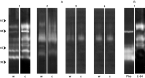
Fig. 3. (A) Detection of cellular proteolytic activities in the wild (w) and cured (c) strains of Blastocrithidia culicis using gelatin (1), casein (2), haemoglobin (3) and BSA (4) as substrates incorporated into SDS-PAGE. Only the proteolytic profile obtained in the BHI culture medium is shown, as it is similar to the one detected in RCM. Gel strips were incubated for 48 h at pH 5·5 supplemented with 2 mM DTT. (B) Gel strips (containing gelatin incorporated) showing the inhibitory pattern by 10 mM 1,10-phenanthroline (Phe) and 10 μM E-64 (E-64). Numbers on the left indicate relative molecular weight markers (in kDa).
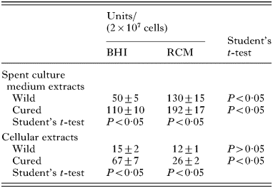
Table 1. Comparison of the proteinase activity in the wild and cured strains of Blastocrithidia culicis
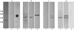
Fig. 4. Western blotting showing the reactivity of polypeptides from Blastocrihtidia culicis wild strain grown in RCM with anti-cruzipain (A), anti-gp63 (B) and anti-CRD (C) antibodies. Lane 1, cellular extract; lane 2, spent culture media from parasites; and lane 3, positive controls: Trypanosoma cruzi (Dm28c strain) cellular extract (A), and Leishmania amazonensis cellular extract (B). Panel D: Supernatant from GPI-PLC-treated cells probed with anti-gp63 (lane 1) and anti-CRD (lane 2); lane 3, supernatant from control cells probed with anti-CRD. For simplicity, the profiles obtained from the wild strain in BHI and from the cured strain in both culture media, which are similar to the wild strain grown in RCM, are not shown. The numbers on the left indicate relative molecular weight markers (in kDa).
 You have
Access
- 26
- Cited by