Abbreviations
ARE- antioxidant response element; BW- body weight; GSH-Px- glutathione peroxidase; HSP- heat shock protein; MDA- malondialdehyde; Nrf2- Nuclear factor-erythroid-2- related factor 2; ROS- reactive oxygen species; SD- stocking density; SelP- selenoprotein P; SOD- superoxide dismutase; SPS2- selenophosphate synthetase 2; Trx- thioredoxin; TrxR- thioredoxin reductase.
Introduction
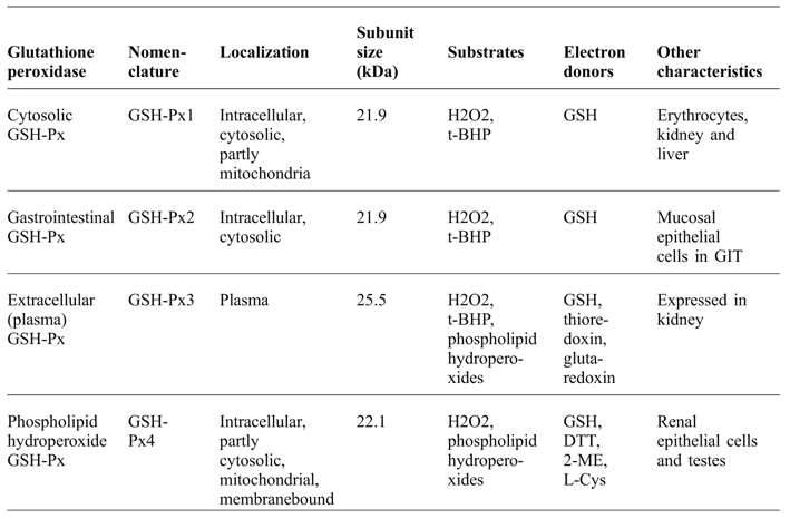
For the last few decades information has been actively accumulated to show that, in poultry, certain stressors are associated with overproduction of free radicals. This includes environmental (Surai and Fisinin, Reference SURAI and FISININ2016a), nutritional and internal stresses (Surai and Fisinin, Reference SURAI and FISININ2016b). Indeed, about 20 billion of free radicals are produced in every cell every day (Chance et al., Reference CHANCE, SIES and BOVERIES1979) and can damage all types of molecules, including lipids, proteins and DNA. During evolution, antioxidant defence mechanisms were developed to deal with such damaging radicals. There are three major levels of antioxidant defence in the cell and body (Surai, Reference SURAI2006). The first level of the antioxidant defence is based on the enzyme activity of superoxide dismutase (SOD), glutathione peroxidase (GSH-Px) and catalase (CAT), which are responsible for the detoxification of superoxide radicals produced in mitochondria. It is important to note that old biological textbooks, written 30 and more years ago, include only one GSH-Px, but in animals, the GSH-Px family includes at least eight members. They differ in molecular weight, substrate specificity, cell distribution and perform a variety of functions. There are four Se-dependent forms of GSH-Px in avian species, namely GSH-Px1, GSH-Px2, GSH-Px3 and GSH-Px4 (Table 1). A PubMed search conducted on October 16th 2017 on the ‘glutathione peroxidase’ gave 33013 publications, including more than 1692 references in 2017, demonstrating intense interest in this subject. The aim of this review paper is to review GSH-Px properties and functions in relation to poultry with emphasis to its role in adaptations to common stress conditions.
Se-dependent GSH-Px characteristics (Adapted from Surai, Reference SURAI2006).

t-BHP: tert-Butyl hydroperoxide; DTT: 1,4-Ditiothreitol; 2-ME: 2_mercaptoethanol; L-Cys: L-Cysteine
Cytosolic glutathione peroxidase (cGSH-Px or GSH-Px1)
Cytosolic GSH-Px (glutathione H2O2 oxidoreductase E.C. 1.11.1.9) was discovered by Mills in 1957, who showed that this enzyme had a protective effect in erythrocytes against H2O2 or ascorbate-induced haemolysis. Sixteen years later it became clear that GSH-Px was a selenoenzyme. In fact, Rotruck et al. (Reference ROTRUCK, POPE, GANTHER, SWANSON, HAFEMAN and HOEKSTRA1973) were the first to show that in rat red blood cells Se was tightly bound to the enzyme and demonstrated the uptake of 75Se by GSH-Px. As mentioned above, GSH-Px is responsible for detoxification of hydroperoxides and hydrogen peroxide in the following reactions:

These reactions employ a so-called ‘ping-pong’ mechanism, for example SeCys in the active centre of the enzyme is oxidised with a selenenic acid formation, which is reduced back by a reaction with two molecules of GSH. The Se atom in the enzyme catalytic site undergoes a redox cycle involving the selenolate anion in its active form, which reduces H2O2 and organic peroxides (Mugesh and Singh, Reference MUGESH and SINGH2000). Recently, an elegant mathematical model and unified catalytic scheme with the incorporation of pH regulation mechanism of GSH-Px have been developed, which confirmed that GSH-Px follows the described ‘ping-pong’ mechanism (Pannala et al., Reference PANNALA, BAZIL, CAMARA and DASH2014). GSH-Px is characterised by high specificity for GSH as a donor of a reducing equivalent (substrate) and catalyses the reduction of a variety of hydroperoxides. It is interesting to note that thioredoxin can be used, beside GSH, as reducing substrate, and GSH-Px4 can use other protein thiols as reducing equivalents (Brigelius-Flohé and Maiorino, Reference BRIGELIUS-FLOHÉ and MAIORINO2013). However, GSH-Px1 activity is related only to free peroxides and it is not able to reduce esterified fatty acid hydroperoxides. Therefore, hydroperoxides in membranes have to be released by other enzymatic systems (e.g. phospholipases) or another member of GSH-Px family (GSH-Px4) can deal with them.
GSH-Px activity is dependent on the Se status of tissues. In fact, dietary Se supplementation has been shown to be effective in increasing GSH-Px in a variety of animal species including rat, mouse, chicken, quail, sheep, cattle, horse, pig, deer and salmon. (Flohe and Brigelius-Flohe, Reference FLOHE and BRIGELIUS-FLOHE2016). There are a range of nutritional means of decreasing GSH-Px activities in various tissues including vitamin E excess, deficiencies of iron, zinc, riboflavin, vitamin B6 or copper as well as consumption of silver, tri-o-cresyl phosphate or doxorubicin (Surai, Reference SURAI2006). In fact, depending on concentration and duration of exposure, various chemical elements and compounds can either decrease or increase GSH-Px activity in tissues. When Se is available, increased GSH-Px activity could provide a compensatory mechanism to deal with stress conditions and oxygen-responsive elements (responsive to the oxygen tension in culture) were identified in the 5I-flanking region of the GSH-Px gene (Cowan et al., Reference COWAN, WEISEL, WILLIAMS and MICKLE1993).
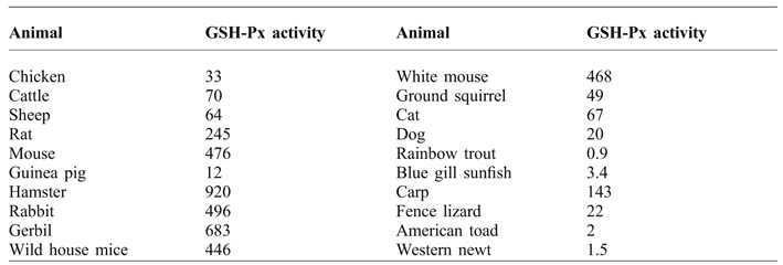
The earliest response in the specific activity of Se-dependent GSH-Px occurs in chicken plasma eight hours and in liver 24 hours after Se administration (Bunk and Combs, Reference BUNK and COMBS1980). Importantly, GSH-Px was found in the chicken egg proteome (Mann and Mann, Reference MANN and MANN2008). GSH-Px activity was shown to have species- and tissue-specificity. For example, GSH-Px in mouse leg muscle was shown to be almost 10-fold higher than that in the chicken muscle (Omaye and Tappel, Reference OMAYE and TAPPEL1974). A comprehensive study of GSH-Px in various tissues of different animals was conducted by Tappel et al. (1982; Table 2). In fact, the total GSH-Px activities found in the study of chicken liver, heart and lung were 33, 27 and 10 nmol NADPH oxidised/min/mg protein, respectively. GSH-Px activity in chicken, duck, turkey, ostrich and lamb muscles were measured (Daun and Akesson, Reference DAUN and AKESSON2004). It was shown that the activity of GSH-Px varied more than five-fold among muscles from different species. The highest activity, found in duck muscles, was significantly greater than that from other species. Moreover, lamb muscles had a significantly higher GSH-Px activity than chicken and turkey breast and ostrich fillet (Daun and Akesson, Reference DAUN and AKESSON2004). It is interesting to note that GSH-Px activity was shown to be 2.5-fold higher in duck embryo liver in comparison to chicken liver (Jin et al., Reference JIN, KENNEDY, DI MUCCIO and MOON2001) or 15-fold higher in the postnatal duck muscle in comparison to chicken muscle (Hoac et al., Reference HOAC, DAUN, TRAFIKOWSKA, ZACKRISSON and ÅKESSON2006). Furthermore, GSH-Px activity in chicken meat was almost two-fold lower than that in camel or cattle meat (Gheisari and Motamedi, Reference GHEISARI and MOTAMEDI2010). In the liver of emperor penguins, GSH-Px activities were up to three times higher than those in other avian species (Zenteno-Savin et al., Reference ZENTENO-SAVIN, ST LEGER and PONGANIS2010). GSH-Px activities in the liver of rat, chicken, lizard and frog were 36,6; 23.2; 14.3 and 9.2 µmol NADPH/min/g respectively (Venditti et al., Reference VENDITTI, DANIELE, BALESTRIERI and DI MEO1999). In comparison to rats, turkey is characterised by a 10-fold decrease in GSH-Px1 activity and increased (10-fold) GSH-Px4 activity in the liver (Sunde and Hadley, Reference SUNDE and HADLEY2010). In accordance with GSH-Px1 activity (IU/g protein) turkey tissues can be placed in the following descending order: kidney>>gizzard>heart>liver>>thigh muscle>>breast muscle (Sunde et al., Reference SUNDE, SUNDE, SUNDE, SUNDE and EVENSON2015). In turkey kidney, GSH-Px3 expression was low while GSH-Px1 expression was comparatively high (Sunde et al., Reference SUNDE, SUNDE, SUNDE, SUNDE and EVENSON2015). Whole blood GSH-Px in chicken reduced by age while the enzymatic activity was constant in the chicken liver at 2, 4 and 6 weeks of age (Chadio et al., Reference CHADIO, PAPPAS, PAPANASTASATOS, PANTELIA, DARDAMANI, FEGEROS and ZERVAS2015). Interestingly, there was a significant decrease in GSH-Px activity in the utero-vaginal junction of the laying hens between 40 and 60 weeks of age (Breque et al., Reference BREQUE, SURAI and BRILLARD2006).
Total GSH-Px activity in the liver of various animals, U/mg protein (Adapted from Tappel et al., Reference TAPPEL, CHAUDIERE and TAPPEL1982).

Studies of GSH-Px1 knockout mice led De Haan et al. (Reference DE HAAN, CRACK, FLENTJAR, IANNELLO, HERTZOG and KOLA2003) to conclude that GSH-Px1 functions as the primary protection against acute oxidative stress, particularly in stress conditions, where high levels of ROS occur. Surai (Reference SURAI2006) indicated that overexpression of GSH-Px is associated with an increased protection against oxidative stress created as a result of various environmental or nutritional manipulations. Indeed, GSH-Px is a well-regulated enzyme and its increased activity can be considered as an important protective mechanism in stress conditions. Most research related to GSH-Px activity in poultry has been related exclusively to GSH-Px1 and only a few studies measured GSH-Px4, while GSH-Px2 and GSH-Px3 data are mainly based on gene expression studies.
In newly hatched chickens the highest GSH-Px activity was found in the liver and kidney, with intermediate activity in the heart, lung and yolk sac membrane (YSM) and comparatively low GSH-Px activity was shown in muscles and brain (Surai et al., Reference SURAI, SPEAKE, NOBLE and SPARKS1999). In all the tissues, Se-dependent GSH-Px was the main enzymic form, comprising from 65% (lung) up to 90% (heart) of the total enzyme activity (Surai et al., Reference SURAI, SPEAKE, NOBLE and SPARKS1999). Similarly, in the chicken liver Se-dependent GSH-Px comprises about a half (48%) of total activity of the enzyme (Engberg et al., Reference ENGBERG, LAURIDSEN, JENSEN and JAKOBSEN1996). The specific activity of GSH-Px in embryonic liver increases continuously during the second half of the in vivo developmental period so that the activity at hatching was three times greater than that at embryonic day 10 (Surai, Reference SURAI1999). The most rapid increase in GSH-Px activity occurred between days 11 and 15 with a much more gradual increase thereafter. Interestingly, by the time of hatching, the specific activity of the enzyme in the liver was 6.1 times greater than that in the brain. GSH-Px activity in the prenatal normoxic lung demonstrated a sharp increase between day 16 and day 18 and remained constant until hatch (Starrs et al., Reference STARRS, ORGEIG, DANIELS, DAVIES and LOPATKO2001). According to GSH-Px activity, tissues of 35 day old chickens can be placed in the following descending order: liver>>kidney>plasma=erythrocytes>>femoral muscle>>pectoral muscle (Arai et al., Reference ARAI, SUGAWARA, SAKO, MOTOYOSHI, SHIMURA, TSUTSUI and KONNO1994).
GSH-Px has been found to be expressed in chicken seminal plasma and spermatozoa (Surai et al., Reference SURAI, BLESBOIS, GRASSEAU, GHALAH, BRILLARD, WISHART, CEROLINI and SPARKS1998a; Reference SURAI, CEROLINI, WISHART, SPEAKE, NOBLE and SPARKS1998b). There are species-specific differences in activity and distribution of GSH-Px in avian semen. For example, in seminal plasma, total GSH-Px activity was the highest in turkey and lowest in duck and goose (Surai et al., Reference SURAI, BLESBOIS, GRASSEAU, GHALAH, BRILLARD, WISHART, CEROLINI and SPARKS1998a). However, in spermatozoa the highest GSH-Px activities were found for goose and duck and lower GSH-Px activity was recorded for guinea fowl, turkey and chicken. In seminal plasma, the activity of GSH-Px was two times greater in the White Koluda ganders than in chickens (Partyka et al., Reference PARTYKA, LUKASZEWICZ and NIŻAŃSKI2012). A process of freezing and thawing fowl semen was associated with increased GSH-Px activity in the seminal plasma (Partyka et al., Reference PARTYKA, ŁUKASZEWICZ and NIŻAŃSKI2012a). It has been shown that despite a high proportion of PUFAs and a low level of vitamin E, duck spermatozoa have the same susceptibility to lipid peroxidation as chicken spermatozoa (Surai et al., Reference SURAI, BRILLARD, SPEAKE, BLESBOIS, SEIGNEURIN and SPARKS2000). It has been suggested that an increased activity of Se-GSH-Px in duck semen compensates for the relatively low concentrations of other antioxidants. If selenium is limited in the diet (which is the case in many countries in the world), then dietary supplementation of this trace element should have a beneficial effect on the antioxidant defence in various tissues including sperm. This was found in other studies where inclusion of Se in the cockerel diet significantly increased Se-GSH-Px activity in the liver, testes, spermatozoa and seminal plasma (Surai et al., Reference SURAI, KOSTJUK, WISHART, MACPHERSON, SPEAKE, NOBLE, IONOV and KUTZ1998c). As a result, a significant decrease in sperm and tissue susceptibility to lipid peroxidation was observed. It is important that an inducible form of the enzyme (Se-GSH-Px) represents more than 75% of the total enzymatic activity in chicken spermatozoa and more than 60% in the testes and liver of cockerels. In layers, increased GSH-Px activity in the utero-vaginal glands compared to other regions of the lower oviduct (vagina, uterus) could be related to a necessity of antioxidant defence during sperm storage in sperm-storage glands (Breque et al., Reference BREQUE, SURAI and BRILLARD2006).
Gastrointestinal glutathione peroxidase (GI-GSH-Px, GSH-Px2)
Gastrointestinal GSH-Px2 was first described in 1993 (Chu et al., Reference CHU, DOROSHOW and ESWORTHY1993) indicating that the enzymatic and physical properties of this enzyme are similar to those of cytosolic GSH-Px. In fact, the authors showed similar substrate specificities for GSH-Px1 and GSH-Px2. Furthermore, GSH-Px2 mRNA was readily detected in human liver and colon, and occasionally in human breast samples, but not in other human tissues including kidney, heart, lung, placenta, or uterus. However, in rodent tissues, GSH-Px2 mRNA was only detected in the gastrointestinal tract, and not in other tissues including liver (Chu et al., Reference CHU, DOROSHOW and ESWORTHY1993). In fact, GSH-Px2 appeared to be the major GSH-dependent peroxidase activity in rodent GI tract where at least three more selenoproteins including plasma GSH-Px, selenoprotein P and thioredoxin reductase (TR) are found (Mork et al., Reference MORK, LEX, SCHEURLEN, DREHER, SCHUTZE, KOHRLE and JAKOB1998). There are several important unique features of GSH-Px2. First of all, GSH-Px2 mRNA is comparatively stable in Se deficiency, ranking this enzyme high in the selenoprotein hierarchy. This indicates a vital importance of this enzyme in the intestine and probably in other tissues. In fact, Se deficiency was associated with increased expression of GSH-Px2 and decreased GSH-Px1 expression in chicken testes (Gao et al., 2017). Secondly, upon re-supplementation of Se after its deficiency, GSH-Px-2 is shown to be synthesized first confirming its priority position among selenoproteins. Thirdly, GSH-Px2 is exclusively located in the crypts of the intestine. Fourthly, in the gut a high expression of GSH-Px2 is related to Paneth cells which are responsible for secretion of antimicrobial defensins upon exposure to bacteria suggesting an important role of this enzyme in gut immunity (Banning et al., Reference BANNING, DEUBEL, KLUTH, ZHOU and BRIGELIUS-FLOHÉ2005).
The data on GSH-Px-2 clearly indicate that this enzyme should be considered as a major antioxidant defence in the intestine. GSH-Px2 knockout mice are shown to be viable and there is a great synergy between GSH-Px2 and GSH-Px1 in their participation in antioxidant defences. In fact, GSH-Px2 could be considered as an effective barrier against hydroperoxide absorption and an important regulator of gut inflammation (Flohe and Brigelius-Flohe, Reference FLOHE and BRIGELIUS-FLOHE2016). GSH-Px2 activity was shown to be detected in both the villus and crypt regions of rat mucosal epithelium and its activity nearly equalled that of GSH-Px1 throughout the small intestine and colorectal segments (Esworthy et al., Reference ESWORTHY, SWIDEREK, HO and CHU1998). It seems likely that induction of GSH-Px2 in other tissues could be an important part of the stress response, since its gene expression is regulated by the antioxidant response element (ARE) and the Keap1/Nrf2/ARE pathway is proven to regulate the gene expression of various enzymes, including antioxidant enzymes (Lubos et al., Reference LUBOS, LOSCALZO and HANDY2011). Indeed, it is well appreciated that activation of ARE is associated with the transcription of a number of antioxidant proteins, detoxifying enzymes and transport proteins. Therefore, GSH-Px-2 is considered to be an important oxidative stress-inducible cellular GSH-Px isoform and its basal and inducible expression is shown to be dependent on Nrf2 (Singh et al., Reference SINGH, RANGASAMY, THIMMULAPPA, LEE, OSBURN, BRIGELIUS-FLOHÉ, KENSLER, YAMAMOTO and BISWAL2006). In fact, by binding to the ARE in the upstream promoter region of genes encoding various antioxidant molecules, Nrf2 regulates the expression of hundreds of cytoprotective genes responsible for synthesis of a range of protective molecules involved in the maintenance and responsiveness of the cellular antioxidant systems (Surai, Reference SURAI2015). This includes enzymes from the first line of antioxidant defence (SOD, GSH-Px and CAT), detoxification enzymes (HO-1, NQO1, and GST), GSH-related proteins (γ-GCS), NADPH-producing enzymes and others stress-response proteins contributing to preventing oxidative and inflammatory damages. In fact, Nrf2 together with other transcription factors, such as NF-κB, orchestrate adaptive responses to diverse forms and levels of stress.
Plasma glutathione peroxidase (pGSH-Px, GSH-Px3)
GSH-Px3 from human plasma was purified to homogeneity by Takahashi et al. (Reference TAKAHASHI, AVISSAR, WHITIN and COHEN1987). This enzyme is a glycoprotein synthesised in the kidney. GSH-Px3 is extracellular enzyme found in blood plasma, eye fluid or amniotic fluid. Furthermore, Maddipati and Marnett (Reference MADDIPATI and MARNETT1987) showed that the human plasma GSH-Px3 is a tetramer of identical subunits of 21.5 kDa molecular mass. Furthermore GSH-Px3 is found to be a selenoprotein containing one selenium per subunit (Maddipati and Marnett, Reference MADDIPATI and MARNETT1987). The protein has a molecular weight of approximately 92,000 Da and containing four Se atoms per molecule (Cohen and Avissar, Reference COHEN and AVISSAR1993).
The biological role of this enzyme remains speculative, since plasma contains comparatively low concentrations of extracellular GSH or reduced thioredoxin. Indeed, GSH-Px3 is considered to have intermediate specificity to peroxides. It can reduce lipid hydroperoxides in LDL, however, it is not active against peroxidised cholesterol esters (Flohe and Brigelius-Flohe, Reference FLOHE and BRIGELIUS-FLOHE2016). In turkey, the highest expression of GSH-Px3 was shown to be in liver, heart and kidney (Sunde et al., Reference SUNDE, SUNDE, SUNDE, SUNDE and EVENSON2015), while transcripts for GSH-Px3 were highly expressed in chicken pectoral muscle at day 42 of age (Yao et al., Reference YAO, ZHAO, ZHAO, FAN, KHOSO, ZHANG, LIU and XU2014). Plasma GSH-Px3 activity in Se-deficient chicks was shown to decrease to 3% of Se-adequate levels (Li and Sunde, Reference LI and SUNDE2016). There are species-specific differences in GSH-Px3 expression. For example, high expression of GSH-Px3 transcript in chicken gizzard and pancreas was identified and these tissues were suggested to secrete and probably participate in regulation of this enzyme in chicken (Li and Sunde, Reference LI and SUNDE2016), while in mammals, kidney is the major source of plasma GPx3 (Flohe and Brigelius-Flohe, Reference FLOHE and BRIGELIUS-FLOHE2016). Indeed, GSH-Px3 is considered to be a redox buffer involved in a regulation of inflammatory reactions and its more detail characterisation in avian species is a priority for future research.
Phospholipid glutathione peroxidase (GSH-Px4, PH-GSH-Px)
In 1985 Ursini and co-workers reported that another form of GSH-Px, which used a phosphatidyl choline hydroperoxide as a substrate, was Se-dependent (Ursini et al., Reference URSINI, MAIORINO and GREGOLIN1985). They showed that the enzyme was a monomer 23 kDa in size. It contained one g-atom Se in the selenol form per 22,000 g protein. The kinetic data of GSH-Px4 action were compatible with a ‘ping-pong’ mechanism, as described for GSH-Px1. It was suggested that this enzyme was active at the interface of the membrane and the aqueous phase of the cell. In fact, GSH-Px4 is distinguished from classical GSH-Px as it is active in a monomeric form and has a different amino acid composition (Sunde, Reference SUNDE1993). There are three forms of GSH-Px4. It is synthesised in a long (L-form; 23 kDa) and a short form (S-form, 20 kDa) from mRNA that is transcribed from two initiation sites in exon Ia of GSH-Px4 genomic DNA (Imai and Nakagawa, Reference IMAI and NAKAGAWA2003). The S-form GSH-Px4 is non-mitochondrial GSH-Px4 and L-form GSH-Px4 is mitochondrial GSH-Px4. Recently, the third form of GSH-Px4, a 34 kDa selenoprotein, was detected in rat sperm nuclei and was termed ‘sperm nuclei GSH-Px’ (snGSH-Px). However, in chicken there is no snGSH-Px (Bertelsmann et al., Reference BERTELSMANN, KUEHBACHER, WESELOH, KYRIAKOPOULOS and BEHNE2007).
GSH-Px4 is unique in its capability of reducing ester lipid hydroperoxides incorporated in biomembranes or lipoproteins. It is well known that GSH-Px4 is widely expressed in normal tissue and is especially high in testes (Imai et al., Reference IMAI, SUMI, HANAMOTO, ARAI and SUGIYAMA1995), where it has an important role in spermatogenesis and sperm function. In these organs, relevant GSH-Px4 activity is strongly linked to mitochondria undergoing differentiation to spermatozoa. In testes mitochondria GSH-Px4 is electrostatically bound to the inner surfaces of the organelle (Roveri et al., Reference ROVERI, MAIORINO, NISII and URSINI1994). Interestingly, GSH-Px4 is found to be localised in the midpiece of spermatozoa in various species including Drosophila melanogaster, frog, fish, cock, mouse, rat, pig, bull, and human (Nayernia et al., Reference NAYERNIA, DIACONU, AUMULLER, WENNEMUTH, SCHWANDT, KLEENE, KUEHN and ENGEL2004). It is important to mention that GSH-Px4 mRNA expression in the male reproductive organs is under oestrogenic control (Nam et al., Reference NAM, BAEK, LEE, IN, JUNG, YON, AHN, KANG, YU and YUN2003). The most important discovery related to GSH-Px4 is its polymerisation and conversion from an active enzyme to the structural protein. Indeed, GSH-Px4 protein was identified as the major constituent of the keratin-like material that embeds the helix of mitochondria in the midpiece of mammalian spermatozoa (Ursini et al., Reference URSINI, HEIM, KIESS, MAIORINO, ROVERI, WISSING and FLOHÉ1999).
In 1991 chick liver GSH-Px activity was separated into three peaks by gel permeation chromatography (Miyazaki, Reference MIYAZAKI1991). The relative molecular weights and enzyme activities indicated that the first peak was Se-GSH-Px1 and the second peak was related to non-Se-GSH-Px. The third peak was the monomeric GSH-Px, later called GSH-Px4. The proportions of the GSH-Px1, non-Se-GSH-Px and GSH-Px4 activities to total liver GSH-Px activity were approximately 30%, 42% and 28%, respectively (Miyazaki and Motoi, Reference MIYAZAKI and MOTOI1992). In chick samples, total GSH-Px activity ranged from 15.3 nmol/min/mg in plasma to 118 nmol/min/mg in kidney. In all tissues except plasma GSH-Px activity was separated into three peaks, while in plasma, only one peak of GSH-Px1 was detected. In terms of percentage of total GSH-Px activity, Se-GSH-Px activity was high in plasma and erythrocytes, intermediate in testes, brain, kidney and liver, and low in duodenum. All the organs examined contained GSH-Px4 in different proportions. Specific GSH-Px4 activity was high in liver, duodenum and kidney, intermediate in testes and low in brain. The high GSH-Px4 activity in bird livers suggests that this enzyme is a major enzymatic system for reducing membrane lipid hydroperoxides in avian species. Se-GSH-Px was the main GSH-Px activity in rat liver while non-Se-GSH-Px was predominant in bovine liver. In avian livers, GSH-Px4 activity ranged from 10% of the total GSH-Px activity in Japanese quail to 28% in chicks. In terms of specific activity toward cumene hydroperoxide, GSH-Px4 activity of mammalian livers was below 6% of the activity of chick liver (Miyazaki and Motoi, Reference MIYAZAKI and MOTOI1992). Later, the same authors purified GSH-Px4 to homogeneity from a broiler chick liver cytosolic fraction using five different column chromatographic methods (Miyazaki and Motoi, Reference MIYAZAKI and MOTOI1996). The molecular weight of the purified enzyme determined by sodium dodecyl sulphate-polyacrylamide gel electrophoresis was 19,500. Therefore, it was suggested that the enzyme protein is a single polypeptide. The isoelectric point of the enzyme was determined to be 7.0 and the optimum pH for the enzyme reaction was 7.0. The purified enzyme catalysed the reduction of hydrogen peroxide, cumene hydroperoxide, tert-butyl hydroperoxide and linoleic acid hydroperoxide. By using an antiserum against the purified enzyme, it was shown that it reacted with the 19.5 kDa polypeptide in the liver cytosol of duck and quail suggesting presence of the enzyme in these avian species (Miyazaki and Motoi, Reference MIYAZAKI and MOTOI1996). GSH-Px4 has been shown to exist as both a 197 amino acid mitochondrial targeting protein and as a 170 amino acid non-mitochondrial protein (Kong et al., Reference KONG, KIM and FOSTER2003). The cDNA encoding the non-mitochondrial chicken GSH-Px (cGSH-Px4) was isolated from a chicken embryonic fibroblast cell line cDNA library. The nucleotide sequence of cGSH-Px4 was shown to be 802 bp in length with an open reading frame that encoded 170 amino acids but lacked the N-terminal domain that encoded the mitochondrial leader sequence. Chicken non-mitochondrial GSH-Px4 was highly expressed in brain and stromal tissues. Research showed that ovarian stromal tissue cGSH-Px4 expression is regulated according to the reproductive status of the bird and its steroid hormone status, suggesting that GSH-Px4 may play an important role in avian reproduction (Kong et al., Reference KONG, KIM and FOSTER2003). GSH-Px4 in avian species is shown to be sensitive to Se status. In fact, the liver had the highest GSH-Px activity in Se-adequate poults, and activity in Se-deficient liver decreased to 5% of the Se-adequate levels (Sunde and Hadley, Reference SUNDE and HADLEY2010). Based on GSH-Px4 activity turkey tissues can be placed in the following descending order: liver>heart>kidney>>gizzard>thigh muscle> breast muscle (Sunde et al., Reference SUNDE, SUNDE, SUNDE, SUNDE and EVENSON2015). It is interesting that liver GSH-Px4 mRNA levels can be down-regulated by excess of Se in chicken diet (Zoidis et al., Reference ZOIDIS, PAPPAS, GEORGIOU, KOMAITIS and FEGGEROS2010).
Se-independent GSH-Px
There are four Se-independent GSH-Px including GSH-Px5, GSH-Px6, GSH-Px7 and GSH-Px8 respectively (Flohe and Brigelius-Flohe, Reference FLOHE and BRIGELIUS-FLOHE2016). From those, human GSH-Px6 is shown to be selenoprotein expressed in olfactory epithelium and probably participating in antioxidant defences. Furthermore, GSH-Px5 is an extracellular Cys-containing GSH-Px located in the epididymis with suggestive regulatory roles in sperm physiology. GSH-Px7, is another Cys-containing GSH-Px, located at the luminal site of the endoplasmic reticulum, detoxifying H2O2 and participating in protein folding. Interestingly, the knockout of the Gpx7 gene is shown to be associated with multiple organ dysfunctions and shortened life span. Finally, GSH-Px8 is a Cys-containing GSH-Px located in the ER and participating in oxidative protein folding and insulin signalling (Flohe and Brigelius-Flohe, Reference FLOHE and BRIGELIUS-FLOHE2016). Indeed, non-Se GSH-Px in avian species awaits further investigations.
GSH-Px ranking
GSH-Px activity depends on Se provision in the diet. In an experiment, chicks produced from hens marginally deficient in Se and vitamin E were used (Kim and Combs, Reference KIM and COMBS1993). The hepatic activity of Se-GSH-Px was significantly greater in Se-adequate chicks than in Se-deficient ones which was about 20% of the control level. When an experiment of the same design was conducted using chicks produced from hens that had been depleted of Se and vitamin E for a longer period of time (nine months), the hepatic activity of Se-GSH-Px for treatment group of chicks was about one-fifth of the activity observed for the same dietary treatment in the previous experiment (Kim and Combs, Reference KIM and COMBS1993).
There are substantial differences among different forms of GSH-Px with regard to response to Se deficiency (Flohe and Brigelius-Flohe, Reference FLOHE and BRIGELIUS-FLOHE2016). The selenoproteins retained in tissues for longer periods during progressive Se deficiency are considered to have higher physiological significance in comparison to those whose activities rapidly decline. In this respect, the main GSH-Px forms rank as follows (Flohe and Brigelius-Flohe, Reference FLOHE and BRIGELIUS-FLOHE2016): GSH-Px2 >GSH-Px4 >GSH-Px3=GSH-Px1. However, GSH-Px ranking is likely species- and tissue-specific. For example, in chicken CNS the rank of GSH-Px is as follows GSH-Px3> GSH-Px4> GSH-Px2> GSH-Px1 (Jiang et al., Reference JIANG, CAO, LI, LI, ZHANG, LIN, LI and LI2017). Furthermore, liver and gizzard GSH-Px activities in Se-deficient chicks were shown to be only 2% and 5%, respectively, of Se-adequate birds (Li and Sunde, Reference LI and SUNDE2016), while GSH-Px4 activities in the same tissues in Se-deficient chicks comprised 10% and 5%, respectively, of the values for Se-adequate birds. At the same time, plasma GSH-Px3 activity in Se-deficient chicks was only 3% of Se-adequate levels (Li and Sunde, Reference LI and SUNDE2016). It seems likely that pancreas is more resistant to Se depletion, since GSH-Px1 and GSH-Px4 activities in Se-deficient chicks decreased to 39 and 25% of the physiological level (Li and Sunde, Reference LI and SUNDE2016). Similarly, in comparison to Se-adequate growing turkeys, Se deficiency has been shown to decrease plasma GSH-Px3, liver GSH-Px1 and liver GSH-Px4 activities to 2, 3, and 7%, respectively (Taylor and Sunde, Reference TAYLOR and SUNDE2016). In fact, recently it has been suggested that GSH-Px, TrxR1, SELP, and SPS2 to play a more important role than the other selenoproteins in poultry (Luan et al., Reference LUAN, ZHAO, YAO, ZHAO, FAN, ZHAO, ZHANG and XU2016). Interestingly, the mRNA levels of GSH-Px2, GSH-Px4 and GSH-Px3 were increased in chicken marrow due to low Se diet (0.028 mg/kg; Jiang et al., Reference JIANG, CAO, LI, LI, ZHANG, LIN, LI and LI2017). Similarly, in chicken kidney GSH-Px1, GSH-Px2 and GSH-Px4 were upregulated in Se deficiency (Zhang et al., Reference ZHANG, XU, HUANG, GAO, CHEN and SHAN2016).
Conclusions
The data clearly indicate that, in 44 years of research, Se-dependent GSH-Px has received substantial attention as a key player in the antioxidant defence system in animals. Indeed, GSH-Px, being an inducible enzyme, participates in poultry adaptations to stress conditions. It was shown that GSH-Px1 can modulate redox-dependent cellular responses and signalling by regulating mitochondrial function (Handy et al., Reference HANDY, LUBOS, YANG, GALBRAITH, KELLY, ZHANG, LEOPOLD and LOSCALZO2009). However, an oxygen-responsive element was identified in the GSH-Px gene (Cowan et al., Reference COWAN, WEISEL, WILLIAMS and MICKLE1993). Indeed, relationships between GSH-Px and various transcription factors deserves more attention. It seems likely that GSH-Px1 plays a vital role in regulating pro-inflammatory pathways, including mitogen-activated protein kinases (MAPK) and transcription factor nuclear factor-κB (NF-κB; Sharma et al., Reference SHARMA, YUEN, HUET, PICKERING, STEFANOVIC, BERNATCHEZ and DE HAAN2016). In particular, NF-κB was shown to be upregulated in the GPx1-/- mouse (Crack et al., Reference CRACK, TAYLOR, ALI, MANSELL and HERTZOG2006). Importantly, NF-κB is known to be a key regulator of cellular death and survival under oxidative stress conditions. The involvement of Nrf2 in basal and inducible expression of GSH-Px2 (Singh et al., Reference SINGH, RANGASAMY, THIMMULAPPA, LEE, OSBURN, BRIGELIUS-FLOHÉ, KENSLER, YAMAMOTO and BISWAL2006) and regulation of redox-sensitive genes by GSH-Px4 (Savaskan et al., Reference SAVASKAN, UFER, KÜHN and BORCHERT2007) warrants further investigation.
In poultry production, only two forms of Se-dependent GSH-Px (GSH-Px1 and GSH-Px4) have received substantial attention as important antioxidant status markers as well as indexes of Se status. Indeed, there is a need to expand research related to roles of GSH-Px2 and GSH-Px3 in poultry. Furthermore, modulation of GSH-Px activity in poultry by various factors will be considered in the second part of the review.
Acknowledgements
This work was supported by a grant of the Government of Russian Federation (Contract No. 14. W03.31.0013).