Introduction
Spermatogenesis is a complex, highly regulated and multistage process of germ cell proliferation and differentiation, starting from spermatogonia to spermatocytes and giving rise to spermatids, the future spermatozoa. It is well known that cell-to-cell interactions play an essential role in the regulation of mammalian spermatogenesis (for review see Jegou, Reference Jegou1992; Griswold, Reference Griswold1998) and that the connections between testicular cells may be mediated through paracrine pathways and direct intercellular communication channels made up of gap junctions and their consti-tutive proteins: the connexins (Cx, for review see Bruzzone et al., Reference Bruzzone, White and Paul1996; Kumar & Gilula, Reference Kumar and Gilula1996). Gap junctions are composed of aggregations of membrane channels, called connexons, joined to similar connexons in adjacent cells to form intercellular pathways for the diffusion of ions and small molecules (Beyer et al., Reference Beyer, Paul and Goodenough1990). Cx are expressed in a tissue-specific manner and various cell types express more than one Cx. Cx43 is the most abundant connexin identified in the mammalian testis and it is the only Cx expressed in Leydig cells (LC) to date (Risley et al., Reference Risley, Tan, Roy and Saez1992; Tan et al., Reference Tan, Roy, Saez, Saez, Paul and Risley1996; Batias et al., Reference Batias, Siffroi, Fenichel, Pointis and Segretain2000; You et al., Reference You, Li and Lin2000; Perez-Armendariz et al., Reference Perez-Armendariz, Lamoyi, Mason, Cisneros-Armas, Luu-The and Bravo Moreno2001). Several lines of evidence indicate that, in rodents, Cx43 is localized between adjacent Sertoli cells, Sertoli cells and germ cells and between LC (Risley et al., Reference Risley, Tan, Roy and Saez1992; Perez-Armendariz et al., Reference Perez-Armendariz, Romano, Luna, Miranda, Bennett and Moreno1994; Batias et al., Reference Batias, Siffroi, Fenichel, Pointis and Segretain2000; Roscoe et al., Reference Roscoe, Barr, Mhawi, Pomerantz and Kidder2001) and that it is essential for normal testicular function (Batias et al., Reference Batias, Defamie, Lablack, Thepot, Fenichel, Segretain and Pointis1999, Reference Batias, Siffroi, Fenichel, Pointis and Segretain2000; Juneja et al., Reference Juneja, Barr, Enders and Kidder1999; Plum et al., Reference Plum, Haalas, Magin, Dombowsky, Hagendoff, Shumacher, Wolpert, Kim, Lamers, Evert, Meda, Trau and Willecke2000; Roscoe et al., Reference Roscoe, Barr, Mhawi, Pomerantz and Kidder2001). In fact, Cx43 is highly expressed in rodent testis and decreases in impaired spermatogenesis showing a decrease in germ cell number (Batias et al., Reference Batias, Defamie, Lablack, Thepot, Fenichel, Segretain and Pointis1999).
Little information has been reported so far concerning the presence of Cx43 in the testis of non-mammalian vertebrates. Recently, we have demonstrated, at an immunocytochemical level, the presence of Cx43 protein in the testis of the frog, Rana esculenta (Palmiero et al., Reference Palmiero, Ferrara, De Rienzo, d'Istria and Minucci2003). Interestingly, Cx43 immunopositivity sharply decreased on day 4 after ethane 1,2-dimethane sulphonate (EDS, a drug with specific cytotoxic action on LC; see Palmiero et al., Reference Palmiero, Ferrara, De Rienzo, d'Istria and Minucci2003) injection, simultaneously with the loss of cellular adhesion between Sertoli cell and germ cells and the signal resumed on day 28 from EDS injection when a new population of LC reappeared in the interstitium.
In this study we report the partial cloning of a Cx43 transcript in the testis of the frog Rana esculenta, a seasonal breeder with a cyclic spermatogenesis and a cystic organization of the testis that favours the study of testicular activity (Rastogi & Iela, Reference Rastogi, Iela and Dallai1992). In addition, here we have analysed the tem-poral and spatial expression of Cx43 transcript and protein by semiquantitative RT-PCR, in situ hybridiza-tion and immunohistochemistry, during the annual spermatogenic cycle and after EDS treatment.
Material and methods
Animals
Adult male frogs, Rana esculenta, were collected monthly (n = 10/month) in the area surrounding Naples by a local dealer. Animals were maintained in plastic tanks (23 × 16 × 11 cm) with food and water available ad libitum. Frogs were killed by decapitation under anaesthesia (MS-222, Sigma); the testes were dissected and quickly frozen by immersion in liquid nitrogen for RNA (10 testes) and protein extraction (seven testes). In addition, three testes/month were rapidly immersed in Bouin's fixative for histological analysis.
This project was approved by the Italian Ministry of Education and Research (MIUR).
EDS treatment
Five frogs, used as an initial control, were immediately sacrificed; 25 animals were treated with a single injection of ethane 1,2-dimethane sulfonate (EDS, 100 mg/kg body wt in DMSO–dH2O 1:3 v/v, 100μl/dose) at day 0. The animals (five animals/group) were sacrificed at different time points: 1, 2, 3, 4 and 28 days after the injection.
Histology and immunocytochemistry
The testes were fixed by immersion in Bouin's fluid and were dehydrated in ethanol, cleared in xylene and embedded in paraffin. Tissue sections (5μm) were stained with haematoxylin–eosin for histological observations. Randomly chosen sections (five sections/animal) of the testis were processed by the peroxidase antiperoxidase technique (PAP). Testes sections were rinsed in phosphate-buffered saline (PBS 0.01 M at pH 7.1) and then were treated for 20 min with H2O2 to block endogenous peroxidase. Sections were incubated at 37 °C for 2 h with primary antiserum (polyclonal anticonnexin-43 raised in goat; Santa Cruz Biotechnology). The antiserum was diluted 1:50–1:400 in PBS containing 3% normal swine serum and 0.1% Tween 20. After washing in PBS, the sections were incubated for 1.5 h at room temperature with a secondary antiserum (goat anti-Ig; DAKO) according to the source of the primary antiserum, diluted 1:80 in the same buffer. Finally the sections were incubated for 1.5 h with goat PAP complex (DAKO) diluted 1:80 in PBS. The antigen was visualized using 3,3′-diaminobenzidine tetrahydrochloride (DAB; Sigma) and 0.3% H2O2 in Tris–HCl 50 mM at pH 7.6. To check the specificity of the immunoreaction, the controls either omitted one step of the reactions or used the primary antibody preabsorbed with an excess (10−6 M) of the cognate peptide (Santa Cruz).
RNA and first strand cDNA synthesis
Total RNA was isolated from R. esculenta testes according to Chomczynski and Sacchi (Chomczynski & Sacchi, Reference Chomczynski and Sacchi1987). The testes were homogenized in 10 ml lysis buffer (guanidinium thiocyanate 4 M, sodium citrate 25 mM pH 7.0, 0.5% sarcosyl and β2-mercaptoethanol 0.1 M) followed by extraction with phenol acid:chloroform:isoamyl alcohol (50:49:1) and precipitation with isopropanol. The resulting RNA pellet was dissolved in water.
Five μg of total RNA were reverse transcribed into cDNA using 40 ng random hexameric primers and 200 U Superscript III RT enzyme (Invitrogen) according to manufacturer's instruction.
PCR cloning and sequencing
Based on published sequences for Cx43 mRNA from different vertebrates, a pair of degenerate primers were designed (Cx43 forward: 5′-GBTTCTGGGTBCTDCAGATCAT-3′; Cx43 reverse: 5′-GGGGMCKDGAVAGGAARCAGTCCAC-3′) to amp-lify Cx43 cDNA from the Rana esculenta testes. PCR reactions (25μl volume) were performed in the presence of 0.4μg of cDNA, 0.3 U Taq polymerase and its buffer (1×), plus MgCl2 1.5 mM, dNTP 0.2 mM and 5 pmol of each primer. PCR amplification was carried out for 35 cycles of denaturation at 94 °C for 30 s, annealing at 58 °C for 30 s and extension at 72 °C for 45 s, followed by a final extension at 72 °C for 7 min. Amplification product was electrophoresed on 1.5% agarose gel and visualized with ethidium bromide staining and UV light. The expected DNA fragment (381 bp) was purified by QIAGEN gel extraction kit (QIAGEN) and cloned into the pGemT Easy Vector (Promega) according to manufacturer's instruction. Sequence determination was performed on both strands using the Sanger dideoxynucleotide chain termination method with [35S]-dATP as precursor. The deduced amino acid sequence (126 aa) was compared with the EMBL GenBank database and the protein sequence alignments were generated using the CLUSTALW Multiple Sequence Alignment Program (http://workbench.sdsc.edu).
PCR and quantification
The expression of frog connexin 43 (fCx43) mRNA levels was evaluated using a semiquantitative PCR system. A pair of specific primers (fCx43 up: 5′-GGTTCTGGGTGCTACAGATCA-3′; fCx43 down: 5′-CCACTTTATGAGGGCATGGA-3′) were designed to obtain a 363 bp fCx43 transcript by PCR reaction. An appropriate region (356 bp fragment) of frog ribosomal protein fP1 (EMBL databank accession no. AJ298875), amplified with specific oligonucleotide primers (fP1 for: 5′-TACGAGCGTCCATCACACAC-3′ fP1 rev: 5′-AGACCAAAGCCCATGTCATC-3′) was used as control. PCR amplification was carried out for 40 cycles with denaturing at 94 °C for 30 s, annealing at 58 °C for 30 s and extension at 72 °C for 45 s, followed by a final extension at 72 °C for 7 min. The expected RT-PCR products were separated by agarose gel (1.2%) electrophoresis and the quantization was performed using Scion Image.
Statistics
Data were expressed as means ± SD. Relative amount of fCx43/fP1 densitometric analysis was obtained using a Gel Doc 1000 Instrument (Biorad). Gaussian distribution of continuous variables was assessed by the Shapiro–Wilk normality test and statistical analysis was performed by the Student's t-test. Probability levels of less than 0.05 were considered statistically significant.
In situ hybridization
Frog Cx43 cDNA was cloned into the pGemT Easy Vector. A clone was linearized with either ApaI (antisense probe) or PstI (sense probe) to produce a template for in vitro transcription using digoxigenin–UTP (Roche Diagnostics) exactly as recommended by the manufacturer. Paraffin sections (5μm) were dewaxed in xylol twice for 5 min each time, rehydrated in graded concentrations of ethanol and rinsed in diethylpyrocarbonate (EDTA)-treated 1× PBS. The sections were fixed in 4% paraformaldehyde in 0.5 M NaCl, 0.1 M MOPS, pH 7.5, for 30 min at room temperature, then washed in 1× PBS. The slides were treated with 10μg/ml proteinase K in 100 mM Tris–HCl and 1 mM EDTA at pH 7.2 for 7 min at room temperature and rinsed in 1× PBS for 5 min, then transferred to 5× SSC twice for 2 min each time. After hybridization performed at room temperature for 30 min in Tris–glycine buffer at pH 7.2, the sections were hybridized in a humidified chamber overnight at 60 °C in a buffer containing 40% deionized formamide, 5× SSC, 1× Denhardt's solution, 100μg/ml sonicated salmon testes, 100μg/ml transfer RNA and 80 ng digoxigenin-labelled complementary RNA probe. After incubation, the slides were washed three times in 5× SSC for 20 min each and then in posthybridization buffer (0.5× SSC, 20% deionized formamide) for 40 min at 60 °C and incubated in NTE (0.5 M NaCl, 10 mM Tris–HCl, 5 mM EDTA, pH 7.0) containing 10μg/ml ribonuclease A for 30 min a 37 °C.
The slides were rinsed in NTE for 15 min at 37 °C, then washed in posthybridization buffer for 30 min at 60 °C and rinsed in 2× SSC for 30 min at room temperature. The sections were incubated in 1% blocking solution (Roche Diagnostics) in MBT buffer (0.1 M maleic acid, 0.15 M NaCl, pH 7.5) for 10 min before incubation overnight at 4 °C with an alkaline phosphatase-conjugated sheep antidigoxigenin antibody (Roche Diagnostics) diluted 1:2000 in MBT buffer. The slides were rinsed four times in Tris-buffered saline (TBS) pH 7.4 for 10 min each and then in solution B (0.1% Tween 20, 0.5 mg/ml levamisole) for 10 min. The colour detection substrate solution was applied (1 ml BM purple, Roche Diagnostics; 10μl of 100× solution B) and the incubation was carried out overnight in the dark at room temperature. The reaction was terminated by rinsing the sections in 1× PBS, 1 mM EDTA for 10 min at room temperature. The slides were dehydrated and mounted.
Results
Temporal and spatial expression of Cx43 in the frog testis
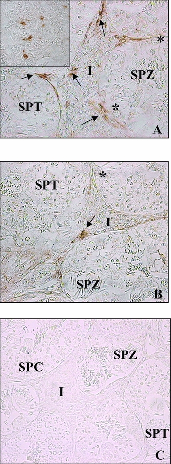
The temporal and spatial expression of Cx43 was immunocytochemically studied during the annual sexual cycle. Cx43 immunoreactivity was localized throughout the year principally between Leydig cells, but also between Sertoli cells and between the germ cells inside the cysts (Fig. 1A, insert). In particular, we report Cx43 localization in the testis of a September frog (Fig. 1A), when Cx43 protein is present at high levels and in the testis of a May frog (Fig. 1B), when Cx43 is present at low levels. No signal was detected in the immunoreaction control (Fig. 1C), obtained by using the primary antibody preabsorbed with excess amount of the cognate peptide of testes collected in September.
Immunohistochemical staining of September (A) and May (B) frog testes using anti-Cx43 antibody. (C), Control reaction. Positive staining is indicated by arrows; I, interstitium; SPC, spermatocytes; SPT, spermatids; SPZ, spermatozoa; *, primary spermatogonia.

Cloning of Cx43 cDNA from the frog testis
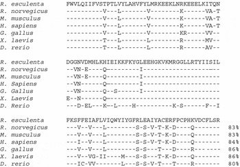
Two degenerate primers raised against the published sequences for Cx43 mRNA of different vertebrates were used to identify Cx43 cDNA in Rana esculenta testis. Subsequently, the cDNA product was cloned and sequenced. The insert was a partial cDNA fragment of 381 bp (fCx43, EMBL databank accession no. AM040672) lacking both start and stop codons (data not shown). The 126 aa sequence encoded from the open reading frame was compared with non-redundant GenBank entries and showed similarly to Cx43 from different species; in particular, the sequence homology was 86% with Gallus gallus and Xenopus laevis, 84% with Homo sapiens, 83% with Rattus norvegicus and Mus musculus and 80% with Danio rerio (Fig. 2).
Multiple alignment of the fCx43 deduced amino acid sequence of Rana esculenta with Rattus norvegicus, Mus musculus, Homo sapiens, Gallus gallus, Xenopus laevis and Danio rerio. Numbers indicate % identity.

Temporal and spatial expression of fCx43 mRNA from the frog testis
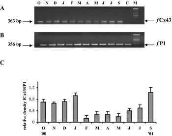
RT-PCR analyses were carried out on total RNA extracted from testes collected monthly during the annual cycle using specific oligonucleotides (fCx43 up and fCx43 down). The expected 363 bp fragment was detected in all the samples, showing high levels of expression from September to January (Fig. 3A) as compared to the levels observed from February to July (Fig. 3A). Quantification of the amount of cDNA in each lane was controlled using specific primers for the frog ribosomal protein fP1 (Fig. 3B). In Fig. 3C the ratio of relative densitometry of fCx43 transcript during the year is reported, normalized with the value of fP1 cDNA.
fCx43 expression in the testis of Rana esculenta during the annual sexual cycle from October 2000 to September 2001. (A, B) Agarose gel electrophoresis of RT-PCR products using fCx43- and fP1-specific primers, respectively. M, molecular weight marker XIV (Roche Diagnostic); C, control PCR. (C) Pattern of relative densitometric amounts, shown as Rana esculenta fCx43:fP1 ratios.

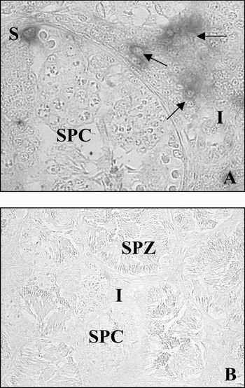
To examine the spatial localization of Cx43 RNA, an in situ hybridization using an antisense RNA probe against fCx43 cDNA, was carried out on September frog testis. The signal was detected particularly in the cytoplasm of interstitial LC and in the germinal compartment particularly in Sertoli cells (Fig. 4A). No signal was detected in sense RNA-probe-treated testis (Fig. 4B).
Localization of fCx43 mRNA by in situ hybridization on testes from October frogs using a fCx43 antisense probe (A) and a fCx43 sense probe (B) as the control reaction. Arrows, Leydig cells; I, interstitium; S, Sertoli cells; SPC, spermatocytes; SPZ, spermatozoa.

Expression of fCX43 in EDS-treated frog testis
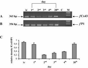
The expression of fCx43 mRNA was analysed in frog testes of collected in October and injected with DMSO (control animals) or with EDS-treated animals (1, 2, 3, 4 and 28 days after a single injection).
RT-PCR analyses carried out on total RNA extracted from EDS-treated animals showed that fCx43 transcript levels were progressively less intense from 1 to 4 days after injection than those observed in the control frogs (Fig. 5A). On day 28 after treatment, the level of transcript product was the same as in the control (Fig. 5A). The amount of cDNA in each lane was controlled using primers specific for fP1 (Fig. 5B). In Fig. 5C the relative densitometric ratio of R. esculenta fCx43 cDNA versus fP1 cDNA is reported.
fCx43 expression in the testis of Rana esculenta after EDS treatment. (A, B) Agarose gel electrophoresis of RT-PCR products using fCx43- and fP1-specific primers, respectively. ic, initial control; M, molecular weight marker XIV (Roche Diagnostic); C, control PCR. (C) Pattern of relative densitometric amount, shown as Rana esculenta fCx43:fP1 ratios.

Discussion
It is well known that testicular cell-to-cell interactions play a principal role in the regulation of mammalian spermatogenesis (Jegou, Reference Jegou1992; Griswold, Reference Griswold1998). In the testis, within the seminiferous tubules, signals between germ cell and germ cell and/or between germ cell and Sertoli cell are mediated through paracrine regulations, direct cell-to-cell interactions and communication via gap junctions. In addition, there is strong evidence that gap junctions are present within seminiferous tubules (Gilula et al., Reference Gilula, Fawcett and Aoki1976; McGinley et al., Reference McGinley, Posalaky and Provaznik1977; Nagano & Suzuky, Reference Nagano and Suzuky1980; Risley et al., Reference Risley, Tan, Roy and Saez1992; Pelletier, Reference Pelletier1995; Tan et al., Reference Tan, Roy, Saez, Saez, Paul and Risley1996; Batias et al., Reference Batias, Siffroi, Fenichel, Pointis and Segretain2000; You et al., Reference You, Li and Lin2000). In mammalian testis, gap-junctional communication between Sertoli cell and germ cell requires connexin channels for postnatal expansion of the male germ line (Roscoe et al., Reference Roscoe, Barr, Mhawi, Pomerantz and Kidder2001). To date, 12 different connexins mRNAs have been reported in the testis, communicating between the different cell types that constitute this organ (Kadle et al., Reference Kadle, Zhang and Nicholson1991; Haefliger et al., Reference Haefliger, Bruzzone, Jenkins, Gilbert, Copeland and Paul1992; Risley et al., Reference Risley, Tan, Roy and Saez1992, Reference Risley2000). However, Cx43 represent the most abundant connexin identified in mammalian testis and, to date, it is the only one expressed by the Leydig cells (LC) (Risley et al., Reference Risley, Tan, Roy and Saez1992; Tan et al., Reference Tan, Roy, Saez, Saez, Paul and Risley1996; Batias et al., Reference Batias, Siffroi, Fenichel, Pointis and Segretain2000; You et al., Reference You, Li and Lin2000) and its expression correlates well with stages of spermatogenesis (Risley et al., Reference Risley, Tan, Roy and Saez1992; Tan et al. Reference Tan, Roy, Saez, Saez, Paul and Risley1996; Batias et al., Reference Batias, Defamie, Lablack, Thepot, Fenichel, Segretain and Pointis1999). In mammals, Cx43 has been localized between neighbouring Sertoli cells, between Sertoli cells and spermatogonia and between early and late spermatocytes, but not between Sertoli cells and spermatids (Decrouy, Reference Decrouy, Gasc, Pointis and Segretain2004).
Little information has been reported so far concerning the presence of Cx in the testis of non-mammalian vertebrates. Recently, Batlouni et al. (2004) have identified gap junctions, composed of Cx32 dimers, between Sertoli cell and Sertoli cell, between Sertoli cell and germ cell and between germ cell and germ cell, in the testis of the catfish Pseudoplatystoma fasciatum. In addition, our previous work, using a polyclonal Cx43 antibody, highlighted, for the first time, the presence of Cx43 protein in the testis of the frog Rana esculenta collected throughout the year (Palmiero et al., Reference Palmiero, Ferrara, De Rienzo, d'Istria and Minucci2003). In particular, Cx43 immunoreactivity is present between LC in the interstitium, between Sertoli cells and primary spermatogonia and between Sertoli cells and germ cells in the tubules (Palmiero et al., Reference Palmiero, Ferrara, De Rienzo, d'Istria and Minucci2003). Interestingly, here we found that Cx43 expression is more abundant in the frog testis from September to January and, in addition, we highlighted a strong Cx43 immunopositivity also between the germ cells inside the cysts. In this context, it is to be remembered that in amphibians, Sertoli cells envelope germ cells creating the cysts in which germ cells differentiate synchronously (Minucci et al., Reference Minucci, De Rienzo, Sena, Cobellis, Meccariello, Pierantoni and Fasano2000; Palmiero et al., Reference Palmiero, Ferrara, De Rienzo, d'Istria and Minucci2003) Thus, it is not surprising that germ cells are interconnected through cytoplasmic bridges. In fact, it seems that germ cell to germ cell gap junctions may be involved in transmitting substances throughout all germ cells in the cyst, in order to co-ordinate mechanisms such as the synchronous development of the cells. In addition, in situ hybridization experiments confirm that the expression of Cx43, in frog testis, is mainly confined in LC and Sertoli cells revealing that Cx43 transcript is present in the cytoplasm of LC and Sertoli cells, while a very little signal is also visible in the cytoplasm of germ cells inside the cysts.
In the present paper we report also, and for the first time to our knowledge, isolation of a Cx43 cDNA fragment of 381 bp from Rana esculenta testis. Its deduced amino acid sequence shows high level of similarity to Cx43 homologues of other vertebrates, with homology values ranging from 80–86% (see Fig. 2) showing that the protein is highly conserved among vertebrates.
Rana esculenta is a seasonal breeder with an annual spermatogenic cycle regulated by endocrine and environmental factors (Rastogi et al., Reference Rastogi, Iela, Delrio, Meglio, Russo and Chieffi1978; Minucci et al., Reference Minucci, De Rienzo, Sena, Cobellis, Meccariello, Pierantoni and Fasano2000), for this reason we focused our interest on the temporal and spatial localization of both Cx43 protein and transcript in the frog testis during the reproductive cycle. Therefore, immunocytochemical observation of Cx43 protein performed on frog testicular sections collected throughout the year showed that Cx43 expression is stronger in the testis of frogs collected September to January, in particular among LC (Fig. 1A, B). RT-PCR analysis, carried out during the frog's annual cycle, demonstrated the presence of Cx43 mRNA with differing expression throughout the year according to the immunocytochemical results. In fact, Cx43 transcript is more abundant from September to January, while it is found at a low level from February to July (Fig. 3). It is of interest to remember that, in Rana esculenta, LC start their testosterone production exactly at this time (Varriale et al., Reference Varriale, Pierantoni, Matteo, Minucci, Fasano, D'Antonio and Chieffi1986), while spermatogenesis resumes from February onwards, showing the highest activity in late summer and early autumn (Minucci et al., Reference Minucci, Fasano and Pierantoni1992). In addition, it has to be highlighted that androgens are necessary for spermatogonial proliferation (February–May) and spermatid formation (September–November), therefore our data may indicate that a high level of expression of both Cx43 mRNA and protein are needed for LC communication in association with the increase of androgen production.
Therefore, to obtain more information concerning the expression of Cx43 in frog LC, we performed experiments using EDS, an alkylating agent that selectively destroys the majority of vertebrate LC (for review see Minucci et al., Reference Minucci, Fasano and Pierantoni1992); in Rana esculenta, LC are destroyed within four days from a single EDS injection and regenerate after 28 days (Minucci et al., Reference Minucci, De Rienzo, Sena, Cobellis, Meccariello, Pierantoni and Fasano2000; Palmiero et al., Reference Palmiero, Ferrara, De Rienzo, d'Istria and Minucci2003). Results coming from EDS treatment confirm that injection provokes LC disruption with consequent severe alterations of the testicular morphology supporting the existence of a Leydig cell–germinal cell communication with the role of maintaining spermatogenesis. In addition, RT-PCR analyses show a progressive decrease of Cx43 expression in EDS-treated testis from day 1 to day 4 after the injection, when the majority of LC are destroyed, while Cx43 expression resumes at normal levels on day 28 when a new population of LC reappear in the interstitium. Present data, obtained by the use of EDS, demonstrate that, in the frog testis, Cx43 is mainly expressed in LC. Interestingly, our data support the results from other studies that indicate that many testicular toxicants may vary the expression of Cx in the testis (Bruzzone et al., Reference Bruzzone, White and Paul1996), inducing gap junction disruption (Herve et al., Reference Herve, Pluciennik, Bastide, Cronier, Verrecchia, Malassine, Joffre and Deleze1996; Chun & Cheng, Reference Chun and Cheng2001); however, the precise mechanisms whereby these toxicants may exert their effects on intercellular junctions are still not entirely known.
In conclusion, in the present study from the testis of the frog Rana esculenta, we isolated a fragment of cDNA and identified a protein homologous to Cx43. The results showed that both are expressed throughout the frog's annual reproductive cycle. In particular, from September to January, we observed the maximum expression of Cx43 transcript and protein. Cx43 transcript is specifically localized in the cytoplasm of LC and Sertoli cells as well as in germ cells inside the cysts. While Cx43 protein is localized between LC, between Sertoli cells and germ cells and between germ cells inside the cysts. These last data strongly indicate that germ cells inside the cysts are not completely free; they are connected to each other and with the Sertoli cell envelope. Gap-junctional communication among LC in the interstitium, between Sertoli cell and germ cells and between germ cells inside the cyst require Cx43 channels, consequently they are probably involved with the synchronization of germ-cell maturation inside the cysts. Lastly, our data also indicate that high levels of expression of Cx43 are needed at the time when LC increase androgen production. Taken together these results indicate that Cx43 has an important role in frog spermatogenesis.
Acknowledgments
We thank Dr R.M. Scharpe (Edinburgh, UK) for the gift of EDS and Dr R. Colacino for revision of the manuscript. This work was supported by grants from MIUR ‘ex 40% PRIN’; ‘ex 60% Ricerca di Ateneo’ and from Regione Campania ‘L.R. n.5, 2003’.