

Introduction
During the early years of polar exploration, before the use of reliable motorised vehicles, dog sledging was an essential form of transportation and dog nutrition was key to dog performance. Malnourished, sick dogs on sledging journeys could lead to disastrous consequences. The British Antarctic expeditions of the early 20th century led by Robert Falcon Scott and Ernest Shackleton included in their provisions large quantities of Spratt’s dog cakes, a commercially produced dog food similar to hardtack or cabin bread. The diet of dogs in Antarctica did not consist exclusively of dog cakes and was supplemented by seal meat and blubber once on the ice and, in some instances, penguins, ponies, and other dogs. This article investigates the Spratt’s dog cakes used by early Antarctic expeditions. It begins with a history of Spratt’s Patent, Ltd., the use of their product in Antarctica, and the reasons why dog cakes were taken. It then moves into an examination of their composition and nutritional value employing destructive (electron microscopy and flash combustion) methods requiring minute amounts of material along with non-destructive spectroscopy (Raman microscopy, Fourier transform Raman, and Fourier-transform infrared (IR) spectroscopy). A comparison of the composition of a Spratt’s dog cake with a modern dog biscuit and published energy requirements of high-performance dogs aims to provide insight into the suitability of dog cakes used by early Antarctic explorers.
Spratt’s in Antarctica
The origin story of Spratt’s Patent, Ltd. recounts that sometime before 1860, American entrepreneur James Spratt observed sailors throwing hardtack to the dogs on the Liverpool docks and this gave him the idea of producing a dry biscuit dog food (Grier, Reference Grier2006, Reference Grier, Belasco and Horowitz2009; Worboys, Strange, & Pemberton, Reference Worboys, Strange and Pemberton2018). In about 1861, he set up a business selling dog biscuits that he marketed as “dog cakes” and stamped with a trademark “X”. He is often recognised as a pioneer in the invention of dry dog food (Grier, Reference Grier2006). However, this product had been around since at least the early 19th century. In 1828, The Sporting Magazine published a letter where the author reports having visited Mr. Smith’s dog biscuit manufactory near Maidenhead (UK), commenting that he was “surprised to find he has been for a long period manufacturing the enormous quantity of five tons a-week!” (The Sporting Magazine, 1828, p. 250). The use of ship biscuits for dog food also predated Spratt. Edward Mayhew’s Reference Mayhew1854 manual Dogs: Their Management advises feeding “favourites”, for example, non-working pet dogs, ship biscuits, or oatmeal (Mayhew, Reference Mayhew1854, p. 55). While it is clear that Spratt was not the inventor of the dog biscuit, his company came to be the leading dog food manufacturer in the United States of America and Britain in the 19th and early 20th centuries. The name of the business and its marketing suggested that its products were patented and although no patent or recipe has been found for dog cakes, Spratt’s did secure patents for some of its other products such as an “Improvement in food for cattle” (United States Patent Office, 1876). By the close of the 19th century, Spratt’s had diversified, producing food for other animals such as horses, fowl, and fish. Recognising a related market in animal maintenance, the company expanded its range to include kennels, poultry houses, feeding hoppers, dog chains, and collars.
The use of Spratt’s dog cakes is well documented in the diaries and reminiscences of many early Antarctic expedition members. On the British National Antarctic Expedition 1901–1904 (Discovery), Robert Falcon Scott took a supply of them in addition to dried Norwegian stockfish (Scott, Reference Scott1907, p. 36; Wilson, Reference Wilson and Savours1966, p. 219). Clarence Harold Hare, steward on the expedition, noted in his diary that for the sledging expedition to Cape Crozier, the dogs were given a “daily allowance of two dog biscuits and half a dried fish” (Hare, Reference Hare1902, 15 March). However, dog cakes were not taken on the Southern journey for the South Pole by Scott, Edward Wilson, and Ernest Shackleton and problems with spoilage of the dried fish contributed to the deaths of all 18 dogs. In his diary, Scott recorded with great misgivings his mistake: “Originally, I had intended to take ordinary dog-biscuits for our animals, but in an evil moment I was persuaded by one who had had great experience in dog-driving to take fish. Fish has been used continually in the north for feeding dogs, and the particular article which we ordered was the Norwegian stock-fish such as is split, dried, and exported from that country in great quantities for human food. There is no doubt about the excellent food-value of this fish, and in every way it seemed well adapted to our purpose; and yet it was this very fish that poisoned our poor animals.” (Scott, Reference Scott1907, p. 22). Recognising the irony of the situation with dogs on the Southern journey, expedition member Louis Bernacchi noted that as those dogs were dying, “piled up on the shore at winter quarters were considerable supplies of Spratt’s cod-liver oil cakes” (Bernacchi, Reference Bernacchi1938, p. 67). He added that expedition members from Shackleton’s Aurora found them 16 years later “still in perfect condition” (Bernacchi, Reference Bernacchi1938, p. 67). No doubt keeping in mind the issues with dog food during the Discovery expedition, Shackleton provisioned the British Antarctic Expedition 1907–1909 (Nimrod) with Spratt’s dog cakes. The company used Shackleton’s success in reaching the furthest south in its advertising, implying that their product played a role despite the fact that Shackleton decided to use four ponies and no dogs (Fig. 1).
Advertisement for Spratt’s Dog Cakes, c. 1909. Private collection.

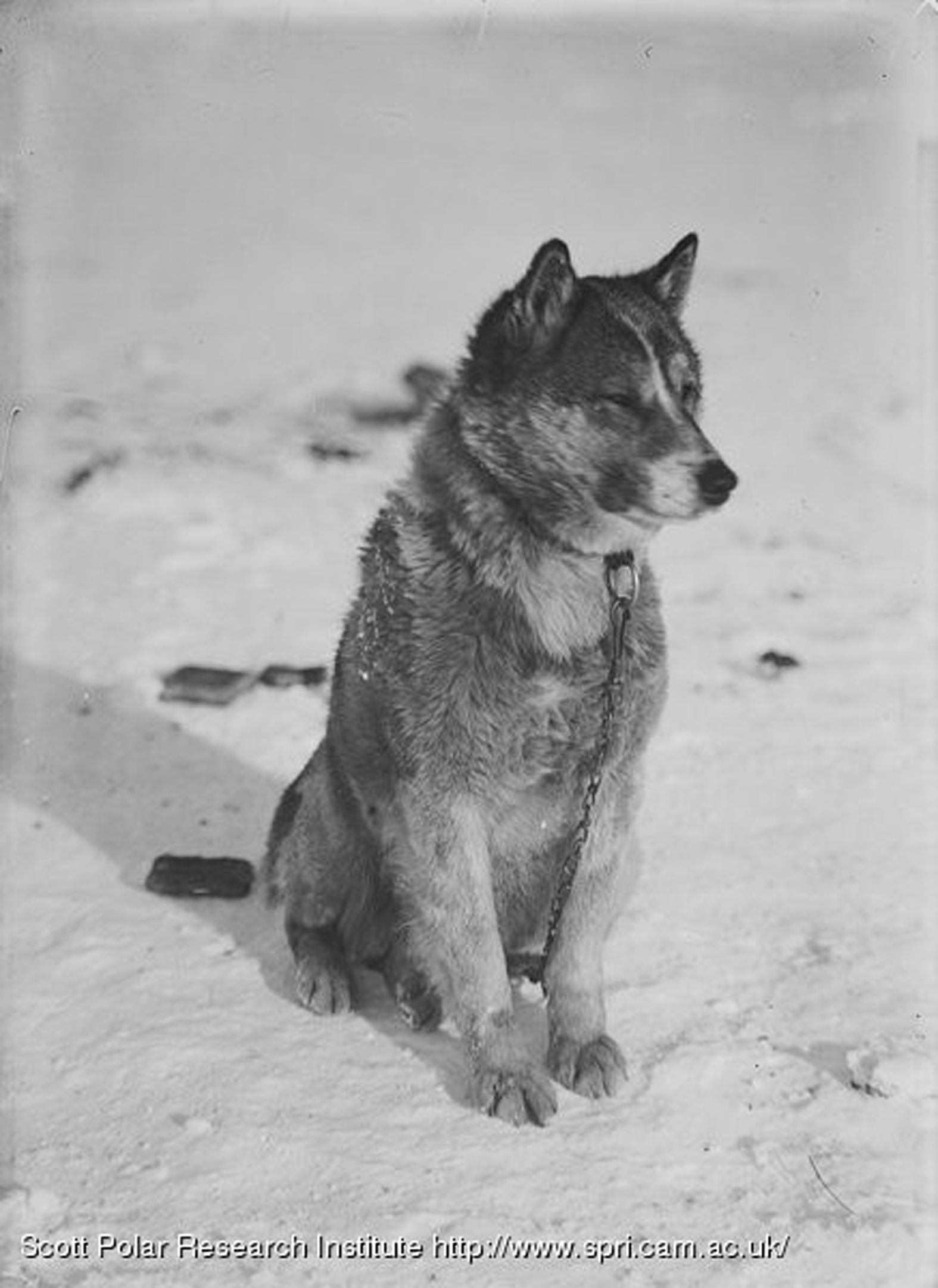
Scott learned from his mistake and instead of taking stockfish for the sledge dogs on the British Antarctic Expedition 1910–1913 (Terra Nova), he took large quantities of Spratt’s dog cakes. In New Zealand alone, wholesalers Mason, Struthers and Co. of Christchurch offered to supply “any quantity of Spratt’s dog biscuits that the expedition may require” (The Press, 1910, p. 10). According to Scott’s diary, about 5 tons were taken south (Scott, Reference Scott1936, p. 4). The expedition’s chief dog handler, Cecil Meares, indicated that although the dogs had been fed on dried salmon in Siberia, dog biscuits were the only food that they would be given in Antarctica (The New Zealand Times, 1910, p. 1). Scott disagreed with Meares’ decision to rely on them but he appears to have initially followed it (Scott, Reference Scott1936, p. 4). At the end of January, Scott thought that Meares would find that the two-thirds pound (0.30 kg) daily biscuit ration per dog was not enough (Scott, Reference Scott1936, p. 114) and in mid-February observed that even though the dogs were being fed “a good allowance of food”, they were “desperately hungry” and eating their excrement (Scott, Reference Scott1936, p. 139). A couple of days later, the dogs had not improved and he noted: “The dogs are as thin as rakes; they are ravenous and very tired. I feel this should not be, and that it is evident that they are underfed. The ration must be increased next year and we must have some properly thought out diet. The biscuit alone is not good enough. Meares is excellent to a point, but ignorant of the conditions here.” (Scott, Reference Scott1936, p. 145). As a result, the dogs’ diet was changed and seal meat added. Apsley Cherry-Garrard remembered that dog sledging rations were 1½ pounds (0.68 kg) of biscuit, occasionally supplemented by a seal-based pemmican for variety (but observed that both foods caused purgation), while at Cape Evans and Hut Point, dogs were given plenty of biscuits and frozen seal (Cherry-Garrard, Reference Cherry-Garrard1951, p. 452). In his 1933 documentary film 90 Degrees South, expedition photographer Herbert Ponting stated that at base, the dogs were fed on seal meat and biscuits on alternate days and observed that the dogs liked the meat days best. A photograph by Frank Debenham of Cresarovitza (also called Krisravitsa or Kris) suggests this and shows a dog seemingly disinterested in the several dog cakes behind him (Fig. 2).
Cresarovitza, October 1912. Scott Polar Research Institute P54/16/753b(1)z/b/16.

It is clear from these observations that Spratt’s biscuits alone were found to be an insufficient diet and they were subsequently supplemented with seal. With the Imperial Trans-Antarctic Expedition 1914–1917 (Endurance), the disadvantage of Spratt’s dog cakes as an exclusive food for dogs was acknowledged and the dogs were fed a diverse diet. Expedition member Frank Hurley noted that each dog received “on one day a pound of seal meat and half a pound of blubber, the next, a pound and a half of dog biscuits and the third a pound of pemmican which, being designed for sledging rations, is very concentrated. This routine was altered to suit the circumstances and the condition of the dogs.” (Hurley, Reference Hurley1925, p. 167).
Why Spratt’s dog cakes?
There were a number of reasons why early British Antarctic expeditions used Spratt’s dog cakes. First and foremost, they were an option that had practical advantages over other forms of dog food. Their uniform size and relatively lightweight compared with meat meant they were easy to transport on ships and sledges. They did not perish, unlike the bundles of dried fish on the Discovery expedition that were found to be “green in the centre,” possibly spoiled from the damp conditions of the ship when passing through the tropics (Armitage, Reference Armitage1905, pp. 210–11). And unlike seal that required preparation such as hunting, killing, and butchering, dog cakes could be fed straight from the packing crate. They were a reliable food that could be cached at depots without concern.
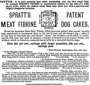
However, the use of dog cakes went beyond these simple practical considerations. The concern was not just about feeding dogs, but feeding them well. When the Discovery dogs began to fail, Scott advised future Antarctic explorers that they should “safeguard their dogs as surely as they do their men” (Scott, Reference Scott1907, p. 37). During the late 19th century, commercially produced dog food was seen as an advanced form of animal nutrition, superior to the practice of feeding dogs table scraps or letting them scavenge for themselves (Grier, Reference Grier, Belasco and Horowitz2009). Food made specifically for dogs was a concept that was readily embraced by middle-class dog owners who increasingly saw their canines as pets with their own special needs and had the discretionary income to spend on them. This attention to dog nutrition also reflected the affective, companionate relationships with dogs that developed in the 19th century (Chez, Reference Chez2017). Spratt’s took advantage of this shift in attitude and the desire of owners to feed and care for their dogs in new ways and marketed their products accordingly. They asserted that dog cakes gave dogs “all the elements for endurance, bone, muscle, and vigorous health”, benefits that caring owners would want for their pets (Stonehenge, 1872, p. 287). Spratt’s dog cakes, advertisements claimed, were free from salt and made from carefully selected ingredients (Fig. 3).
British Veterinary Journal, 1876.

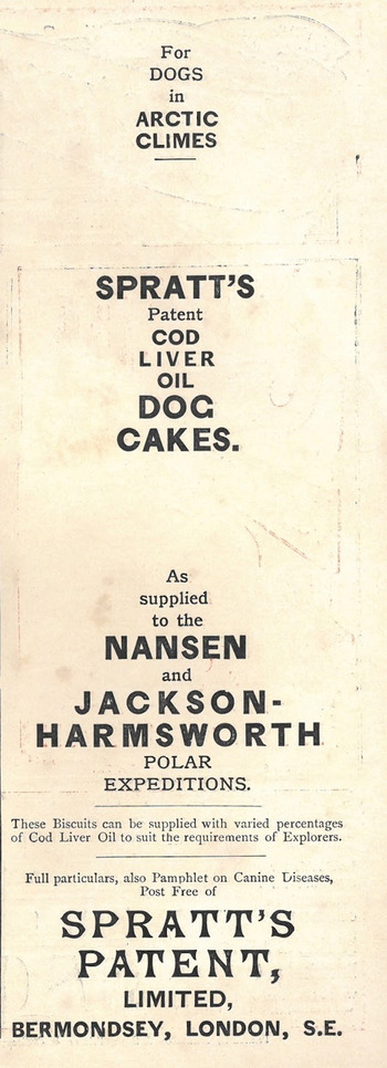
Such ingredients included not only meat but also fibrine (fibrous protein), dates, and vegetables such as beetroot, which Spratt’s promoted as having “nutrient” and “anti-scorbutic” (anti-scurvy) qualities (Spratt’s, c. 1880; Stonehenge, 1872, p. 287). The idea of a scientific dog food would have been of interest to expedition planners who were already concerned with the nutritional requirements of expedition members. Spratt’s strengthened this association of their product with science by including in their marketing endorsements by dog health experts such as Charles Rotherham, Queen Victoria’s canine veterinarian (Worboys et al., Reference Worboys, Strange and Pemberton2018). Scott and Shackleton might also have been swayed by the use of Spratt’s dog cakes in the Arctic. According to an advertisement in the 1898 serialised version of Fridtjof Nansen’s book Farthest North, Spratt’s Patent Cod Liver Oil Dog Cakes were used on the Nansen and Jackson-Harmsworth Arctic polar expeditions (Rauner Special Collection Library, 2016). The advertisement stated that the percentage of cod liver oil would be adjusted to suit the requirements of explorers, suggesting that this particular ingredient influenced sledge dog nutrition (Fig. 4).
Rauner Special Collections Library, Dartmouth Collection, Stef G700 1893.N22 (“Spratt’s Patent Cod Liver Oil Dog Cakes,” 2016).

This option to adjust the amount of cod liver oil in dog cakes reflected a move towards developing breed-specific nutrition by the company. By 1872, Spratt’s was experimenting with meat proportions for different varieties of dogs and by the 1880s had begun developing formulas for different breeds and life stages (Grier, Reference Grier, Belasco and Horowitz2009; Spratt’s, c. 1880; Stonehenge, 1872). However, much of this was motivated by commercial gain rather than dog welfare. Charles Cruft, founder of the Crufts dog show, worked for Spratt’s and led the company’s promotional activities; his connections with breeders provided Spratt’s with new breed-specific markets to tap (Worboys et al., Reference Worboys, Strange and Pemberton2018). Dog cakes that were once sold as food for any and all canines were now specialised for greyhounds, puppies, and with an adjustment to the amount of cod liver oil, sledge dogs. In addition to food, Spratt’s showed a wider interest in products aimed at dog health. In 1877, the company opened a hospital for dogs (Worboys et al., Reference Worboys, Strange and Pemberton2018), and in 1911, dogs destined for Douglas Mawson’s Australasian Antarctic Expedition 1911–1914 were quarantined there (Riffenburgh, Reference Riffenburgh2014). Spratt’s also sold a number of medications for dogs, ranging from cough and purging pills to liniment for sprains and rheumatism to a complete medical chest (Spratt’s, c. 1880). One of these chests, now in the collection of Canterbury Museum in Christchurch, New Zealand, was taken on the Discovery expedition.
We have taken the opportunity to examine the recovered Spratt’s dog biscuit using non-destructive analytical methods to complement the historical insight behind the item. We chose Raman and IR spectroscopy because these are complementary techniques that inform on the bonding in molecules, through fundamental vibrational modes, and thus on the composition of complex samples (Fraser-Miller, Saarinen, & Strachan, Reference Fraser-Miller, Saarinen and Strachan2016; Gordon & Fraser-Miller, Reference Gordon and Fraser-Miller2016; Ozaki & Šašic, Reference Ozaki and Šašic2008). IR spectroscopy reveals the nature of molecules through the absorption of IR photons, with energies associated with vibrations that exhibit a change in dipole moment (e.g. O–H bonds). The wavenumber (energy of the photons) is related to the type of bond and atoms involved – as such it gives a fingerprint for the particular molecules present (such as those in bone) (Boskey & Camacho, Reference Boskey and Camacho2007). Raman spectroscopy is an inelastic scattering method that also informs on bonds and molecules, but is active for vibrations that exhibit a change in polarisability (e.g. C = C bonds) modes (Gordon & Fraser-Miller, Reference Gordon and Fraser-Miller2016). While Raman and IR spectroscopy both look at fundamental vibrational transitions, the vibrations that are observed are complementary and give a rounded picture of the constituents within a sample.
Raman and IR spectroscopic methods have been used to non-destructively examine historical items for the purpose of identification, conservation, and authentication (Derrick, Stulik, & Landry, Reference Derrick, Stulik and Landry2000; Edwards & Chalmers, Reference Edwards and Chalmers2005; Kavkler & Demšar, Reference Kavkler and Demšar2011; Lowe, Smith, Fraser-Miller, Paterson, Daroux, Ngarimu-Cameron, Ford, & Gordon, Reference Lowe, Smith, Fraser-Miller, Paterson, Daroux, Ngarimu-Cameron, Ford and Gordon2017; Samanali, Paasi, Lowe, Smith, Fraser-Miller & Gordon, Reference Samanali, Paasi, Lowe, Smith, Fraser-Miller and Gordon2020; Smith & Clark, Reference Smith and Clark2001). Raman microscopy has been particularly insightful for the identification of pigments and inks (Buzzini & Suzuki, Reference Buzzini and Suzuki2016; Smith & Clark, Reference Smith and Clark2001). Whereas, IR is sensitive in the assessment of cellulosic materials such as historical paper and textiles (Lowe et al., Reference Lowe, Smith, Fraser-Miller, Paterson, Daroux, Ngarimu-Cameron, Ford and Gordon2017; Trafela, Strlič, Kolar, Lichtblau, Anders, Mencigar, & Pihlar, Reference Trafela, Strlič, Kolar, Lichtblau, Anders, Mencigar and Pihlar2007). The use of these techniques to examine historical foods (for animals or humans) has not been previously reported. However, these techniques have been applied to modern foodstuffs for authentication and product quality control purposes (Lohumi, Lee, Lee, & Cho, Reference Lohumi, Lee, Lee and Cho2015; O’Donnell et al., Reference O’Donnell, Fagan and Cullen2014).
In addition to non-destructive methods, micro-calorific (flash combustion) and scanning electron microscopy (SEM) were also undertaken. Microscopy and calorific analysis may aid the identification of possible excipients of the dog cakes and also provide an estimation of the daily energy provided to dogs used by early Antarctic expeditions. While these techniques are destructive, resulting in the complete mineralisation or physical degradation respectively of samples, they only require small amounts of material (in the order of a few milligrams) for analysis.
Materials and methods
Materials
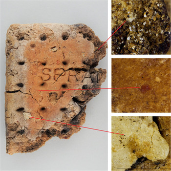
Canterbury Museum in Christchurch, New Zealand, holds four Spratt’s dog cakes: three complete cakes (accessions 1982.147.2, A182.24 and 19XX.4.344) and one partial cake (accession UD2018.5.331). Two of the dog cakes were recovered from Ross Island, Antarctica (probably Hut Point) before 1982. The other two dog cakes, including the partial one, have no provenance information but are part of the museum’s Antarctic collection, indicating that they had Antarctic provenance when they were acquired. They are believed to have been supplied for Robert Falcon Scott’s and/or Ernest Shackleton’s expeditions to Antarctica, making them more than 100 years old. The partial dog cake was analysed as a whole using Raman spectroscopy. Particles shed from it were collected and analysed by IR spectroscopy, light microscopy, SEM, and flash combustion for elemental (carbon, hydrogen, nitrogen, and sulphur) and micro-calorific content. These particles were selected according to their appearance and grouped as “bone”, “matrix” (the main bulk material of the dog cake), and “meat”. For Raman spectroscopy, the regions studied were believed to represent the various components of the sample, as shown in Figure 5. The dimensions and weight of the partial and three complete dog cakes were also measured.
Photograph of the partial dog cake studied with the measurement locations labelled. Left – partial dog cake, top right – interior matric, middle right – meat, bottom left – bone. Scale 0.5 mm.

A series of four model dog cakes (and individual excipients) made using material sourced from a local supermarket, as well as a commercially available dog biscuit (TUX® Adult Original Biscuit, Nestlé Purina PetCare Ltd., New Zealand) were compared with the partial dog cake from Antarctica using Raman and IR spectral analysis. These materials were analysed alongside the partial dog cakes to provide reference spectra from samples of known composition and processing. Reference spectra from Spratt’s dog cake excipients would have been preferred, however, no patent or recipe has been found for dog cakes.
The model dog cake recipes were (M1) 25g plain white flour, 5 mg baking soda, 5 mg salt, and 18 ml cold water; (M2) with the addition of 250 mg dripping; (M3) with the addition of 4 g mutton, and; (M4) with the addition of 250 mg dripping and 4 g mutton. The model dog cakes were prepared and baked as follows. The dripping and flour were rubbed together to form a fine consistent crumb, then the baking soda and salt were added and mixed. Water was added a little at a time and the mixture kneaded to a soft pliable dough. The dough was placed on a lightly floured surface and rolled to approximately 1 cm in thickness and cut into rectangles. The surfaces were lightly pricked with a fork, put on a baking tray, and then placed in a preheated oven (Blue Seal Turbofan 32max convection oven, Blue Seal Ltd., UK) at 190 °C. After 25 min, the model dog cakes were removed from the oven to cool on the bench.
Additional materials were sourced from a local supermarket and brewing supplies company (malted grains) for comparison of spectra. These include vegetable shortening, wheat flour, rolled oats, polenta, rice flour, potato flour, quinoa, tapioca flour, malted barley (AA), malted barley (WC), malted oat, malted wheat, and malted rye. The polenta, quinoa, and all malted materials were processed with additional gentle grinding prior to analysis.
Methods
Light microscopy
Images of the partial dog cake and dislodged particles were recorded using Nikon NIS Elements software and a Nikon DS-Ri1 camera attached to a Nikon AZ100M stereomicroscope.
Scanning electron microscopy
Images of particles that had dislodged from the partial dog cake were recorded using a JEOL JSM 7000F field emission, high-resolution scanning electron microscope.
Raman microscopy
Raman microscopic measurements were collected using an Alpha 300 R confocal Raman microscope equipped with 785 and 532 nm lasers (WITec, Ulm, Germany). Spectra were collected using the 785 nm laser at 5 mW power and a 10× objective to give a sample spot of 2 µm diameter. Spectra were collected using Control Four software (WITec, Ulm, Germany) with 0.5 s integration time and 100–1000 co-additions.
FT-Raman
Spectra were collected using a MultiRam FT-Raman spectrometer (Bruker Optic, Ettlingen, Germany) equipped with a liquid N2 cooled Ge detector. The sample was measured with a downward-looking objective and the defocused aperture setting gave a sample spot size of 1–2 mm. Spectra were collected using Opus 7.5 (Bruker Optic, Ettlingen, Germany) with 4 cm−1 resolution. Dislodged particles from the partial dog cake that appeared to be bone fragments were measured with 150 mW power and the bulk cake with 80 mW power with 1000 co-added scans per spectrum. The model dog cakes were measured in triplicate for each portion with 80 mW laser power and 200 co-added scans per spectrum.
FTIR
FTIR measurements were performed on a dry air purged Vertex 70 FTIR spectrometer (Bruker Optics, Ettlingen, Germany) fitted with a GladiATR diamond attenuated total reflectance (ATR) accessory (Pike Technologies, Madison, WI, USA). Spectra were acquired using OPUS 7.5 (Bruker Optics, Ettlingen, Germany). A background was acquired before each sample spectrum. Samples were measured in triplicate with each spectrum consisting of 128 or 256 co-added scans at a 4 cm−1 spectral resolution between 4000 and 400 cm−1.
Multivariate data analysis
Principal component analysis (PCA) with full cross-validation was carried out on the FT-IR and FT-Raman spectra after spectral preprocessing. Rubber band baseline correction (carried out using Orange canvas v.3.25 (Demšar et al., Reference Demšar, Curk, Erjavec, Gorup, Hočevar, Milutinovič and Štajdohar2013)) followed by standard normal variate (SNV) transformation were carried out over the spectral windows 370–1800 and 2650–3200 cm−1 for the FT-Raman spectra. Linear baseline correction and SNV transformation was carried out over the spectral region 400–1800 cm−1 for the FTIR data. SNV and PCA were carried out using the Unscrambler X 10.3 (Camo Analytics, Oslo, Norway).
Elemental composition and micro-calorific analysis
Dislodged particles from the partial dog cake less than 1 mm in size were collected and separated according to appearance corresponding to possibly “meat”, “bone”, and “matrix”. Samples were analysed for elemental (carbon, hydrogen, nitrogen, and sulphur) composition and also gross heating value (GHV) and net heating value (NHV) by the Campbell Microanalytical Laboratory (University of Otago, New Zealand) using flash combustion. Analyses were performed using a Thermo Flash 2000 Elemental Analyzer (Thermo Fisher Scientific, Bremen, Germany).
Results and discussion
Detailed results presenting PCA and flash combustion for elemental (carbon, hydrogen, nitrogen, and sulphur) and micro-calorific content are available in the Supplemental Material. A summary of the findings is discussed here.
Measurements, light microscopy, and scanning electron microscopy
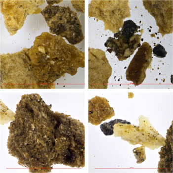
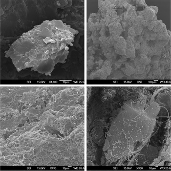
The three complete dog cakes had an average weight of 144.2 g with average lengths, widths, and heights of 107 × 99 × 25 mm, respectively. The partial dog cake was 25 mm high and had no obvious intact length or width (Fig. 5) and weighed 57.7 g. Particles shed from the partial dog cake were easily classified by appearance when examined by light microscopy (Fig. 6). The appearance of particles shed from the partial dog cake examined by SEM was consistent with their designated descriptions of “bone”, “matrix”, and “meat”. Interestingly, contamination of the partial dog cake by mites was discovered (Fig. 7). How these mites came to be present in the sample has not been determined.
Photograph of the partial dog cake crumbs. From top left clockwise: matrix with bone fragments (large lower item), meat (central dark item), bone (central item), matrix. Scale 0.5 mm.

SEM of partial dog cake crumbs. From top left clockwise: bone fragment, matrix, mite, meat.

Spectroscopic assessment
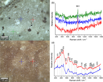
Raman microscopy was used to selectively interrogate specific domains on the dog cake (Fig. 8). Spectra collected from the white fleck designated as likely to be bone were consistent with that of bone, albeit with a poor signal to noise ratio resulting in only the ν1 PO4 3− band at 961 cm−1 being distinguished from the noise (Morris & Mandair, Reference Morris and Mandair2011; Penel, Leroy, Rey, & Bres, Reference Penel, Leroy, Rey and Bres1998; Thomas, Fordyce, Frew, & Gordon, Reference Thomas, Fordyce, Frew and Gordon2007), see Figure 8(a) and 8(b). The spectra collected from the partial dog cake bulk matrix had a higher contribution of fluorescence than the white fleck. As such, after baseline correction, curvature of the spectra is still observed, Figure 8(c) and 8(d). The bands at 479 and 1125 cm−1 are not inconsistent with a starch such as wheat flour (Kizil, Irudayaraj, & Seetharaman, Reference Kizil, Irudayaraj and Seetharaman2002).
(a) Bright-field image of the white spot under the 10× objective with spectral acquisition points marked and (b) the associated spectra collected from the marked locations after linear baseline correction. (c) Bright-field image of the bulk matrix under the 10× objective with spectral acquisition points marked and (d) the associated spectra collected from the marked locations after linear baseline correction.

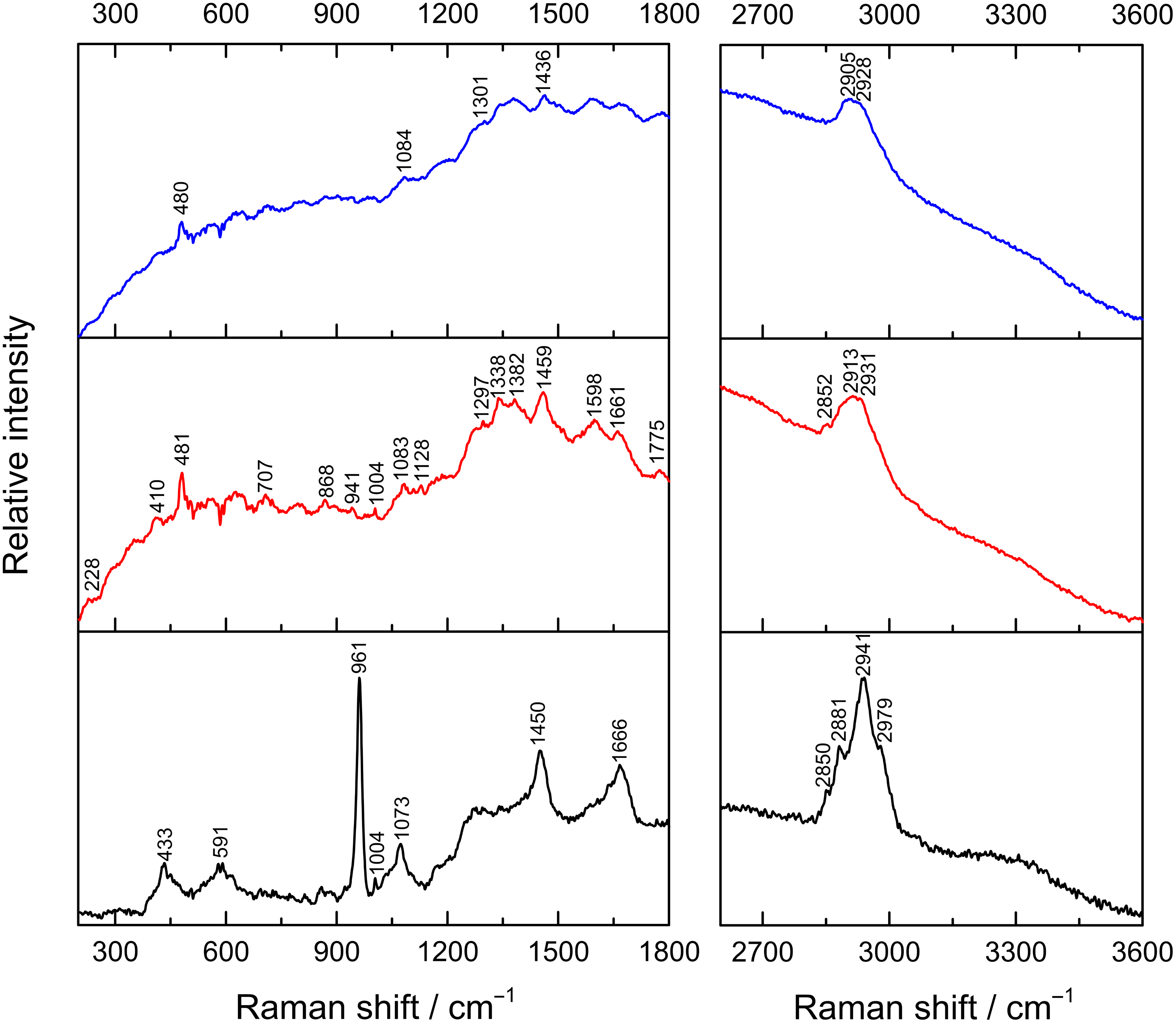
FT-Raman spectra collected from different sites on the partial dog cake indicate different constituents (Fig. 9). Spectra collected from the white fleck (Fig. 8(a)) are consistent to that observed with bone (Morris & Mandair, Reference Morris and Mandair2011; Penel et al., Reference Penel, Leroy, Rey and Bres1998; Thomas et al., Reference Thomas, Fordyce, Frew and Gordon2007). The FT-Raman spectra collected from the bulk matrix have an emissive background which partially obstructs the Raman signature, however, some features were observed that are not inconsistent with starches such as wheat flour (Kizil, Irudayaraj, & Seetharaman, Reference Kizil, Irudayaraj and Seetharaman2002). This appears to be the dominant constituent of the sample based on the Raman spectra. The exact starch source is not identified as the differences between flour types are minor (small changes in relative intensities of peaks) and would require multivariate analysis methods and reference flours. Spectral features could be attributed to blood; however, it is uncertain as other expected spectral features are not observed (Atkins, Buckley, Blades, & Turner, Reference Atkins, Buckley, Blades and Turner2017; Ozaki, Mizuno, Sato, Kawauchi, & Muraishi, Reference Ozaki, Mizuno, Sato, Kawauchi and Muraishi1992). This may suggest the presence of blood is in levels too low to clearly detect in the presence of other components with higher concentrations or that this is absent in the partial dog cake. Butter (Uysal, Boyaci, Genis, & Tamer, Reference Uysal, Boyaci, Genis and Tamer2013) and beetroot carotenoids (de Oliveira, Castro, Edwards, & de Oliveira, Reference de Oliveira, Castro, Edwards and de Oliveira2010) could not be identified in these spectra, but this could be due to the dominance of the starch signals masking other components. It is also possible that the carotenoids have degraded with time due to their reactive nature.
FT-Raman spectra collected from three sites on the partial dog cake. The spectra were collected from “bone” (black), surface “matrix” (red), and interior “matrix” (blue).

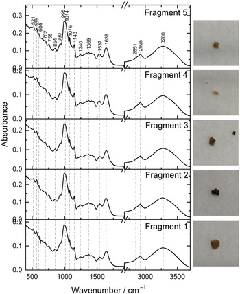
The FTIR spectra collected from loose fragments of partial dog cake, thought to originate from the bulk or interior portion of the cake, were fairly uniform between the fragments, as shown in Figure 10. The spectral features observed are consistent with polysaccharides (Kizil et al., Reference Kizil, Irudayaraj and Seetharaman2002; Wiercigroch et al., Reference Wiercigroch, Szafraniec, Czamara, Pacia, Majzner, Kochan and Malek2017). However, the subtle differences observed between different flours and starches means that multivariate analysis methods such as PCA, are required to help identify the starch present in this dog cake.
FTIR spectra collected from each of the five fragments of dog cake.

PCA was employed to compare the partial dog cake spectra to the model dog cakes and a commercially available dog biscuit (Tux) to help identify its contents. The PCA of the Raman data suggests the presence of bone fragments within the dog cake and a composition more similar to the commercial Tux biscuits than the model dog cakes (see supplementary information for detailed analysis and Fig. S1).
The IR spectra from the partial dog cake were compared using PCA to the model dog cakes, a commercial Tux biscuit and reference flour and grains. All the partial dog cake spectra clustered tightly amongst the model dog cakes and flour samples indicating the IR data was dominated with information associated with the starch component of the sample (see supplementary information for detailed analysis and Fig. S2).
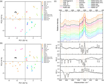
PCA was repeated using the spectra collected from only the matrix area of the partial dog cake and the reference flours and grains to try to gain further insight into the differences between the historical, model, and modern-day dog biscuits (Fig. 11). Some grains and flours included in this analysis, such as quinoa and tapioca, are not expected to have been readily available in England at the time the partial dog cake was manufactured.
PCA of the FTIR spectra collected from partial dog cake samples and a set of different flours and starches. (a) PC1 versus PC2 scores, (b) PC1 versus PC3 scores, and (c) the associated loadings and exemplar spectra.

PC1 provided 52 % of the explained spectral variance and separated based on features at 980, 920 (COH and CH bending, COC stretching in glycosidic linkages), and 860 cm−1 (COC stretching, ring breathing, amylopectin, and amylose) (Wiercigroch et al., Reference Wiercigroch, Szafraniec, Czamara, Pacia, Majzner, Kochan and Malek2017) into PC1 space and features at 1640 (C = O stretching, amide I) and 1540 cm−1 (CN stretching and NH bending, amide II) (Kong & Yu, Reference Kong and Yu2007) in negative PC1 space. This can be described as separating based on the relative abundance of the carbohydrate (polysaccharides) (positive PC1) versus protein (negative PC1) content. The partial dog cake is clustering in negative PC1 space (higher relative protein content) along with rolled oats and wheat samples. PC2 appears to separate some minor differences in the baseline curvature from the dominant contents of the flour signals (lipid, protein, and carbohydrate in nature). The dominant separating features in PC3 are associated with subtle differences in the carbohydrates within the flour and cereals. The positioning of the partial dog cake samples in PC1 versus PC2 scores space (Fig. 11(a) and 11(b)) could indicate it contains a mixture of wheat and oat flours or that the flour/cereal used is spectrally similar to a mixture of the two (Fig. 11(c)).
The IR spectra from the particles from the partial dog cake do not exhibit any lipidic features observed in many reference grains including rolled oats and wheat flour and the relative abundance of protein compared to carbohydrate signals is greater than that of the majority of flours measured here. It is plausible that the absence of the lipidic feature is associated with lipid degradation (Doblado-Maldonado, Pike, Sweley, & Rose, Reference Doblado-Maldonado, Pike, Sweley and Rose2012) within the flour/cereal. Differences in reference grains compared with the partial dog cake could be associated with changes over time due to selective breeding of grains for specific traits (e.g. breeding for increased gluten protein content in wheat) over the last 100 years, different processing of the grains, or the degradation of different components in the dog cakes occurring at different rates over time.
Elemental composition and micro-calorific analysis
The elemental analysis of the carbon, hydrogen, and nitrogen percent content of the particles of the partial dog cake designated “matrix”, “bone”, and “meat” are consistent with those reported for food waste (Zhang et al., Reference Zhang, Arnold, Paavola, Vaz, Neiva Correia, Cavinato and Heaven2013)) and meat and bone (Chaala & Roy, Reference Chaala and Roy2003). It is not surprising the carbon content of particles from the partial dog cake thought to be bone has a much lower carbon content than the matrix and meat particles since the bone has significant calcium and therefore skews the proportion of carbon to a low content.
The gross energy (GE) of the dry matter of cow feeds has been reported as ranging from 16203 kJ/kg/kg for low GE alfalfa silage to 21520 kJ/kg for high GE distiller’s grains (Weiss & Tebbe, Reference Weiss and Tebbe2018). The matrix and meat net (dry) heating values (21227 and 20467 kJ/kg respectively) of the partial dog cake appear similar to that of a high-energy grain. The possible addition of some meat and/or bone along with the likely carbohydrate components of wheat and oats (as identified by spectroscopic analysis) perhaps explains this similarity of energy content. Interestingly, modern crackers contain 21353 kJ/kg (USDA National Nutrient Database for Standard Reference, 2016)
During heavy exertion, sledge dog metabolic energy (ME) intake has been reported to be 44600 KJ/d in the form of a feed containing ME of 21057.8 kJ/kg (Hinchcliff, Reinhart, Burr, Schreier, & Swenson, Reference Hinchcliff, Reinhart, Burr, Schreier and Swenson1997). This was provided using a mixed dry ration of a proprietary racing diet (chicken by-product meal, chicken, chicken fat, corn, fish meal, lamb, corn starch, rice flour, animal fat, beet pulp, vegetable oil, chicken liver meal, fish oil, and glucose polymers) and a commercial dog food (chicken by-product meal, ground corn, rice flour, chicken, and chicken fat). These dogs weighed an average of 23 kg and consumed an average of 2.2 kg of food/day with a daily energy expenditure of 47100 (kJ/d). The GE energy content of 71 commercial dry dog foods averaged 24660 kJ/kg and the digestible energy (DE) content 86% (i.e. 21310 kJ/kg) (Hervera, Castrillo, Albanell, & Baucells, Reference Hervera, Castrillo, Albanell and Baucells2012). Therefore, the DE content of the partial dog cake might be approximately 21000 × 0.86 = 18060 kJ/kg. With an energy expenditure of 47100 kJ/d (assuming an average dog weight of 23 kg). an Antarctic expedition dog would need to have been fed 2.6 kg/d of dog cakes. However, this estimate is based on the possible DE content of the dog cake, which is greater than the ME content. If the DE content of dog cakes converts to the ME in a similar ratio as for an animal able to extract energy from food in a highly efficient manner (such as the cow), then ME might be only 82% of DE (Weiss & Tebbe, Reference Weiss and Tebbe2018), but it is likely this conversion is lower for the dog. The daily dog cake intake required may have therefore been in the order of more than 3.2 kg, or 1.45 times the amount of a modern commercial dry dog food.
While it was reported by expedition members that the dogs preferred fresh seal meat over dog cakes, the energy available from foods traditionally fed to sledge dogs from animal sources (being wet) is far lower than a dry food. For example, whale contains 4640 kJ/kg (0% fat), walrus 11800 kJ/kg (24.1% fat), seal 5960 kJ/kg (3.2% fat), and cod 3430 kJ/kg (0.6% fat) (Gerth, Redman, Speakman, Jackson, & Starck, Reference Gerth, Redman, Speakman, Jackson and Starck2010). It is likely that a traditional dog food diet would have been less suitable than dog cakes for providing the daily energy requirements, especially as they will have experienced exceptionally heavy energy demands through work. While Scott’s and Shackleton’s expeditions each used a low moisture, high-energy content feed in the form of pemmican, it was only used occasionally as a supplement or every three days (Cherry-Garrard, Reference Cherry-Garrard1951; Hurley, Reference Hurley1925). However, after 1930, “Bovril” dog pemmican became the basis of sledging rations of British expeditions (Taylor, Reference Taylor1957) at 1–2 lb per day (James, Reference James1947; Orr, Reference Orr1965, Orr, Reference Orr1966; Reece, Reference Reece1954). With an energy content of 21000–23000 kJ/kg (Lewis, Masterton & Ward, Reference Lewis, Masterton and Ward1957; Taylor, Reference Taylor1957), Bovril dog pemmican compares very favourably in term of energy content with a modern high-performance dog food (21000 kJ/kg) such as that reported by Hinchcliff, et al., Reference Hinchcliff, Reinhart, Burr, Schreier and Swenson1997. The dog cake, while not comparable with a modern dog food in terms of energy content, might still have been a technological advance on a traditional dog diet.
Conclusions
Despite Spratt’s claims that dog cakes were a complete food for dogs, anecdotal evidence from the early British Antarctic expeditions suggests they weren’t. Scott had suspected this at the beginning of the Terra Nova expedition and questioned Meares’ decision to feed the dogs exclusively on dog cakes. Subsequently, he monitored the dogs’ health and made the decision to supplement their diet with seal. During the Endurance expedition, dog cakes, seal meat, and blubber as well as pemmican were fed to them. Given the limited understanding of dog nutrition during the 19th century, especially the requirements of working dogs, it is not surprising that there was a degree of experimentation with the dogs’ diet.
The analysis of the partial Spratt’s dog cake from Canterbury Museum’s collection indicates that its contents (oat, wheat, and bone) are similar to modern commercially made dog biscuits. This suggests that the issue that the early British Antarctic expeditions encountered using dog cakes exclusively was not because they were an incomplete food. Instead, the issue was that an insufficient number of calories were being fed to the dogs through the dog cake rations. The additions of seal meat and pemmican simply contributed variety or some additional calories to the dogs’ diet. To provide sufficient energy, the daily intake of dog cakes would need to have been in the order of more than 3.2 kg, but the dogs were only being fed around 0.5 kg. The dog cake, while not up to a modern dog food in terms of energy content, was still a technological advance on a less energy-dense traditional dog food.
Acknowledgements
Weiyi Liu, Department of Agricultural Sciences, LincolnUniversity, Lincoln, New Zealand, providing SEM analysis.
Supplementary material
To view supplementary material for this article, please visit https://doi.org/10.1017/S0032247421000103.