INTRODUCTION
Scuticociliatosis, an emergent systemic disease caused by histiophagous ciliates, leads to high mortality in intensively cultured marine flatfish (Yoshinaga and Nakazoe, Reference Yoshinaga and Nakazoe1993; Dyková and Figueras, Reference Dyková and Figueras1994; Sterud et al. Reference Sterud, Hansen and Mo2000; Iglesias et al. Reference Iglesias, Paramá, Álvarez, Leiro, Fernández and Sanmartín2001; Kim et al. Reference Kim, Cho, Kim, Nam and Kim2004). Philasterides dicentrarchi (Ciliophora, Scuticociliatia) is a causative agent of scuticociliatosis in the farmed turbot Scophthalmus maximus in Galicia (NW Spain) (Iglesias et al. Reference Iglesias, Paramá, Álvarez, Leiro, Fernández and Sanmartín2001). Possible strategies for the control of turbot scuticociliatosis necessarily include erradication from the external environment and destruction of the endoparasitic stage of the parasite (Leiro et al. Reference Leiro, Arranz, Paramá, Álvarez and Sanmartín2004). The elimination of scuticociliates from water is feasible, via the application of chemical disinfectants such as formalin and hydrogen peroxide (Iglesias et al. Reference Iglesias, Paramá, Álvarez, Leiro and Sanmartín2002; Paramá et al. Reference Paramá, Iglesias, Álvarez, Leiro, Aja and Sanmartín2003; Budiño et al. Reference Budiño, Pata, Leiro and Lamas2012; Harikrishnan et al. Reference Harikrishnan, Balasundaram and Heo2010; Jin et al. Reference Jin, Harikrishnan, Moon, Kim, Kim, Balasundaram and Heo2010); however, at present there are no drugs available to treat systemic infections in fish. There are also numerous legal restrictions regarding the use of drugs in fish in relation to food safety and environmental impacts (Hedley and Huntington, Reference Hedley and Huntington2009). There is currently increasing interest in the use of natural phytochemical antioxidants to prevent pathologies derived from oxidative stress (Young and Woodside, Reference Young and Woodside2001). The natural polyphenol resveratrol (RESV) is known to exert antiparasitic effects in vitro against P. dicentrarchi (Leiro et al. Reference Leiro, Arranz, Paramá, Álvarez and Sanmartín2004), an effect that is at least partly related to alterations in mitochondrial electron transport in the parasite (Lamas et al. Reference Lamas, Morais, Arranz, Sanmartín, Orallo and Leiro2009; Morais et al. Reference Morais, Lamas, Sanmartín, Orallo and Leiro2009, Reference Morais, Piazzon, Lamas, Mallo and Leiro2013). Antioxidants are also widely used as additives in the food industry to delay, retard and prevent oxidative deterioration (Rathore et al. Reference Rathore, Suthar, Pareek and Gupta2011). Propyl gallate (PG) is the n-propyl ester of gallic acid (3,4,5-trihydroxybenzoic acid). This phenolic antioxidant is generally recognized as safe (GRAS) and has been extensively used in food since 1948 to protect fats, oils and fat-containing food from the rancidity resulting from the formation of peroxides. Use of PG for human consumption as a food additive (code E310) is approved in Europe (International Journal of Toxicology, 2007; Garrido et al. Reference Garrido, Garrido and Borges2012). PG is a well-known inhibitor of lipoxygenase, a cyanide-resistant dioxygenase found in most plant tissues (Parrish and Leopold, Reference Parrish and Leopold1978; Peterman and Siedow, Reference Peterman and Siedow1983), and it is also an inhibitor of alternative oxidase (AOX) (Siedow and Girvin, Reference Siedow and Girvin1980; Costa et al. Reference Costa, Mota, Cambursano, Lauxman, Nogueira de Oliveira, Silva Lima, Orellano and Fernandes de Melo2010; Sircar et al. Reference Sircar, Cardoso, Mukherjee, Mitra and Arnholdt-Schmitt2012). AOX is a mitochondrial enzyme involved in the alternative respiratory pathway (AP), which occurs in mitochondria of higher plants and is characterized by cyanide-resistant oxygen consumption (in contrast to the conventional pathway (CP), which utilizes CN-sensitive cytochrome oxidase) (Genevois, Reference Genevois1929; Henry and Nyns, Reference Henry and Nyns1975). AOX catalyses cyanide- and antimycin-A (AMA)-resistant oxidation of ubiquinol and the reduction of oxygen to water, thus short-circuiting the mitochondrial electron-transport chain prior to proton translocation by complexes III and IV and thereby dramatically reducing ATP formation (Moore et al. Reference Moore, Shiba, Young, Harada, Kita and Ito2013). In addition to being present in plants, AOX is also present in fungi (Lambowitz et al. Reference Lambowitz, Sabourin, Bertrand, Nickels and McIntosh1989; Sakajo et al. Reference Sakajo, Minagawa, Komiyama and Yoshimoto1991, Reference Sakajo, Minagawa and Yoshimoto1997; Jarmuszkiewicz Reference Jarmuszkiewicz2001), in several protists such as the amoebozoans Acanthamoeba castellanii and Dictyostelium discoideum (Jarmuszkiewicz et al. Reference Jarmuszkiewicz, Wagner, Wagner and Hryniewiecka1997, Reference Jarmuszkiewicz, Sluse-Goffart, Hryniewiecka and Sluse1999, Reference Jarmuszkiewicz, Behrendt, Navet and Sluse2002; Czarna et al. Reference Czarna, Mathy, MacCord, Dobson, Jarmuszkiewicz, Sluse-Goffart, Leprince, De Pauw and Sluse2010; Woyda-Ploszczyca et al. Reference Woyda-Ploszczyca, Koziel, Antos-Krzeminska and Jarmuskiewicz2011), in the heterokont Blastocystis hominis (Williams et al. Reference Williams, Elliot, Burri, Kido, Kita, Moore and Keeling2010) and in the trypanosome Trypanosoma brucei (Clarkson et al. Reference Clarkson, Bienen, Pollakis and Grady1989; Chaudhuri et al. Reference Chaudhuri, Ott and Hill2006). In the alveolates, it is found in the apicomplexan Cryptosporidium, Plasmodium falciparum and some other distantly related alveolates, including some ciliates (Young Reference Young1983; Murphy et al. Reference Murphy, Doeller, Hearn and Lang-Unnasch1997; Roberts et al. Reference Roberts, Roberts, Henriquez, Akiyoshi, Samuel, Richards, Milhous, Kyle, McIntosh, Hill, Chaudhuri, Tzipori and McLeod2004; Eisen et al. Reference Eisen, Coyne, Wu, Wu, Thiagarajan, Wortman, Badger, Ren, Amedeo, Jones, Tallon, Delcher, Salzberg, Silva, Haas, Majoros, Farzad, Carlton, Smith, Garg, Pearlman, Karrer, Sun, Manning, Elde, Turkewitz, Asai, Wilkes, Wang, Cai, Collins, Stewart, Lee, Wilamowska, Weinberg, Ruzzo, Wloga, Gaertig, Frankel, Tsao, Gorovsky, Keeling, Waller, Patron, Cherry, Stover, Krieger, del Toro, Ryder, Williamson, Barbeau, Hamilton and Orias2006; Seeber et al. Reference Seeber, Limenitakis and Soldati-Favre2008). The presence of AOX in some parasites has been interpreted as a metabolic adaptation to their host; for example, the form of the protozoan parasite T. brucei that is infective to mammals only uses the AP, but the procyclic form, which resides in the insect gut, utilizes both the AP and CP (Walker et al. Reference Walker, Saha, Hill and Chaudhuri2005). It has also been hypothesized that AOX is used in antioxygen defence by transporting to oxygen the excess of reducing equivalents (Purvis and Shewfelt, Reference Purvis and Shewfelt1993).
In previous studies, we have shown that mitochondrial respiration of scuticociliate P. dicentrarchi is resistant to cyanide, thus providing functional evidence for the presence of a cyanide-insensitive oxidase (Mallo et al. Reference Mallo, Lamas and Leiro2013a ). The oxidase reacts with antibodies raised against plant AOX and, furthermore, inhibitors of this enzyme such as salicylhydroxamic acid (SHAM) significantly inhibit in vitro growth of the ciliate (Morais et al. Reference Morais, Piazzon, Lamas, Mallo and Leiro2013; Mallo et al. Reference Mallo, Lamas and Leiro2013a ). Cyanide-insensitive trypanosome AOX has been targeted for the development of anti-trypanosomal drugs because it does not exist in the host (Nihei et al. Reference Nihei, Fukai and Kita2002). As turbot do not possess AOX, AP inhibitors could be used as a novel anti-scuticociliate therapy (Mallo et al. Reference Mallo, Lamas and Leiro2013a ).
The main aims of the present study were to investigate the antiparasitic effects of RESV, and AOX inhibitors, such as PG and also to elucidate the effect of RESV on AOX activity. We evaluated the potential in vitro activity of PG on AP respiration by ciliates under different conditions of oxygen availability. We also analysed the effects of the AOX inhibitors on the induction of parasite mitochondrial dysfunction, which may be a useful target for developing antiparasitic drugs.
MATERIALS AND METHODS
Ciliate isolation and culture
Specimens of P. dicentrarchi (isolate I1; Budiño et al. Reference Budiño, Lamas, Pata, Arranz, Sanmartín and Leiro2011) were collected under aseptic conditions from ascitic fluid removed from the intraperitoneal cavity of experimentally infected turbot, as previously described (Paramá et al. Reference Paramá, Iglesias, Álvarez, Leiro, Aja and Sanmartín2003). The ciliates were cultured at 21 °C in complete sterile L-15 medium, as previously described (Iglesias et al. Reference Iglesias, Paramá, Álvarez, Leiro, Ubeira and Sanmartín2003). To ensure normoxic conditions, the ciliates were cultured in tissue culture flasks fitted with vented caps that allowed aeration of the culture medium. To ensure hypoxic conditions, L-15 culture medium was bubbled with argon (to displace gases, including oxygen, dissolved in the medium) for 10 min before adding the ciliates and sealing the flasks (Mallo et al. Reference Mallo, Lamas and Leiro2013a ).
Oxygen uptake
Oxygen uptake was measured polarographically with a Clark electrode (Hansatech Instruments, UK), as previously described (Morais et al. Reference Morais, Lamas, Sanmartín, Orallo and Leiro2009; Mallo et al. Reference Mallo, Lamas and Leiro2013a ). To study the functioning of the mitochondrial respiratory chain, ciliates were permeabilized with digitonin (DIG, Sigma). Aliquots of 3×10⁶ ciliates per mL, obtained at the exponential phase of growth, were centrifuged at 700 g for 5 min, washed twice with L-15 medium and resuspended in 15 mL of the same medium. The protease inhibitor phenylmethylsulfonyl fluoride (PMSF; Sigma-Aldrich) was then added to the medium, to a final concentration of 1 mm, along with 100 μL of 10 mm DIG. The tubes were shaken gently for one min, centrifuged at 700 g for 5 min, at 4 °C, and the cells were then resuspended in 0·1 mL of L-15 medium. During this time, the oxygraph chamber was emptied, refilled with 500 μL of L-15 medium, 50 μL of 50 mm ADP and 100 μL of permeabilized cells, and maintained at 25 °C. After stabilization of the baseline, 50 μL each of 1 m glutamate (G) and 1 m succinate (S) stock solutions (in PBS) were added with the aid of a Hamilton syringe. The final concentration of the inhibitors (AMA, RESV and PG) was 100 μ m. All assays were performed in triplicate, and O2 consumption was analysed by Oxygraph Plus software (Hansatech, UK). The DMSO used to solubilize the inhibitors did not affect the O2 consumption at the maximum concentration used.
Anti-ciliate activity
The anti-ciliate activity was assayed, as previously described (Lamas et al. Reference Lamas, Morais, Arranz, Sanmartín, Orallo and Leiro2009). The PG, RESV and AMA (Sigma-Aldrich) were prepared in 100 mm stock solutions, in dimethyl sulphoxide (DMSO), and maintained at −20 °C in the dark until use. To investigate the anti-ciliate effects, the compounds were added to the wells of 24-well sterile culture plates (Corning), each containing 15×103 ciliates, to final concentrations of 6·25, 12·5 and 25 μ m (for PG) and 50 μ m (for RESV and AMA). The plates were then incubated for 3 or 4 days at 21 °C under normoxic or hypoxic conditions. Aliquots (25 μL) of medium were removed from each of five replicate wells, after prior inactivation of the ciliates with 0·25% glutaraldehyde, to quantify the number of ciliates by counting in a haemocytometer (Iglesias et al. Reference Iglesias, Paramá, Álvarez, Leiro and Sanmartín2002). To rule out any possible effects of the DMSO, L-15 medium containing the highest concentration of DMSO used (100 μ m) was added to five replicate wells in each culture plate.
SDS-PAGE and immunoblotting
Aliquots of 3×10⁶ ciliates were resuspended in 100 μL of reduction buffer (62 mm Tris-HCl buffer, pH 6·8, containing 2% SDS, 10% glycerol and 0·02 m dithiothreitol (DTT) and 1 mm PMSF) and denatured by incubation for 5 min at 100 °C. The aliquots were electrophoresed on a 5% polyacrylamide stacking gel and a 12·5% polyacrylamide resolving gel at 20 mA, as previously described (Iglesias et al. Reference Iglesias, Paramá, Álvarez, Leiro, Ubeira and Sanmartín2003). After separation by SDS-PAGE, reduced proteins were immunoblotted at 15 V for 35 min on to Immobilon-P transfer membranes (0·45 μm, Millipore, USA) in a trans-blot s.d. transfer cell (Bio-Rad, USA) with the electrode buffer containing 48 mm Tris, 29 mm glycine, 0·037% SDS and 20% methanol, pH 9·2. Membranes were washed with Tris buffer saline (TBS; 50 mm Tris, 0·15 m NaCl, pH 7·4) and stained with Ponceau S to verify transfer. They were then blocked for 2 h at room temperature (RT) with TBS containing 0·2% Tween 20 and 3% bovine serum albumen (BSA, Sigma-Aldrich), before being washed with TBS and incubated overnight at 4 °C with rabbit anti-AOX1/2 plant AOX (Agrisera, Sweden) and with rabbit polyclonal antibody raised against amino acids 342–641 mapping at the C-terminus of heat shock protein-70 (HSP 70) of human origin (Santa Cruz Biotechnology, USA) (the antibodies were both diluted 1 : 500). The membranes were then incubated for 1 h at RT with peroxidase-conjugated goat anti-rabbit Ig (Dakopatts; dilution 1:800), visualized with enhanced luminol-based chemiluminescent substrate (Pierce ECL Western Blotting Substrate, Thermo Scientific, USA), and photographed with a FluorChem® FC2 imaging system (Alpha Innotech, USA). In experiments investigating the effects of PG, RESV and AMA, ciliates were incubated for 4 h. Control wells containing DMSO at the maximum concentration used in the assays were also included in each culture plate.
Mitochondrial transmembrane electrical potential (ΔΨm)
The ΔΨm of P. dicentrarchi was determined by using the JC-1 kit (Invitrogen, USA), with the cationic fluorescent probe 5,5′,6,6′-tetrachloro-1,1′,3,3′ tetraethyl-benzimidazol-carbocyanine iodide (JC-1), as previously described (Mallo et al. Reference Mallo, Lamas and Leiro2013b ). Aliquots of 5×10⁵ P. dicentrarchi trophozoites in 100 μL were suspended in L-15 medium containing PG (at concentrations of 0, 6·25, 12·5 and 25 μ m) or RESV (at concentrations of 25, 50 and 100 μ m) and incubated in 96-well cell culture plates for 2 h at 25 °C. After this period, 10 μL of the probe (diluted 1 : 10 in L-15 medium) was added to each well, and the trophozoites were incubated at 25 °C in darkness for 30 min. The PG and RESV were then removed by centrifuging the plates (200 g for 5 min); the trophozoites were then washed twice with 200 μL of L-15 medium and suspended in 100 μL of the same medium. Finally, emitted fluorescence was measured immediately in a microplate fluorometer (FL×800, BioTek, USA) to detect fluorescein isothiocyanate (FITC) (excitation/emission 485/535 nm). In all experiments, the fluorescence produced by spontaneous oxidation of the probe was subtracted from the fluorescence values obtained for the experimental samples.
Reverse transcription-quantitative PCR (RT-qPCR)
Total RNA from P. dicentrarchi trophozoites (107 cells/sample) was isolated using a NucleoSpin RNA kit (Macherey-Nagel, Germany), in accordance with the manufacturer's instructions. The RNA was dried and dissolved in diethylpyrocarbonate (DEPC)-treated RNase-free water at a concentration of 1 μg mL−1. cDNA synthesis (25 μL/reaction mixture) was achieved with 1·25 μ m random hexamer primers (Roche), 250 μ m each deoxynucleoside triphosphates (dNTPs), 10 mm dithiothreitol (DTT), 20 U of RNase inhibitor, 2·3 mm MgCl2, 200 U of Moloney murine leukaemia virus (MMLV) reverse transcriptase (Promega) in 30 mm Tris and 20 mm KCl (pH 8·3), and 2 μg of simple RNA. qPCR was performed with gene-specific primers for the Hsp70 gene (forward/reverse primer pair: 5′-GCTATTGCCTACGGATTGGA-3′/5′-TTAAGGCTCTGGGGTTTCCT-3′). A parallel qPCR with the primers for the β-tubulin gene (forward/reverse primer pair: 5′-ACCGGGGAATCTTAAACAGG-3′/5′-GCCACCTTATCCGTCCACTA-3′) was used as a reference for quantitative RT-qPCR. Primer sets were designed and optimized using the Primer 3Plus program (http://www.bioinformatics.nl/cgi-bin/primer3plus/primer3plus.cgi). PCR mixtures (20 μL) Maxima SYBR green qPCR Master Mix (Thermo Scientific), the primer pair at 300 nm, 1 μL of cDNA, and RNase-DNase -free water. PCR mixtures were heated at 95 °C for 5 min, followed by 40 cycles at 95 °C for 10 s and 60 °C for 30 s. This was followed by melting-curve analysis at 95 °C for 15 s, 55 °C for 15 s, and 95 °C for 15 s. The specificity and size of PCR products for each gene were confirmed by gel electrophoresis. All PCRs were performed in an Eco real-time PCR system (Illumina). Relative quantification of gene expression was determined by the 2−ΔΔCq method (Livak and Schmittgen, Reference Livak and Schmittgen2001) by using software conforming to MIQUE (minimum information for publication of quantitative real-time PCR experiments) guidelines (Bustin et al. Reference Bustin, Benes, Garson, Hellemans, Huggett, Kubista, Mueller, Nolan, Pfaffl, Shipley, Vandesompele and Wittwer2009).
Statistical analysis
The results shown in the figures are expressed as mean values±s.e. Significant differences (α = 0·05) were determined by one-way analysis of variance (ANOVA) followed by the Tukey–Kramer test for multiple comparisons.
RESULTS
Effects of antioxidants and cytochrome pathway inhibitors on mitochondrial respiration
The effects of the addition of PG, RESV and the CP inhibitor AMA (all at a concentration of 100 μ m) on oxygen uptake in DIG-permeabilized ciliates obtained from cultures in the early exponential growth phase (day 2) under normoxic and hypoxic conditions are shown in Fig. 1. AMA completely inhibited respiration of the ciliates under normoxia and partly inhibited this activity under hypoxia (Fig. 1c). RESV completely inhibited O2 consumption in trophozoites cultured under normoxic (Fig. 1b) and hypoxic conditions (Fig. 1d). The antioxidant PG partly inhibited respiration under normoxia (Fig. 1e) and almost totally inhibited respiration under hypoxia (Fig. 1f). PG also completely blocked O2 consumption in trophozoites grown under normoxic conditions after being pre-incubated with AMA (Fig. 1c).
Polarographic analysis of O2 consumption by ciliates (C) (Philasterides dicentrarchi) cultured for 2 days in the exponential growth phase under normoxic (a, b and e) and hypoxic (c, d and f) conditions. Assays were carried out on digoxigenin-permeabilized ciliates, which were stimulated to respire by adding the respiratory substrates succinate (S), glutamate (G) and ADP and after the addition of inhibitors antimycin A (AMA), resveratrol (RESV) and propyl gallate (PG) (all at a concentration of 100 μ m). The values indicated on the graphs show the mean O2 consumption (nmol mL−1) obtained from three individual assays.

Growth of scuticociliates in presence of the antioxidants PG and RESV and CP inhibitor AMA
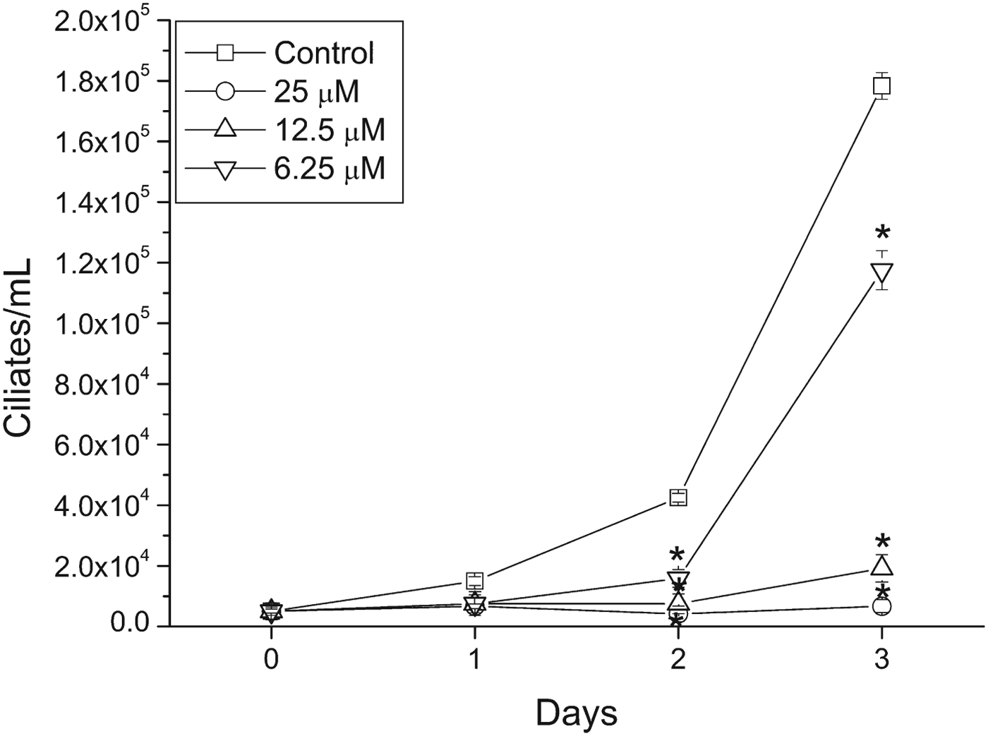
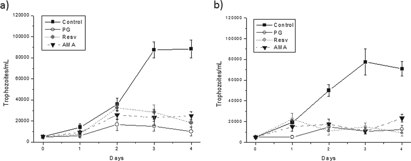
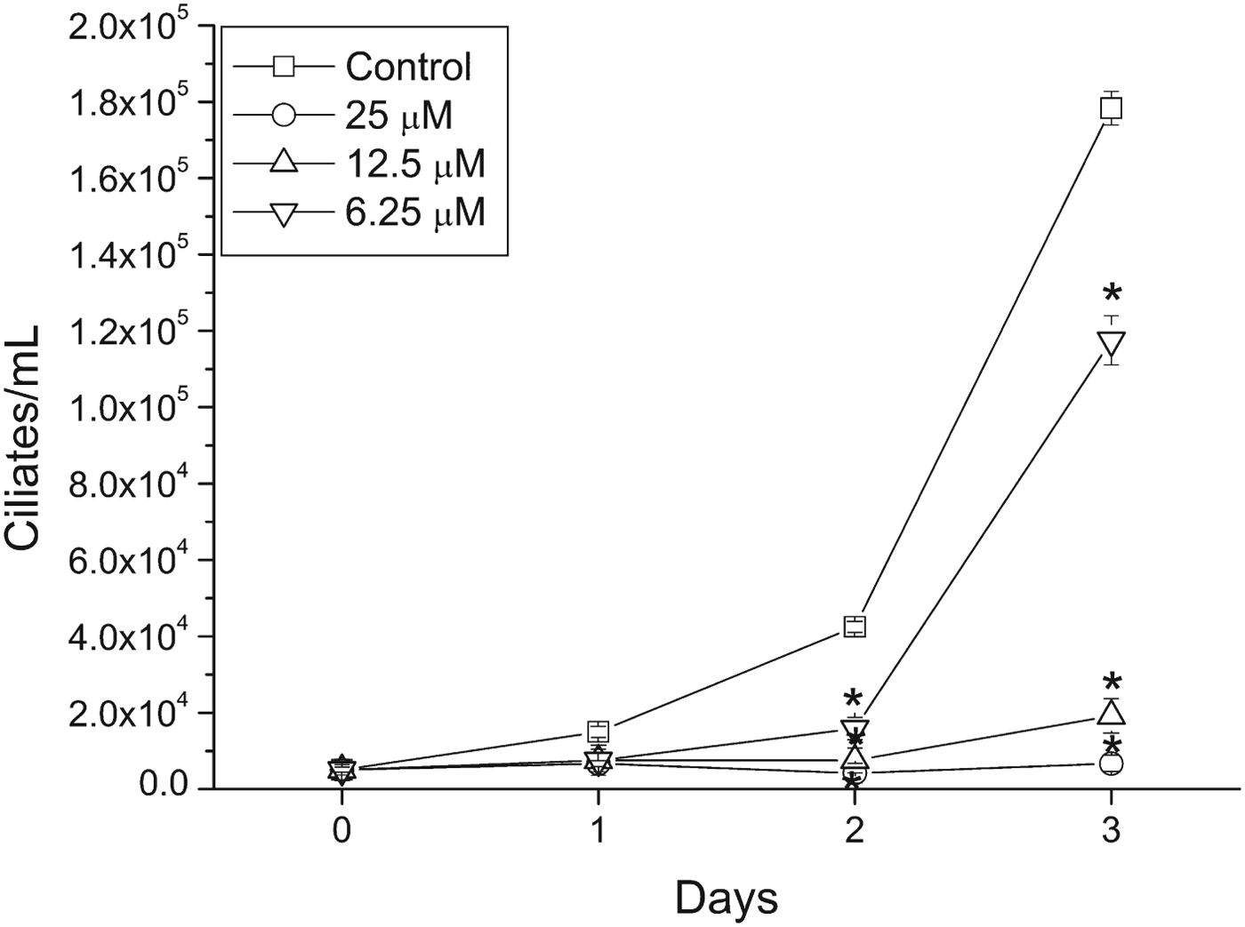
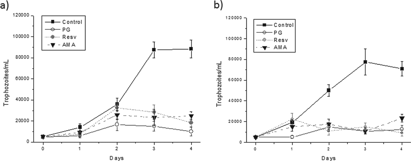
Incubation of ciliates in the presence of various concentrations of PG (6·25, 12·5 and 25 μ m) and under normoxic conditions led to dose-dependent inhibition of ciliate growth from the second day of culture (Fig. 2). Under hypoxic culture conditions, in vitro growth of the ciliate also decreased significantly in the presence of 25 μ m PG (Fig. 3b). Furthermore, both RESV and AMA significantly inhibited ciliate growth under normoxic and hypoxic conditions from the 3rd day of incubation (Fig. 3).
Effect of propyl gallate on the in vitro growth of Philasterides dicentrarchi trophozoites in normoxic cultures. The values shown are the means±s.e. *P<0·01 relative to controls (n = 5).
Growth curves for P. dicentrarchi trophozoites obtained after in vitro culture for 4 d in the presence of 25 μ m propyl gallate (PG), 50 μ m resveratrol (RESV) or antimycin A (AMA) under conditions of (a) normoxia and (b) hypoxia. The values shown are the means±s.e. (n = 5). Asterisks indicate significant differences relative to controls (*P<0·01).
Effect of antioxidants PG and RESV and the CP inhibitor AMA on AOX expression
Ciliate extracts from 3-day-old in vitro cultures (i.e. at exponential growth phase) maintained under normoxic (Fig. 4a) and hypoxic (Fig. 4b) conditions and incubated for 4 h with the antioxidants PG and RESV or with the standard inhibitor of CP, AMA, had a differing level of recognition with a polyclonal anti-AOX1/2 of plant AOX depending on the concentration of O2 present in these cultures. Under normoxic conditions, AOX expression increased in ciliates treated with inhibitors (Fig. 4a). By contrast, under hypoxic conditions, the level of regulation of AOX by PG, RESV and AMA was very low or non-existent (Fig. 4b).
Western blot analysis, with a polyclonal anti-AOX, of total proteins of P. dicentrarchi obtained after 3 d (in exponential phase) culture in (a) normoxic and (b) hypoxic conditions and incubated for 4 h in presence of 100 μ m propyl gallate (PG), antimycin A (AMA) or resveratrol (RESV). The box indicates the band corresponding to the alternative oxidase. Mw, molecular weight protein marker in kDa.

Effect of antioxidants PG and RESV on ΔΨm
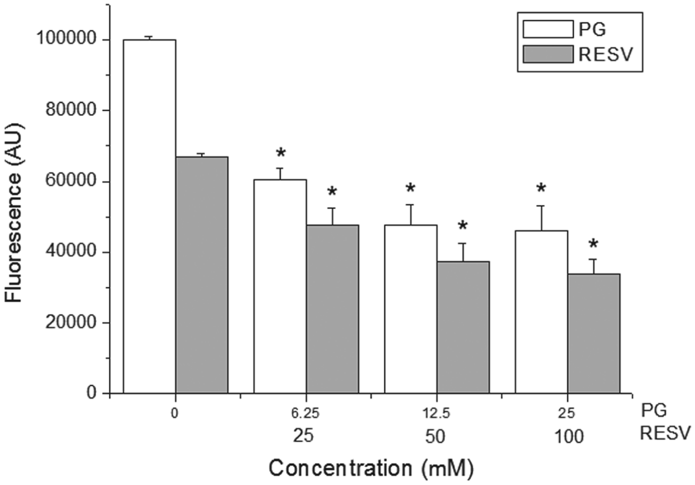
Trophozoites from normoxic cultures in the exponential growth phase (day 3) were incubated with several different doses of PG (between 6·25 and 25 μ m) and RESV (between 25 and 100 μ m) for 2 h, after which the effect on ΔΨm was determined using the fluorescent probe JC-1. There was a significant dose-dependent decrease in the ΔΨm of trophozoites incubated with both antioxidants (Fig. 5).
Effect of propyl gallate (PG), at concentrations of 6·25, 12·5 and 25 μ m, and resveratrol (RESV), at concentrations of 25, 50 and 100 μ m, on the membrane potential of P. dicentrarchi trophozoites cultured for 3 days in normoxic conditions, quantified using the lipophilic cationic probe 5,5′,6,6′-tetrachloro-1,1′,3,3′ tetraethyl-benzimidazol-carbocyanine iodide (JC-1); the results are expressed in arbitrary fluorescence units (AU). Histogram bars show means±s.e. (n = 5). Asterisks indicate statistically significant differences between the treated groups and the untreated controls: *P<0·01.

Effect of antioxidants on Hsp70 expression
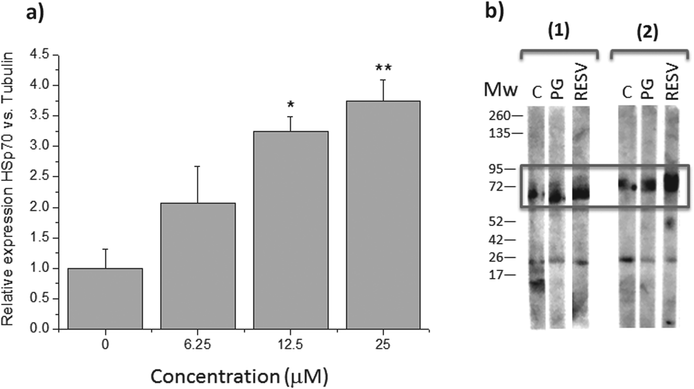
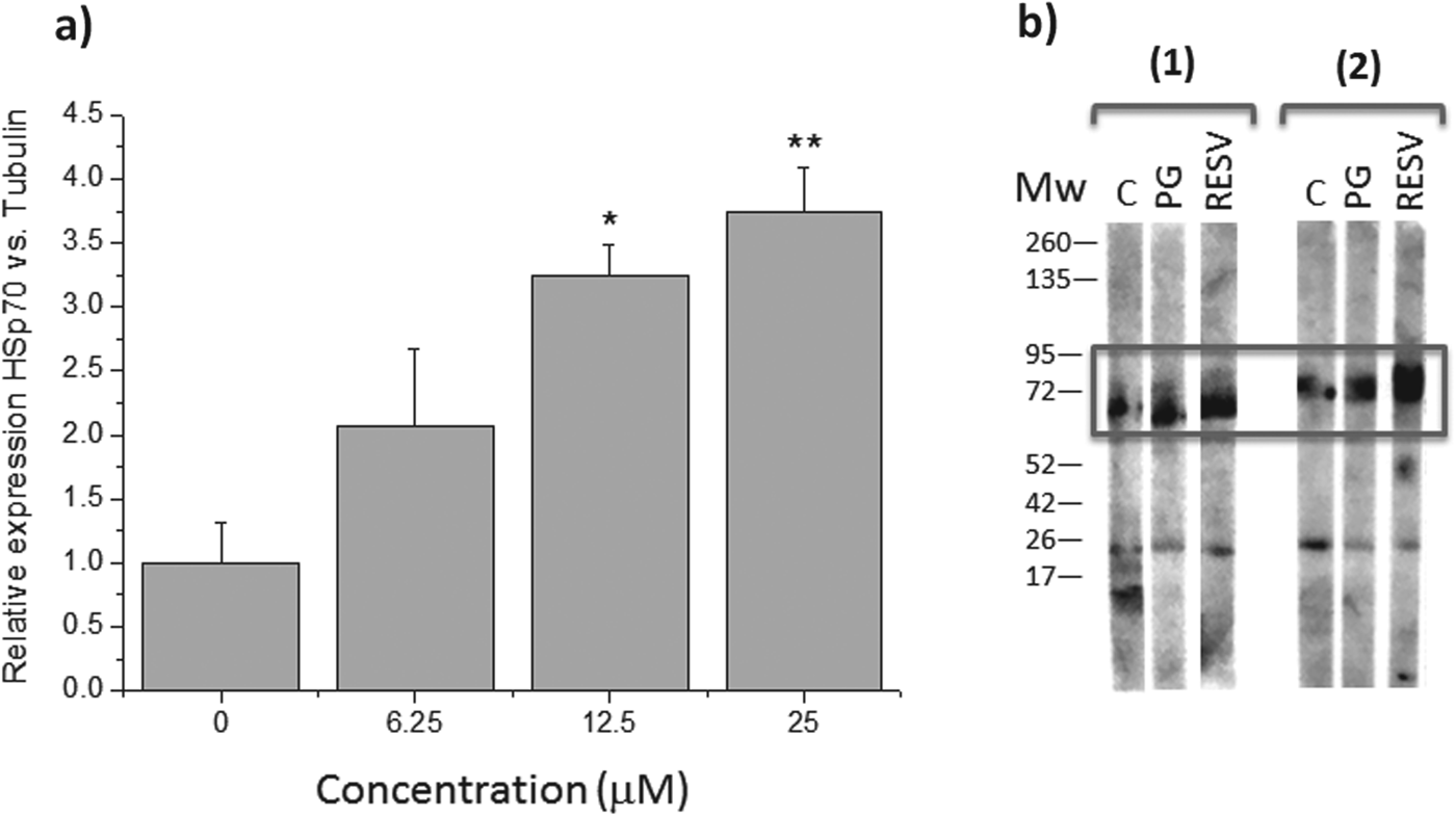
We used RT-qPCR to determine the effect of PG, at concentrations between 6·25 and 25 mm, on the expression of the gene encoding the heat shock protein Hsp70 in ciliates growing in normoxic conditions on the third day of culture (Fig. 6a). We also used western blotting to analyse the effects of PG (at concentration of 25 μ m) and RESV (at a concentration of 100 μ m) on expression of the Hsp70 protein (Fig. 6b) in trophozoites cultured under normoxic (Fig. 6b, 1) and hypoxic conditions (Fig. 6b, 2). Incubation with PG caused a significant dose-dependent increase in Hsp70 gene expression from a concentration of 12·5 μ m (Fig. 6a). An increase in Hsp70 protein expression was also detected in the trophozoite extracts incubated with PG and RESV under normoxic (Fig. 6b, 1) and hypoxic (Fig. 6b, 2) conditions.
(a) Expression of the heat shock protein 70 (Hsp70) gene after exposure to propyl gallate (PG, 6·25–25 μ m), measured by retrotranscriptase-real time PCR (RT-qPCR) using the β-tubulin gene as a reference for normalization of gene expression. Relative gene expression was analysed by the 2−ΔΔCq method, and each vertical bar represents the mean±s.e. Asterisks indicate statistically significant differences between the treated groups and the untreated controls: *P<0·05; **P<0·01; (b) Western-blot analysis of cell lysates of P. dicentrarchi incubated with anti-Hsp70 antibodies. Ciliates were obtained after in vitro culture for 3 days under (a) normoxia and (b) hypoxia, and they were then incubated for 4 h with 100 mm resveratrol (RESV) or 25 μ m propyl gallate (PG), or untreated (controls, C). The box indicates the band corresponding to the Hsp70. Mw, molecular weight protein marker in kDa.
DISCUSSION
Cyanide-resistant non-phosphorylating respiration is known to occur in mitochondria in plants, fungi and microorganisms, although it is clearly absent from vertebrates and arthropods (Vanlerberghe, Reference Vanlerberghe2013). It has therefore been suggested as a potential drug target (Moore and Albury, Reference Moore and Albury2008; McDonald et al. Reference McDonald, Vanlerberghe and Staples2009). In protozoan parasites, cyanide-resistant respiration might be due to a non-heme oxidase that is functionally very similar to plant AOX and may play a role in the energetic status of the cell by decreasing the yield of ATP synthesis and in attenuating production of reactive oxygen species (ROS) (Jarmuszkiewicz et al. Reference Jarmuszkiewicz, Sluse-Goffart, Hryniewiecka, Michejda and Sluse1998; Czarna and Jarmuskiewicz, Reference Czarna and Jarmuszkiewicz2005; Chaudhuri et al. Reference Chaudhuri, Ott and Hill2006; Woyda-Ploszczyca et al. Reference Woyda-Ploszczyca, Sluse and Jarmuskiewicz2009). We recently demonstrated that the mitocondrial respiratory chain of the scuticociliate P. dicentrarchi possesses both cyanide and AMA-resistant AOX and respires by the CP (Mallo et al. Reference Mallo, Lamas and Leiro2013a ). In the present study, we sought to demonstrate the potential antiparasite agent target of the AP in this ciliate. It has been suggested that AOX may play a role in maintaining oxygen homoeostasis by reducing the free oxygen concentration, thereby decreasing ROS production inside the mitochondria without deregulating the energy status of the cell (Gupta et al. Reference Gupta, Zalbalza and Van Dongen2009; Skutnik and Rychter, Reference Skutnik and Rychter2009). In the present study, the ciliates grown at the early stages of exponential growth in a normoxic environment respired by the conventional CP, as shown by the complete inhibition of O2 consumption by AMA. In contrast, in the same growth phase and under conditions of low oxygen availability, the ciliates utilized AMA-resistant respiration, which is completely inhibited by PG, a known inhibitor of the AOX (Sircar et al. Reference Sircar, Cardoso, Mukherjee, Mitra and Arnholdt-Schmitt2012), thus confirming the existence of an alternative AOX activity in P. dicentrarchi. Although the availability of O2 appears to be a factor in modulating CP and AP respiration, the cell growth stage has also been found to have a significant impact on mitochondrial bioenergetics (Czarna et al. Reference Czarna, Sluse and Jarmuszkiewicz2007; Woyda-Ploszczyca et al. Reference Woyda-Ploszczyca, Koziel, Antos-Krzeminska and Jarmuskiewicz2011; Mallo et al. Reference Mallo, Lamas and Leiro2013a ). Regarding RESV, our results indicate that this phytoalexin is capable of inhibiting both CP and AP respiration in ciliates. In a previous study, we found that RESV caused a dose-dependent inhibition of mitochondrial electron transport and O2 consumption in scuticociliates permeabilized with DIG, probably via inhibition of mitochondrial complex III (Zini et al. Reference Zini, Morin, Bertelli and Tillement1999; Morais et al. Reference Morais, Piazzon, Lamas, Mallo and Leiro2013). In this study, we have found that RESV also blocks O2 consumption under hypoxic conditions during AP respiration, indicating that this polyphenol may also be an inhibitor of the AOX. RESV is known to inhibit the respiratory chain by affecting the rotary mechanism of F1-ATPase of the facultative phytopathogenic fungus Botrytis cinerea (Hart, Reference Hart1981; Sbaghi et al. Reference Sbaghi, Jeandet, Bessis and Leroux1996). Thus, because this fungus possesses AOX (Tamura et al. Reference Tamura, Mizutani, Yukioka, Miki, Ohba and Masuko1999), it has been hypothesized that F1-ATPase may be a target for the antifungal activity of RESV (Caruso et al. Reference Caruso, Mendoza, Castro, Cotoras, Aguirre, Matsurhiro, Isaacs, Rossi, Viglianti and Antonioletti2011).
Several studies have demonstrated the antibacterial effect of PG on human bacteria in the buccal cavity (Jordan et al. Reference Jordan, Bowler and Berger1961), on intestinal bacterial strains (Chung et al. Reference Chung, Lu and Chou1998; Kubo et al. Reference Kubo, Fujita and Nihei2002) and on food-borne bacteria (Chung et al. Reference Chung, Stevens, Lin and Wei1993). There are also numerous references concerning the antifungal effect of PG (Kubo et al. Reference Kubo, Xiao and Fujita2001; Fujita and Kubo, Reference Fujita and Kubo2002; Hsu et al. Reference Hsu, Chen, Chang and Chang2009), showing that AOX inhibitors exert a fungistatic, rather than a fungicidal, action (Inoue et al. Reference Inoue, Tsurumi, Ishii, Park and Ikeda2012). However, PG not yet been tested as an antiparasitic agent. In the present study, PG exerted a clear dose-dependent cytostatic effect on the in vitro growth of P. dicentrarchi, which increased from day 3 of culture coinciding with a parallel increase in AOX expression (Mallo et al. Reference Mallo, Lamas and Leiro2013a ). The AMA antibiotics comprise a series of nine membered dilactones that are isolated from different strains of Streptomyces (Atta and Ahmad, Reference Atta and Ahmad2009). These antibiotics display antifungal activity, inhibition of enzymatic activity and also the ability to induce the death of cancer cells (Shiomi et al. Reference Shiomi, Hatae, Hatano, Matsumoto, Takahashi, Jiang, Tomoda, Kobayashi, Tanaka and Omura2005). AMA, a specific classical inhibitor of the complex III, cytochrome bc 1, of the mitochondrial respiratory chain (Barrow et al. Reference Barrow, Oleynek, Marinelli, Sun, Kaplita, Kim, Esser, Hossain, Xia, Yu, Rizo, Helm and Deisenhofer1999), also displays antiparasitic activity as it inhibits the in vitro growth of cultured ciliates under both normoxia and hypoxia. Previous studies have found that AMA and RESV, which are both inhibitors of complex III of the classical mitochondrial pathway, significantly inhibited parasite growth during the exponential and stationary phase (Lamas et al. Reference Lamas, Morais, Arranz, Sanmartín, Orallo and Leiro2009; Morais et al. Reference Morais, Piazzon, Lamas, Mallo and Leiro2013). However, the presence of AMA-mediated inhibition under hypoxia (in which the alternative pathway would be operating) may indicate that the antiparasitic activity of this antibiotic is not confined exclusively to blocking CP but that it may also act at another level of parasite cell metabolism.
Immunological analysis of P. dicentrarchi AOX indicates that this enzyme is constituted by a single monomeric protein present in the mitochondrial membrane, of approximate size 42 kDa, and that its expression is inducible under hypoxia or during experimental infections by CP inhibitors such as cyanide and AMA or by AOX inhibitors such as SHAM (Mallo et al. Reference Mallo, Lamas and Leiro2013a ). Here, we found that under normoxic conditions, in which the ciliate primarily respires by the CP, AMA and RESV (both inhibitors of this pathway), as well as PG, were potent activators of the expression of AOX, although the activating capacity of PG on AOX expression appears to be lower. By contrast, under hypoxic conditions, in which AP dominates, neither AMA nor RESV seemed to affect AOX expression, and PG appeared to increase AOX expression. In previous studies, we have demonstrated that both RESV and AMA induce overproduction of mitochondrial ROS in P. dicentrarchi (Morais et al. Reference Morais, Piazzon, Lamas, Mallo and Leiro2013). ROS may therefore act as sensitive indicators signalling changes in the mitochondrial redox state to the nucleus and thus serving to modulate expression of AOX (Vanlerberghe and McIntosh, Reference Vanlerberghe and McIntosh1997). Inhibition of CP electron transport by AMA and AOX (or by cyanide overexpressing AOX) has been found to play a role in lowering mitochondrial ROS formation by providing a pathway that releases constraints on the CP and prevents depletion of the ubiquinone pool, a major source of superoxide (Maxwell et al. Reference Maxwell, Wang and McIntosh1999; El-Khoury et al. Reference El-Khoury, Dufour, Rak, Ramanantsoa, Grandchamp, Csaba, Duvillié, Bénit, Gallego, Gressens, Sarkis, Jacobs and Rustin2013; Mallo et al. Reference Mallo, Lamas and Leiro2013a ). It has also been shown that specific AOX are strongly induced at the protein level by complex III or complex IV dysfunction (Vanlerberghe and McIntosh, Reference Vanlerberghe and McIntosh1994; Karpova et al. Reference Karpova, Kuzmin, Elthon and Newton2002), suggesting that AOX expression is highly responsive to insufficient CP capacity downstream of the ubiquinone pool (Vanlerberghe, Reference Vanlerberghe2013).
Mitochondria constantly respond to changes in substrate availability and energy utilization to maintain cellular ATP supplies and, at the same time, control ROS production (Kadenbach et al. Reference Kadenbach, Ramzan and Vogt2013). The mitochondrial respiratory chain, particularly in Complexes I and III, is considered the main origin of ROS, especially under conditions of high ΔΨm (Lenaz, Reference Lenaz2012). ΔΨm is critical for maintaining the physiological function of the respiratory chain to generate ATP and a significant loss of ΔΨm renders energy-depleted cells and subsequent cell death (Joshi and Bakowska, Reference Joshi and Bakowska2011). The antifungal effects of gallate compounds in yeast cells expressing AOX, assumed to be due to the pro-oxidant action and not to their antioxidant capacity, induce a morphological change and collapse of the ΔΨm (Sierra-Campos et al. Reference Sierra-Campos, Valdez-Solana, Matuz-Mares, Velázquez and Pardo2009). It is well known that AOX inhibitors (such as SHAM) increase mitochondrial ROS production in organisms that possess AOX and that this increase is deleterious to the cells (Mlejnek, Reference Mlejnek2013). There is a clear relationship between ΔΨm and the rate of ROS formation (Suski et al. Reference Suski, Lebiedzinska, Bonora, Pinton, Duszynski and Wieckowski2012): pro-oxidants are generally considered to trigger the mitochondrial permeability transition (MPT), which non-selectively increases the permeability of the inner membrane of mitochondria to small solutes, causing collapse of the ΔΨm associated with Ca+2 efflux (Kushnareva and Sokolove, Reference Kushnareva and Sokolove2000). RESV also induces a high level of pro-oxidant activity in P. dicentrarchi, causing a significant increase in ROS accompanied by ΔΨm collapse and a significant increase in intracellular Ca+2 levels (Morais et al. Reference Morais, Piazzon, Lamas, Mallo and Leiro2013).
Mitochondria synthesize only a small number of proteins in their matrix; most different mitochondrial proteins are produced on cytosolic ribosomes and are imported into the organelle (Kutik et al. Reference Kutik, Guiard, Meyer, Wiedemann and Pfanner2007). Mitochondria-localized chaperones include an Hsp70 family member, mtHsp70, which resides in the mitochondrial matrix and is encoded by nuclear genes that promote proper protein folding (Neupert and Herrmann, Reference Neupert and Herrmann2007). This member (mtHsp70) is the central component of a translocase-associated motor that completes preprotein translocation in the matrix and binds unfolded preproteins in an ATP-regulated manner (Kutik et al. Reference Kutik, Guiard, Meyer, Wiedemann and Pfanner2007). The presence of protein-altering ROS can negatively affect the mitochondrial protein-folding environment, thus inducing cellular apoptosis (Haynes and Ron, Reference Haynes and Ron2010). The presence of mtHsp70 may suppress programmed cell death by maintaining mitochondrial ΔΨ m and inhibiting amplification of ROS (Qi et al. Reference Qi, Wang, Zou, Liu, Liu, Wang and Zhang2011). Over-expression of mtHsp70 preserves mitochondrial function, decreases oxidative stress and subsequent apoptosis, and inhibits cytochrome c release (Mosser et al. Reference Mosser, Caron, Bourget, Meriin, Sherman, Morimoto and Massie2000; Williamson et al. Reference Williamson, Dabkowsk, Dillmann and Hollander2008). In this study, we demonstrate that in vitro treatment with PG induces dose-dependent over-expression of the gene encoding the chaperone Hsp70 and an increase in expression of this protein under both normoxia and hypoxia. Increased Hsp70 has been shown to enhance cell survival and prevent apoptosis during a wide variety of stress conditions including direct oxidative damage, as well as other types of stress in which generation of ROS is implicated in the cytotoxicity or oxidative damage of proteins, DNA and lipids (Martindale and Holbrook, Reference Martindale and Holbrook2002).
In conclusion, the present results confirm that, in addition to cytochrome c oxidase, the mitochondria of the scuticociliate P. dicentrarchi possess a cyanide- and AMA-resistant AOX, which operates preferentially under conditions of hypoxia that occur during the endoparasitic phase of natural infection. The natural polyphenol RESV and PG, a known inhibitor of AOX, both modulate expression of this protein by inhibiting in vitro growth of the parasite. The antiparasitic activity of polyphenols appears to be due primarily to the pro-oxidant effect on respiratory metabolism, which affects both the CP and AP and generates ciliate mitochondrial dysfunctions that adversely affect development of the organisms.
FINANCIAL SUPPORT
This study was financially supported by grants AGL2010-21219/ACU and AGL2011-29927-C02-01 from the Comisión Interministerial de Ciencia y Tecnología (CICYT; Spain), and grant INCITE07/PX1203039ES via the Aquagenomics project funded through the Consolider-Ingenio 2010 Programme, by the Spanish Ministerio de Economía y Competitividad.