Introduction
The effort to establish neurobiology-based criteria for psychopathology is currently a central focus in psychiatric research (Cuthbert & Insel, Reference Cuthbert and Insel2013). Major depressive disorder (MDD), being the leading cause of disability, is particularly challenged by its diagnostic heterogeneity (Nemeroff, Reference Nemeroff2007). Among several proposed clinical subtypes of MDD, the melancholic form is arguably the most widely accepted (Parker et al. Reference Parker, Fink, Shorter, Taylor, Akiskal, Berrios, Bolwig, Brown, Carroll, Healy, Klein, Koukopoulos, Michels, Paris, Rubin, Spitzer and Swartz2010). Melancholia has long been considered to have a biological weighting, with efforts to differentiate melancholic and non-melancholic depression evident since the third century (Day & Williams, Reference Day and Williams2012). As psychiatry strives towards a nosology based upon genetic, behavioural and neurobiological criteria, defining the biological signatures of MDD subtypes becomes an ever more pressing issue.
Non-invasive neuroimaging technologies permit in vivo characterization of the anatomical, physiological and neurochemical correlates of MDD (Drevets et al. Reference Drevets, Price and Furey2008a ). Using carefully designed task and resting-state paradigms, neuroimaging research in MDD has identified disturbances in brain circuits modulating emotional behaviour, encompassing ventromedial prefrontal cortex (vmPFC), anterior cingulate cortex (ACC), insula and closely related subcortical structures (Anand et al. Reference Anand, Li, Wang, Wu, Gao, Bukhari, Mathews, Kalnin and Lowe2005; Drevets et al. Reference Drevets, Price and Furey2008a ). In particular, subgenual ACC (sACC) and vmPFC are frequently reported to present altered activation and functional connectivity in depression (Drevets et al. Reference Drevets, Price, Simpson and Todd1997; Zald et al. Reference Zald, Mattson and Pardo2002; Greicius et al. Reference Greicius, Flores, Menon, Glover, Solvason, Kenna, Reiss and Schatzberg2007; Kross et al. Reference Kross, Davidson, Weber and Ochsner2009; Walter et al. Reference Walter, Henning, Grimm, Schulte, Beck, Dydak, Schnepf, Boeker, Boesiger and Northoff2009; Myers-Schulz & Koenigs, Reference Myers-Schulz and Koenigs2012). The details of findings, however, are difficult to reconcile. While some have reported increased functional connectivity among these regions (Anand et al. Reference Anand, Li, Wang, Wu, Gao, Bukhari, Mathews, Kalnin and Lowe2005; Bluhm et al. Reference Bluhm, Williamson, Lanius, Théberge, Densmore, Bartha, Neufeld and Osuch2009; Cullen et al. Reference Cullen, Gee, Klimes-Dougan, Gabbay, Hulvershorn, Mueller, Camchong, Bell, Houri, Kumra, Lim, Castellanos and Milham2009; Veer et al. Reference Veer, Beckmann, van Tol, Ferrarini, Milles, Veltman, Aleman, van Buchem, van der Wee and Rombouts2010; Lui et al. Reference Lui, Wu, Qiu, Yang, Kuang, Chan, Huang, Kemp, Mechelli and Gong2011), others have reported decreased connectivity (Greicius et al. Reference Greicius, Flores, Menon, Glover, Solvason, Kenna, Reiss and Schatzberg2007; Sheline et al. Reference Sheline, Price, Yan and Mintun2010). Furthermore, the ecological validity of these findings remains untested. For example, do these circuitry changes underlie the emotional disturbances experienced by people with MDD in real life?
The study of cognitive processes during realistic, natural conditions is an emerging trend in neuroimaging, offering an ecologically valid way to probe functional circuitry in the brain. Despite the seemingly uncontrolled nature of such paradigms, natural conditions, such as free viewing of films, evoke highly consistent responses in many cortical regions across subjects (Hasson et al. Reference Hasson, Malach and Heeger2010). Compared to resting-state, natural-viewing conditions provide a more engaging medium, and evoke distinctive topological changes in functional connectivity networks (Betti et al. Reference Betti, Della Penna, de Pasquale, Mantini, Marzetti, Romani and Corbetta2013). Importantly, natural-viewing paradigms have been effectively used to probe the emotional processes in the brain (Gross & Levenson, Reference Gross and Levenson1995; Nummenmaa et al. Reference Nummenmaa, Glerean, Viinikainen, Jääskeläinen, Hari and Sams2012), offering a promising avenue to unveil the neural underpinnings of affective disorders such as MDD.
Here, we sought a comprehensive comparison of functional brain networks during both natural-viewing and resting-state conditions between patients with melancholia, patients with non-melancholic depression and healthy participants. We used voxel-wise degree centrality, a graph theoretical measure at the voxel level, to examine the topology of functional connectivity network (Liao et al. Reference Liao, Xia, Xu, Dai, Cao, Niu, Zuo, Zang and He2013). This approach recapitulates functional connectivity in the whole brain at the finest scale permissible with functional magnetic resonance imaging (fMRI; Stanley et al. Reference Stanley, Moussa, Paolini, Lyday, Burdette and Laurienti2013). We hypothesized that patients with melancholia would show diminished reactivity to film stimuli, and exhibit distinctive changes in the functional brain topology to those with non-melancholic MDD.
Materials and method
Participants
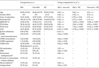
Seventeen patients (nine females) with melancholic MDD, and 17 patients with non-melancholic MDD (nine females) were recruited through the specialist depression clinic at the Black Dog Institute in Sydney, Australia. Study participants comprised those meeting criteria for a current major depressive episode, but without lifetime (hypo)mania or psychosis as identified during the MINI case-finding interview. Subtyping of melancholia was made by clinic psychiatrists using previously detailed criteria (Supplementary material) (Parker et al. Reference Parker, Fink, Shorter, Taylor, Akiskal, Berrios, Bolwig, Brown, Carroll, Healy, Klein, Koukopoulos, Michels, Paris, Rubin, Spitzer and Swartz2010). Patients reported treatment with a variety of antidepressants, mood stabilizers and/or antipsychotics (Table 1). Twenty-three right-handed age-matched healthy controls (HC; 12 females; aged 22–75 years) who disavowed lifetime or current mood and/or psychotic illness were recruited from the local community by way of advertisement.
Demographic and clinical characteristics

Mel, Melancholia; HC, healthy controls; n.s., not significant; QIDS-SR, Quick Inventory of Depressive Symptomatology – Self-report; STAI, State-Trait Anxiety Inventory; GAF, Global Assessment of Functioning; SSRI, serotonin selective reuptake inhibitor.
a For example, serotonin noradrenaline reuptake inhibitor.
b For example, lithium or valproate/divalproex.
Exclusion criteria for all participants were current or past drug or alcohol dependence, neurological disorder (i.e. neurodegenerative conditions, stroke, CNS infection, tremor), brain injury (i.e. neurotrauma from haemorrhage, hypoxia), invasive neurosurgery, and/or an estimated full-scale IQ (WAIS-III) score <80 as assessed by the Wechsler Test of Adult Reading (WTAR). ECT in the past 6 months was an additional exclusion criterion for the clinical participants. The study was approved by the University of New South Wales Ethics Committee (HREC 08077), and all subjects provided written informed consent prior to participation.
Clinical assessment
Depression severity in the clinical group was quantified with the Quick Inventory of Depressive Symptomatology (QIDS-SR; Rush et al. Reference Rush, Trivedi, Ibrahim, Carmody, Arnow, Klein, Markowitz, Ninan, Kornstein, Manber, Thase, Kocsis and Keller2003). To evaluate anhedonia, we selected clinical measures that best capture anhedonia from a 32-item Q-sort, which assesses the relative weighting of melancholic and non-melancholic prototypic features (Parker et al. Reference Parker, Fletcher, Hyett, Hadzi-Pavlovic, Barrett and Synnott2009). Six items were selected: (1) loss of interest in normally enjoyable activities; (2) inability to obtain pleasure from pleasurable activities; (3) inability to be cheered up when something nice occurred; (4) inability to be cheered up by friends; (5) loss of capacity to laugh at humorous things; and (6) not being able to look forward to taking part in usual pleasurable activities. The sum of the Q-sort weightings (−4 to +4) across these six items was used as a measure of anhedonia for each patient.
Natural stimuli – film clips
For the positive condition, participants viewed a stand-up comedy routine by Bill Cosby (taken from ‘Bill Cosby, Himself’). For the negative condition, participants viewed a scene from the drama film ‘The Power of One’, which depicts the inhumane treatment of prisoners during the apartheid era. Participants watched the films through an MRI-compatible monitor, and were provided with brief instructions (displayed on the screen) prior to the onset of each movie (‘Video will begin soon. Please relax and watch’.). High-quality audio from the movies was provided through insert earphones using an MRI-compatible system (Sensimetrics Model S14, USA).
Data analysis
Voxel-wise degree centrality
To examine functional connectivity we computed the degree centrality based on whole-brain voxel-wise connectivity (Liao et al. Reference Liao, Xia, Xu, Dai, Cao, Niu, Zuo, Zang and He2013). We first generated a group grey-matter mask,that encompassed all grey-matter voxels, both cortical and subcortical, across all subjects in our fMRI data. Then, we constructed a voxel-wise functional connectivity network for each subject, where functional connections (edges) between two voxels in this map were estimated using the Pearson's correlation coefficient between their BOLD signals. The ensuing fully connected functional graphs were thresholded to determine the presence or absence of connections between voxels. To generate weighted adjacency matrices, each suprathreshold edge retained its correlation coefficient as its edge weight, whereas subthreshold edges were assigned values of 0. To ensure robustness to the threshold chosen, we studied a broad range of sparsities (T = 0.1, 0.3, 0.5 fraction of total edges retained in each subject). Finally, voxel-wise degree maps were generated by computing the degree centrality for each voxel, i.e. the sum of weights over all suprathreshold edges for that voxel.
Statistical analyses of whole-brain functional imaging data
The degree centrality map for each subject was entered into a second-level, random-effects analyses to identify between-group differences using one-way ANOVA F tests and two-sample t tests. These group comparisons considered positive and negative viewing conditions equally as film viewing conditions. Two additional ANOVA F tests were conducted to examine whether positive and negative viewing conditions had any effect on the group differences between HC and melancholic depression, and between non-melancholic and melancholic depression groups. The resultant statistical images were thresholded using the joint probability distribution method to correct for multiple comparisons. Unless otherwise stated, all group differences in degree centrality maps (two-sample t tests) were evaluated with a height threshold of p < 0.001 and an extent threshold of p < 0.05, false discovery rate (FDR)-corrected. Regression analysis was evaluated with a threshold of p < 0.05 for height and p < 0.05, FDR-corrected for extent. We use cluster-wise thresholds as these have relatively high sensitivity, while still preserving robust family-wise error control (Friston et al. Reference Friston, Holmes, Worsley, Poline, Frith and Frackowiak1994; Smith & Nichols, Reference Smith and Nichols2009).
As cortical thinning has been observed in patients with MDD (van Tol et al. Reference van Tol, Li, Metzger, Hailla, Horn, Li, Heinze, Bogerts, Steiner, He and Walter2014), we further tested whether the observed group differences in functional maps were correlated with grey-matter volume. We re-calculated the two sample tests between HC and melancholic depression, and between non-melancholic and melancholic depression groups, with voxel-wise grey-matter intensity maps added as covariates, using the Biological Parametric Mapping (BPM) toolbox (Casanova et al. Reference Casanova, Srikanth, Baer, Laurienti, Burdette, Hayasaka, Flowers, Wood and Maldjian2007).
Ethical standards
The authors assert that all procedures contributing to this work comply with the ethical standards of the relevant national and institutional committees on human experimentation and with the Helsinki Declaration of 1975, as revised in 2008.
Results
Graph theoretical analyses of our data were performed at three different thresholds (T = 0.1, 0.3, 0.5). The findings were consistent across these thresholds (Supplementary material). Therefore we here focus on T = 0.3. Results from the sparser (T = 0.1) and denser (T = 0.5) graphs are presented in the Supplementary Figures.
All the findings presented below remain significant after controlling for grey-matter intensity.
Natural viewing alters the topology of functional connectivity in healthy brains
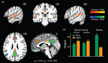
We first examined how functional connectivity changed between resting-state and natural-viewing conditions in healthy participants. Default mode network (DMN) regions, including precuneus, posterior cingulate cortex (PCC), vmPFC and bilateral angular gyrus (Ang), exhibited significantly higher degree centrality during resting-state than natural-viewing conditions (Fig. 1, Supplementary Fig. S1, green). This finding is consistent with prior research that has established that the DMN deactivates during task conditions and becomes more active at rest (Anticevic et al. Reference Anticevic, Cole, Murray, Corlett, Wang and Krystal2012). On the other hand, brain regions associated with goal-directed cognition and sensory integration – including the dorsal anterior cingulate cortex (dACC), superior temporal sulcus (STS), and superior temporal gyrus (STG) – showed significantly higher degree centrality during natural viewing (Fig. 1, Supplementary Fig. S1, orange). These association cortices become highly connected hub regions, presumably to integrate and process the dynamic multimodal stimuli. The same analyses performed on the two films separately revealed qualitatively similar results (Supplementary Fig. S2).
(a) Whole brain difference maps in degree centrality between natural-viewing and resting-state conditions in healthy controls (p < 0.05 for peak height and false discovery rate (FDR)-corrected p < 0.05 for spatial extent). (b) Average degree centrality at PCC (green) and dACC (orange) in healthy controls across natural-viewing and resting-state conditions. dACC, Dorsal anterior cingulate cortex; PCC, posterior cingulate cortex; Precu, precuneus; STG, superior temporal gyrus; vmPFC, ventral medial prefrontal cortex.

Lost network topography in melancholia
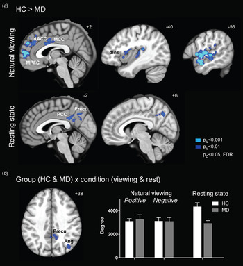
We then studied network topology in melancholia, and in particular, evaluated whether the state-selective patterns observed in health were preserved. We found a robust decrease in connectivity, particularly around those hub regions, during both natural-viewing and rest conditions in melancholia. During natural-viewing conditions, higher-order brain regions in both hemispheres, including bilateral STS, STG, dorsal anterior insula, dACC and medial PFC, showed decreased degree centrality in patients with melancholia compared to HC (Fig. 2 a, upper panel, Supplementary Fig. S3, left panel; p < 0.05, FDR-corrected). These group differences were most strongly expressed in the hub regions evident during film viewing in the healthy cohort, including bilateral STG and dACC (Figs 1 and 2 a). Furthermore, a formal contrast between film viewing conditions showed that the disconnectivity at the dmPFC was even stronger during the viewing of positive film than the negative film (Supplementary Fig. S4). A similar reduction in hub regions was also evident in the resting-state data, where the corresponding hub regions, including precuneus and PCC, showed significantly decreased connectivity in melancholia compared to HC (Fig. 2 a, lower panel; Supplementary Fig. S3, middle panel, p < 0.05, FDR-corrected). These findings suggest that melancholia is associated with functional disconnectivity, particularly in the highly connected hub regions of functional brain networks.
(a) Group difference maps between healthy controls (HC) and patients with melancholic depression (MD) during natural-viewing (top panel) and resting-state (bottom panel) conditions (p < 0.001 (light blue) or p < 0.01 (dark blue) for peak height and false discovery rate (FDR)-corrected p < 0.05 for spatial extent). (b) Group (HC and MD) × condition (natural viewing and resting state) interaction map (p < 0.01 for peak height and FDR-corrected p < 0.05 for spatial extent), and the average degree centrality of significant voxels across conditions in HC (white) and MD (grey). Ang, Angular gyrus; dACC, dorsal anterior cingulate cortex; dIns, dorsal insula; MCC, midcingulate cortex; PCC, posterior cingulate cortex; Precu, precuneus; mPFC, medial prefrontal cortex.

Notably, a significant group × condition interaction was observed in the core regions of the DMN, including precuneus, PCC and right angular gyrus (Fig. 2 b; p < 0.05, FDR-corrected). While these DMN regions switched from high to low connectivity between resting-state and natural-viewing conditions in healthy participants, they showed minimal change in melancholia (Fig. 2 b, left panel, Supplementary Fig. S5a). This non-reactivity of network topology to context mirrors the clinical symptoms that characterise melancholia, such as a non-reactive affect and anhedonia (Parker et al. Reference Parker, Hadzi-Pavlovic, Wilhelm, Hickie, Brodaty, Boyce, Mitchell and Eyers1994). To formally investigate this, we correlated the functional imaging findings with our clinical measure of anhedonia in patients with melancholic depression (see Supplementary material for details). Intriguingly, the group × condition differences in these DMN regions were significantly correlated with anhedonia (one-tailed Pearson correlation, r = −0.55, p < 0.01), such that diminished reactivity in DMN was associated with greater anhedonia (Supplementary Fig. S5b).
Distinct changes in sACC connectivity between depression subtypes at rest
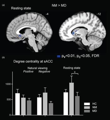
We next examined whether the two subtypes of MDD are associated with distinct patterns of functional connectivity. The ventral emotional circuitry showed distinct and highly significant differences between the melancholic and non-melancholic clinical groups. In particular, significant between-group differences were found in the sACC, ventral striatum (VS) and caudate during resting-state condition (Fig. 3 a; p < 0.05, FDR-corrected). On average, these ventral regions showed an elevated connectivity in patients with non-melancholic depression, and a reduction in patients with melancholia as compared to HC (Fig. 3 b). On the other hand, most of the regions with decreased connectivity in melancholia (Fig. 2 a) also showed a trend of reduction in non-melancholic depression, resulting in an intermediate level of connectivity between the HC and melancholic depression groups (Supplementary Fig. S3). No significant differences between the two clinical groups were found to be specific to the positive or negative conditions. These findings support the notion that different subgroups of MDD may be associated with distinct neurobiological underpinnings.
(a) Group difference maps between patients with non-melancholic depression (NM) and patients with melancholic depression (MD) during resting-state condition (p < 0.01 for peak height and false discovery rate (FDR)-corrected p < 0.05 for spatial extent). (b) Degree centrality at sACC across conditions in HC (white), NM (light grey) and MD (dark grey). sACC, Subgenual anterior cingulate cortex; Cau, caudate.

vmPFC connectivity correlates with depression severity in melancholia
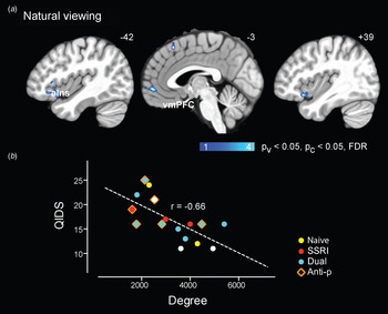
We finally examined whether network connectivity patterns correlated with depression severity. We performed regression analyses between degree centrality and QIDS scores in the melancholic group, separately for film-viewing and resting-state conditions. During film viewing, the degree centrality of vmPFC and bilateral anterior insula was significantly correlated with depression severity (Fig. 4 a, b; p < 0.05, FDR-corrected). For the resting-state condition, the degree centrality at vmPFC correlated with depression severity, although this cluster did not survive multiple comparison correction for the whole brain (Supplementary Fig. S6; p < 0.05). Since patients are on a variety of medications, we inspected how these data points distribute according to the medication classes. There were no specific clusters suggesting a bias associated with medications (Fig. 4 b).
(a) Correlation maps between degree centrality and Quick Inventory of Depressive Symptomatology (QIDS) depression severity scores in patients with melancholic depression during natural-viewing conditions (p < 0.05 for peak height and false discovery rate (FDR)-corrected p < 0.05 for spatial extent). (b) QIDS plotted against degree centrality at vmPFC during film viewing (average between positive and negative conditions). Data points are colour coded according to the types of medication taken by each patient. aIns, Anterior insula; vmPFC, ventral medial prefrontal cortex.

Discussion
We report distinct patterns of neurobiological changes associated with the melancholic and non-melancholic subtypes of major depressive disorder across different behavioural conditions. Melancholia presents a general reduction in degree centrality, particularly at highly connected hub regions including dACC, STG, precuneus and PCC. Intriguingly, the marked changes in functional connectivity of the DMN between resting-state and natural-viewing conditions in health were markedly muted in those with melancholia. A direct comparison between the two subtypes of MDD revealed distinct changes in the ventral emotional circuitry, with increases in degree centrality in non-melancholic and decreases in those with melancholic depression. This divergence might underlie the distinct clinical profiles associated with these two MDD subtypes: while anhedonia and psychomotor retardation are characteristic for melancholia, non-melancholic depression often presents with elevated anxiety, worrying, and agitation (Parker et al. Reference Parker, Hadzi-Pavlovic, Wilhelm, Hickie, Brodaty, Boyce, Mitchell and Eyers1994; Day & Williams, Reference Day and Williams2012). These findings highlight the importance of refining MDD subtypes in exploring the biological underpinnings of MDD.
The topology of functional connectivity networks in our healthy cohort show characteristic changes between natural-viewing and resting-state conditions, such as the bilateral STG and dACC ‘switching on’ while the DMN regions ‘switch off’ during natural viewing. In patients with melancholia, these hub regions are in general much less functionally connected (Fig. 2 a). Some of these regions showed a robust reduction during natural-viewing conditions. A non-significant trend was also present during resting state in areas, such as STG, dACC and midcingulate cortex (MCC), consistent with recent reports of comparable effects in the MCC (Supplementary Fig. S3) (Zhang et al. Reference Zhang, Li, Qin, Demenescu, Metzger, Bogerts, Yu and Walterin press). In contrast, the core nodes of the posterior DMN responded differently to the behavioural conditions, revealing a failure in reacting to contextual changes in melancholia (Fig. 2 b). Post-hoc analyses revealed that the reduction in DMN reactivity was significantly correlated with clinical measures of anhedonia, suggesting a neurobiological signature of the anhedonia and non-reactive mood that characterize melancholia.
Correlation analyses with QIDS revealed that disconnectivity at the bilateral anterior insula correlates significantly with depression severity in melancholia (Fig. 4). The anterior insula respond robustly to emotionally salient stimuli, such as pain and disgust, and are thought to serve as a hub that integrates intero- and exteroceptive information and generates the subjective experience of emotion (Craig, Reference Craig2009; Nguyen et al. Reference Nguyen, Breakspear, Hu and Guo2015). Clinical studies have also demonstrated that injuries to the anterior insula contribute to deficits in emotion recognition, empathy and self-awareness in neuropsychiatric disorders (Sliz & Hayley, Reference Sliz and Hayley2012; Guo et al. Reference Guo, Gorno-Tempini, Gesierich, Henry, Trujillo, Shany-Ur, Jovicich, Robinson, Kramer, Rankin, Miller and Seeley2013; Kumfor et al. Reference Kumfor, Sapey-Triomphe, Leyton, Burrell, Hodges and Piguet2014). This correlation effect at the anterior insula, together with the group differences at dACC, suggests that the salience network, an intrinsic connectivity network anchored in the anterior insula and ACC (Seeley et al. Reference Seeley, Menon, Schatzberg, Keller, Glover, Kenna, Reiss and Greicius2007), is disturbed in melancholia; an impaired salience network could contribute to deficiency in orienting to and mapping emotional salience in these patients (Horn et al. Reference Horn, Yu, Steiner, Buchmann, Kaufmann, Osoba, Eckert, Zierhut, Schiltz, He, Biswal, Bogerts and Walter2010; Menon, Reference Menon2011; Sliz & Hayley, Reference Sliz and Hayley2012).
Our study identified distinct functional connectivity changes associated with the two subtypes of MDD in sACC and adjacent striatum structures, with heightened connectivity in non-melancholic depression, and reduced connectivity in melancholia. sACC activations are frequently observed in functional neuroimaging studies of emotion modulation, and have been found to be positively correlated with elevated anxiety symptoms in affective disorders (Osuch et al. Reference Osuch, Ketter, Kimbrell, George, Benson, Herscovitch and Post2000). Furthermore, post-mortem and structural imaging studies have shown that mean grey-matter volume of sACC is reduced in patients with MDD (Drevets et al. Reference Drevets, Savitz and Trimble2008b ). Functional imaging studies, however, have reported inconsistent findings thus far: while some studies observed increased functional connectivity or activation, others reported decreased functional connectivity or activation. Given most of these previous studies did not differentiate MDD subtypes, these conflicting reports could be due to the heterogeneity of patient cohorts. Additionally, the inter-subject variability of connectivity measures in these regions is higher in non-melancholic group than melancholic and HC groups (Fig. 3 b), suggesting that the non-melancholic group likely constitutes patients with diverse pathogenesis and may deserve further biologically informed sub-classification.
It is possible that our findings are confounded by medication – patients with melancholia were on a mixture of antidepressants, mood stabilisers and antipsychotics. However, clinical research on melancholic depression is challenged by a number of barriers to control for this – the severity of the disorder prohibits withholding of medication for research purposes, whilst the variety of classes and pharmacological targets prevents the possibility to estimate a class-invariant dosage equivalent. We have visualised the distribution of data points from patients using various medications (Fig. 4 b), and did not find any evidence that our results were driven by medication effects.
In summary, our study demonstrates that functional neuroimaging with natural, emotionally salient stimuli offers an effective paradigm with which to probe neural processes during dynamic, naturalistic experiences and characterise its disruption in depressive disorders. With minimum training or in-scanner performance required, this approach enjoys similar advantages to resting-state acquisitions and can hence be readily conducted in patient populations, minimising anxiety associated with completing difficult or repetitive tasks. On the other hand, natural stimuli put ecologically relevant constraints on neuronal processes and are likely to be more effective in selectively engaging brain networks of interest than resting-state acquisitions. Certainly, in our study, many of the results were more robust during natural-viewing than resting-state paradigms, such as the correlation between degree centrality and depression severity. During natural viewing, degree centrality of bilateral anterior insula and vmPFC showed a significant correlation with clinical assessments of depression severity (Fig. 4), consistent with the known contribution of these limbic regions to depression. In contrast, these relationships were much weaker or absent at rest (Supplementary Fig. S6). In addition, the comparison of changes in brain activity between resting-state and natural-viewing conditions offers unique insights that would not be obvious with data only from either condition alone. Therefore, we advocate an integrative approach that combines both a resting-state acquisition with a stimuli-driven condition with ecological validity.
Supplementary material
For supplementary material accompanying this paper visit http://dx.doi.org/10.1017/S0033291716000179.
Acknowledgements
This work was supported by grants from the National Health and Medical Research Council of Australia (Program Grants 510135 and 1037196) (M.P.H., G.B.P., M.J.B.) and Queensland Health (M.P.H., M.J.B.).
Declaration of Interest
None.