Introduction
MicroRNAs (miRNAs) are tiny (~22 nucleotides), endogenous and non-coding RNAs that negatively regulate gene expression by binding to mRNA target sequences in eukaryotes (Legeai et al., Reference Legeai, Rizk, Walsh, Edwards, Gordon, Lavenier and Jaubert-Possamai2010). miRNAs have become a major class of post-transcriptional regulatory molecules, which play a key role in diverse biological processes such as development, metabolism and apoptosis in organisms (Ambros, Reference Ambros2004; Miska, Reference Miska2005). In addition, miRNAs have also been recognized as a trustworthy data set to resolve phylogenetic problems in metazoan systematics due to several properties: (i) new miRNA families are continually added to metazoan genomes; (ii) mature sequence of miRNA accumulates mutations very slowly and (iii) the rate of miRNA acquisition outweighs the rate of miRNA losses in most metazoan taxa (Campbell et al., Reference Campbell, Rota-Stabelli, Edgecombe, Marchioro, Longhorn, Telford and Pisani2011). Since the earliest member of miRNAs lin-4 discovered in 1993 (Lee et al., Reference Lee, Feinbaum and Ambros1993), miRNAs have been found in a variety of eukaryotic organisms ranging from sponges to mammals (Wheeler et al., Reference Wheeler, Heimberg, Moy, Sperling, Holstein, Heber and Peterson2009). In recent years, next generation sequencing technology, such as Illumina sequencing and 454 sequencing has been robust methods to identify numerous miRNAs in animals and plants (Ou et al., Reference Ou, Meng, Li, Xiu, Du, Gu and Wang2012). More than 28,645 miRNAs are involved in the latest miRBase database release 21.0 in June 2014 (http://www.mirbase.org/). Although insects account for a large proportion of known animals, so far, miRNAs for insects have mainly restricted to Diptera (Drosophila, Anopheles gambiae, Aedes aegypti, Culex quinquefasciatus), Hymenoptera (Nasonia, Apis mellifera), Coleoptera (Tribolium castaneum), Orthoptera (Locusta migratoria), Lepidoptera (Bombyx mori, Heliconius melpomene) and Homoptera (Acyrthosiphon pisum).
The genus Qinlingacris (Orthoptera: Acrididae: Catantopinae) was first published in 1979, and is endemic to Taibai Mountain in Shaanxi Province, China (Yin & Chou, Reference Yin and Chou1979). To date, there are three known species within this genus. Qinlingacris elaeodes is the dominant species at the altitude over 3200 m and is a representative species of Qinlingacris. Since its first report in 1979, scarcely any researchers have paid attention to it except for Li et al. (Reference Li, Liu and Zheng2010). Interestingly, Yin & Chou (Reference Yin and Chou1979) found ‘this species of grasshopper is more specific, its fore wings are very astable and are in great variations, from with wings at left and right sides to with wing at left or right side, even to wingless at all sides’. They discussed that, from a user into useless retreat point of view, the fore wing of this grasshopper is becoming vestigial. So they believed that the wings certainly develop to wingless. This species is considered to be a transitional species because some individuals are wingless and the others are short-winged. In fact, Catantopinae is an interesting family in grasshoppers mainly due to polymorphic wings. For example, Patanga japonica is a typical long-winged species, which is good at flying, while the genus Kingdonella are totally wingless and are unable to fly. For Q. elaeodes, the fore wings in Q. elaeodes are likely to be significantly variable, which is easily regarded as wing mutant phenotypes or wings dysplasia. The individuals with a unilateral wing form (seven individuals) accounted for one-third of all collected specimens (21 individuals), and some individuals of both sexes are unilateral-winged. Indeed, this case of Q. elaeodes is rare in insects and even in animals. Therefore, a unilateral wing formation of male and female adults in Q. elaeodes makes this species an interesting model system to study the evolution and development of insect wings.
The wing polymorphism of Q. elaeodes is the result of this species of adaption to its environment and moreover, this adaptative capacity can be explained by phenotypic plasticity (Legeai et al., Reference Legeai, Rizk, Walsh, Edwards, Gordon, Lavenier and Jaubert-Possamai2010). Given the scarcity of genetic information for Q. elaeodes, we largely focus on the post-transcriptional level which is closely related to phenotypic plasticity. In the present study, we described a miRNA library from mixed thoraxes of Q. elaeodes using Illumina sequencing combined with bioinformatics analyses. We identified and characterized 49 conserved miRNAs and 69 species-specific miRNAs for Q. elaeodes. These could contribute to better understanding of the post-transcriptional regulation of wing polymorphism in insects.
Materials and methods
Ethics statement
This species of grasshopper in this study is a common insect. Collections of samples were allowed by the authority of Taibai Mountain National Forest Park. For collection locations, no specific permits were required for the described field studies because sample collection did not involve endangered or protected plant species or privately-owned locations.
Sample collection and RNA isolation
Adults and third to fifth instar larvae of Q. elaeodes were collected from Nantianmen, Taibai Mountain, Shaanxi, China. For Illumina sequencing, 5 for every instar larvae and 20 adult individuals (wingless, female n = 5, male n = 5; short-winged, female n = 5, male n = 5) were pooled together, and total RNA was extracted from mesothorax of pooled individuals using the Total RNA Purification Kit (LC Sciences, Houston, USA), according to the manufacturer's protocol. The quality and quantity of total RNA were measured using a NanoDrop ND-2000 spectrophotometer (Nano Drop Technologies, Wilmington, DE, USA).
Illumina sequencing
The sequencing procedure was once described by Ma et al. (Reference Ma, Huang, Gong, Zhuang, Li, Yang and Cao2011). In brief, the purified small RNAs (14–30 bases, bp) were ligated with a pair of adaptors (Illumina, San Diego, CA, USA) to their 5′ and 3′ ends. Then the small RNAs were amplified using the adaptor primers for 17 cycles, and fragments of 70–90 bp (small RNA + adaptors) were excised from gels. The purified DNA fragments were utilized directly for cluster generation and sequencing analysis by an Illumina/Illumina G1 sequencer.
Bioinformatic analysis of Illumina sequencing data
After masking of vector and adaptor sequences and removal of contaminated reads, the clean reads were then filtered for miRNA prediction with the proprietary ACGT101-miR v4.2 package (LC Sciences, Houston, USA). First, the raw sequences matching to metazoan mRNA, rRNA, tRNA, snRNA, snoRNA, repeat sequences and other ncRNAs deposited in the National Center for Biotechnology Information (NCBI) databases (http://www.ncbi.nlm.nih.gov/), RFam (http://rfam.janelia.org/) and Repbase (http://www.girinst.org/repbase/) were discarded. Next, the remaining 18–25 nucleotide reads were mapped onto the expressed sequence tags (ESTs) of other grasshoppers (http://www.ncbi.nlm.nih.gov/) and the D rosophila melanogaster genome (http://flybase.org/). Then these mapping sequences (three reads or more) were compared with pre-miRNA (mir) and mature miRNA (miR) deposited in the miRBase 21.0. Finally, Mfold software (http://mfold.rna.albany.edu/) using the default folding condition was used for secondary structure prediction of these miRNA precursor sequences.
Polymerase chain reaction (PCR) verification of miRNA precursors
We used a PCR-based method to verify the miRNA precursors. Genomic DNA was extracted from a hind leg of an individual following standard phenol–chloroform procedures (Sambrook et al., Reference Sambrook, Fritsch and Maniatis1989). For conserved miRNA/ miRNA* candidates, one primer pair was designed using Primer Premier 6.0 software (PREMIER Biosoft, Canada), while for the novel miRNA candidates, we designed two primer pairs because mature miRNAs might locate in either arm of the precursors. The length of amplification products was examined on 3.5% agarose gels after PCR. Fragments between 55 and 70 nucleotides in length were then subcloned into pMD19-T vector (Takara, Dalian, China) for sequencing analysis.
Quantification of conserved miRNA*s by real time PCR
For conserved miRNA*s expression studies by quantitative real time (qRT)-PCR, 400 ng of total RNA from the pools prepared for libraries were reverse transcribed using the SYBR PrimeScriptTM miRNA RT-PCR Kit (TaKaRa) following the manufacturer's protocols. Amplification reactions were performed using the same kit and the following protocol: 95°C for 2 min, and 40 cycles of 95°C for 15 s and 60°C for 30 s, in a Rotor-Gene Q (Qiagen). After the amplification phase, a dissociation curve was made to ensure that there was only one product amplified. All reactions were run in triplicate. Statistical analysis of relative expression results was given as copies of RNA per 1000 copies of U6. Primer sequences are available on request.
Results
Overview of the Illumina sequencing data
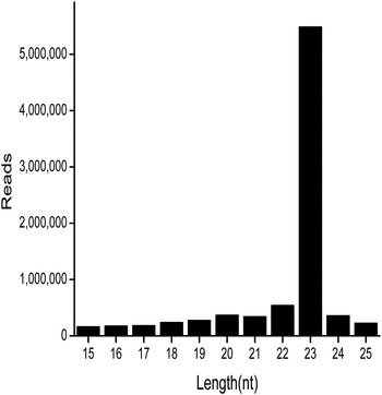
Total RNA was extracted from the mesothorax of Q. elaeodes. Low-quality sequences and those shorter than 15 nucleotides and sequences with no more than three reads were removed from our data, 8,358,988 mappable reads were retained. The length distribution of these mappable reads is shown in fig. 1. A peak was obviously at the 23 nt, and over 76% reads were at the range from 21 to 23 nt (table S1), which is the typical length of miRNAs. Furthermore, we discarded the certain known types of RNA sequences, such as mRNA, rRNA, tRNA, snRNA, snoRNA and repetitive sequences. Finally, 854,580 high-quality reads were of primary interest for the following analysis. All of the raw RNA-Seq data have been submitted to the NCBI database (http://trace.ncbi.nlm.nih.gov/Traces/sra/sra.cgi?view=run_browser) under accession number SRR1598791 and SRR1611786.
Size distribution of mappable reads related to miRNA in the library.

Conserved miRNAs
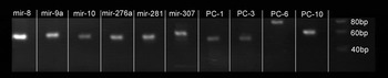
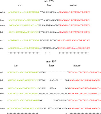
After mapping to D. melanogaster genome and ESTs of L. migratoria and other grasshoppers, a total of 49 miRNAs were identified and divided into two groups (table S2): (i) 34 miRNAs corresponding to the known fruit fly (D. melanogaster) pre-miRNAs were also mapped to the genome and ESTs. (ii) 15 miRNAs corresponding to other known miRBase insects’ pre-miRNAs could not be mapped to the reference genome or ESTs, so they were labelled with PN, which denotes they are homologous to other insects (Ou et al., Reference Ou, Meng, Li, Xiu, Du, Gu and Wang2012). Based on the fact that there is no genome and EST for our species in the present study, the reference genome and ESTs considered truly have differences to some extent. We then treated the miRNAs of group 1 and 2 as conserved miRNAs. They belong to 41 families and these families shared the same ‘seed regions’ except for miR-10. For miR-10, the mature sequence has an additional adenosine at the 5′ end, which was consistent with that of L. migratoria (Wei et al., Reference Wei, Liu, Feng and Chang2011). This might be grasshopper-specific in insects. Previous studies have revealed that an extra U at 5′ end will not change the targets of miRNAs (Ruby et al., Reference Ruby, Stark, Johnston, Kellis, Bartel and Lai2007). Although miRNA* is considered to be less conserved than miRNA, we detected seven miRNA*s in our library. To test whether the miRNA and its miRNA* came from the same precursor, we expected a 55–70 bp PCR product from the genomic DNA. As expected, we gained PCR products of 55–70 bp for six miRNA*s with the exception of miRNA*−71 (fig. 2, fig. 3 and table S3) and inferred an unconventional precursor sequence for miR-71 in Q. elaeodes. The secondary structure of each sequence was predicted by Mfold and all were properly folded into the typical hairpin structure (text S1), which demonstrated that the miRNA pairs may have come from the same precursor.
Conservation of miRNAs in Q. elaeodes. Electrophoretic analysis of miRNA precursor PCR products.

Sequence alignment analysis of two miRNA precursors that have both conserved star and mature sequences. The alignment of mir-276a and mir-307 in different insects shows high conservation of their mature miRNA and miRNA*. The green nt represent miRNA* sequence and the red represent mature miRNA sequence. The asterisks indicate the conserved sites among these species. qel, Q. elaeodes; bmo, B. mori; dme, D. melanogaster; lmi, L. migratoria; tca, T. castaneum; ame, A. mellifera; aga, A. gambiae.

Species-specific miRNAs
Sixty-nine miRNAs corresponded with new candidate pre-miRNAs, which were predicted new RNA hairpins derived from the reference genome of D. melanogaster and ESTs of L. migratoria and other grasshoppers. These miRNAs were labelled with PC, which denotes sequences that have only selected species genomic cognates, but without any homology to the known metazoan miRNAs in miRBase 21.0 (table S4). The results suggest that the PC miRNAs are species-specific. When we compared these PC miRNAs with all 185 novel miRNAs of L. migratoria reported by Wei et al. (Reference Wei, Liu, Feng and Chang2011) using a local Blast program, no positive hit was found, which further proved their specificity.
Among these 69 candidate miRNAs, seven were miRNA duplex-like pairs (PC-1, PC-3, PC-4, PC-6, PC-7, PC-9, PC-10). To test whether they were true miRNA duplexes, a method described above was carried out for four of miRNA duplex-like pairs. As a result, we amplified corresponding fragments from the genomic DNA for tested pairs and they could be folded in standard hairpin structure using Mfold after sequencing (text S1).
The phylogenetic evolution of miRNAs
MiRNAs have been regarded as one of the outstanding markers in phylogenetics despite of their short lengths (Heimberg et al., Reference Heimberg, Cowper-Sallari, Semon, Donoghue and Peterson2010). We sorted 41 conserved miRNA families and 2 miRNAs (PN-qel-miR-193 and PN-qel-miR-750) identified in our library into four groups based on their phylogenetic distribution (table 1). Four families (let-7. mir-1, mir-34 and mir-124) are present in insects, vertebrates and nematodes. Twelve families and PN-qel-miR-750 are found in insects, vertebrates, but not in nematodes. Five families are restricted to insects and nematodes, and the remaining 20 families and PN-qel-miR-193 are insect-specific. Our results were congruent with the previous report of L. migratoria (Wei et al., Reference Wei, Liu, Feng and Chang2011). Importantly, five families (mir-252, mir-137, mir-971, mir-2796 and mir-2765) were first reported in grasshoppers and thus further enhanced their abundance. Moreover, nine families of L. migratoria (mir-29, mir-31, mir-33, mir-219, mir-283, mir-375, mir-929, mir-965 and mir-993) were not detected in our library. It is recognized that a majority of miRNAs are highly evolutionarily conserved throughout different animal taxa, but sequence variations including single nucleotide polymorphism and length difference still exist. We then analyzed the sequences of miRNAs (text S2) and they were divided into three categories: (i) 5′ ends variations; (ii) variations next to the ‘seed regions’ and (iii) 3′ ends variations (Wei et al., Reference Wei, Liu, Feng and Chang2011).
Phylogenetic distribution of Q. elaeodes conserved miRNA families. A plus (+) symbol represents this miRNA family exists in the species named on the left, and a minus (−) symbol means it is absent in that species.

Quantitative value of conserved miRNA*s by real time PCR
The number of reads resulting from deep sequencing is often considered a reliable quantification of miRNA expression. However, this is a hypothesis priori which has not been empirically tested yet. In order to assess whether our Illumina sequencing data have a quantitative value, we carried out qRT-PCR analysis of the seven conserved miRNA*s, using the same RNA samples to construct the libraries.
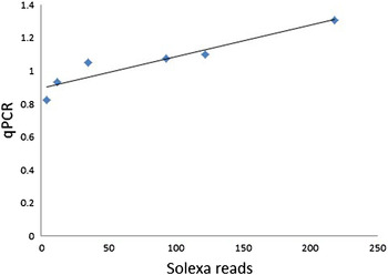
We used Pearson's correlation to test if the numbers of Illumina reads and qRT-PCR experiments were correlated. The results indicated that when miR-8* was considered into the analysis, there was no significant correlation between the reads and qRT-PCR data, as the miR-8* is the most abundant miRNA* and with 9722 reads. However, a significant positive correlation was observed when we considered the other miRNA*s with the reads (R = 0.94, P-value ≤ 0.05) (fig. 4, table S5). These results suggest that Illumina sequencing data with thousands of reads can only roughly represent the relative abundance of miRNA*s.
Quantitative value of Solexa reads. The number of reads of Solexa sequencing and fold change of qPCR analysis (miRNA copies per 1000 copies of U6) are correlated. Squares are the miRNA*s with less than 1000 Solexa reads. Solid lines are trend line for correlation with 1000-less Solexa reads.

Discussion
Since the first miRNA was detected in Caenorhabditis elegans (Lee et al., Reference Lee, Feinbaum and Ambros1993), thousands of miRNAs have been well identified (Kozomara et al., Reference Kozomara and Griffiths-Jones2011). It is still being difficult to determine their functions for species-specific miRNAs. Being a native grasshopper to China, Q. elaeodes is an ideal species to study the evolution of wing pattern diversity within the Acridoidea. The wingless type is regarded as a kind of adaption to the long period of high-altitude living, while the short-winged type may be a transitional form during this process. To further understand the regulation mechanism, we identified the miRNAs for Q. elaeodes, which were basic and important.
In our library, we identified 49 conserved miRNAs and 7 miRNA*s. The reads ranged from 4 to 209,891, which revealed that miRNAs with low expression level could also be identified by Illumina technology. The most abundant miRNA was miR-184-3p with over 200,000 reads. It was followed by miR-1a, miR-275-3p, miR-34, ranging from 6000 to 50,000. Some miRNAs have been the most abundant in other hemimetabolous, such as miR-1, miR-275, miR-276 and miR-8 in L. migratoria (Wei et al., Reference Wei, Liu, Feng and Chang2011); miR-1, let-7, miR-31a, miR-275 and miR-276a in Blattella germanica (Cristino et al., Reference Cristino, Tanaka, Rubio, Piulachs and Belles2011). Concerning the functions of these abundant miRNAs, miR-1a is evolutionary conserved, which plays an important role in muscle development and the expression of miR-275 also show some degree organ-difference in B. mori (Nguyen et al., Reference Nguyen and Frasch2006; Liu et al., Reference Liu, Gao, Zhang, Yin, Xiang and Xia2010). MiR-184 controls germline stem cell differentiation in Drosophila (Iovino et al., Reference Iovino, Pane and Gaul2009) and miR-34 has been shown to be regulated by ecdysone as well as by juvenile hormones (Sempere et al., Reference Sempere, Dubrovsky, Dubrovskaya, Berger and Ambros2002). Importantly, miR-34 is considered as a putative regulator involved in reproductive polyphenism in aphids (Legeai et al., Reference Legeai, Rizk, Walsh, Edwards, Gordon, Lavenier and Jaubert-Possamai2010).
The seven miRNA*s have extremely high sequence similarity in Insecta. The function of miRNA* has been previously ignored because they are usually regarded as important primarily for maintaining the miRNA precursor secondary structure (Okamura et al., Reference Okamura, Phillips, Tyler, Duan, Chou and Lai2008). Nowadays, researchers have gradually focused on their important role in regulating gene expression. MiR-8* is the most abundant miRNA*s and with 9722 reads. And the miR-8* sequencing data with thousands of reads can only roughly represent the relative abundance of miRNA*s, while other six miRNA*s with hundreds of reads are consistent with the sequencing data (fig. 4). Theoretically, miRNA*s will be degraded soon after being exported to cytosol, so they are less abundant compared with their corresponding mature miRNAs (Bartel, Reference Bartel2004). Various miRNA* sequences with high amounts have also been identified in other libraries by deep sequencing (Liu et al., Reference Liu, Gao, Zhang, Yin, Xiang and Xia2010; Cristino et al., Reference Cristino, Tanaka, Rubio, Piulachs and Belles2011; Wei et al., Reference Wei, Liu, Feng and Chang2011). This phenomenon may result from the evolutionary process called ‘arm switching’, which was previously mentioned in the fly (Marco et al., Reference Marco, Hui, Ronshaugen and Griffiths-Jones2010), red flour beetle and worms (de Wit et al., Reference de Wit, Linsen, Cuppen and Berezikov2009). The molecular mechanisms of arm switching are not fully understood and deserve a further discovery. Meanwhile, we should not ignore the fact that some unexpected miR*/miR ratios can be influenced by the adapter and library preparation of high-throughput sequencing (Toedling et al., Reference Toedling, Servant, Ciaudo, Farinelli, Voinnet, Heard and Barillot2012).
The reads for the majority of species-specific miRNAs are lower than 10 and they are much lower than that for the conserved miRNAs. To our surprise, several novel miRNAs such as PC-13-3p, PC-14-5p and PC-8-5p have more than 2000 reads or even reaches 21,655 reads. This is consistent with previous studies which indicate that non-conserved miRNAs are often expressed with an organ- or developmental-specific pattern (Rathjen et al., Reference Rathjen, Pais, Sweetman, Moulton, Munsterberg and Dalmay2009; Jagadeeswaran et al., Reference Jagadeeswaran, Zheng, Sumathipala, Jiang, Arrese, Soulages and Sunkar2010; Fu et al., Reference Fu, Shi, Wu, Zhang, Jia and Chen2011; Li et al., Reference Li, Liu and Zheng2010; Sun et al., Reference Sun, Zhong, Li, Min, Zhao, Sun and Lu2011; Chen et al., Reference Chen, Deng, Qiao, Zheng and Chai2012; Surridge et al., Reference Surridge, Lopez-Gomollon, Moxon, Maroja, Rathjen, Nadeau and Jiggins2011). We actually only identified a small portion of novel miRNAs in Q. elaeodes due to the fact that the small RNA library was constructed from thoraxes of individuals. The low abundance of novel miRNAs might suggest a specific role for these miRNAs under various circumstances, in specific organs, or during certain developmental stages. Whether these low-abundant miRNAs are expressed at higher levels in other developmental stages or whether they are regulated by environmental stress, has yet to be investigated.
The miRNA database of L. migratoria was available in 2009. Although two databases were constructed with the same high-throughput sequencing procedure, this other database was generated from the whole body except the midgut, while ours only from thoraxes. It is not surprising that differences exist in both conserved and species-specific miRNAs owing to several reasons. First, as mentioned before, many miRNAs are organ- and stage-specific. Therefore, different organs used for sequencing are responsible for the miRNA differences. In theory, our database should have a much lower coverage of miRNAs than that of L. migratoria, but the fact is not so serious. Given the fact that the high-throughput sequencing for L. migratoria was carried out in 2008, which was then just beginning for the next generation sequencing, the relatively low data volume obviously becomes an obstacle for a wide-scale identification of miRNAs. Next, Q. elaeodes and L. migratoria belong to Catantopidae and Acrididae, respectively, and a certain genetic distance must exist between these two species. As known to all, although miRNAs are thought to be evolutionarily conserved, they are actually during the process of continuous addition and rarely secondary loss along with the species evolution. Thus, a part of difference for these two miRNA datasets should result from species evolution.
To determine the extent of nucleotide variations in miRNAs, the mature sequences of known miRNAs in our library were analyzed here. The 5′ end ‘seed region’ of miRNAs (2–8 bases) binds to those target mRNAs by perfect complementarity in most of the miRNA–target interactions. Thus, it is just so conserved that any base substitution occurring at ‘seed region’ may lead to the change of targets. In our analyses, uridines and adenosines are the preferred bases at the initial position of 5′ end, which is ubiquitous in the miRNA world. Base addition and base deletion at the beginning of 5′ end was thought to have profound affects in the past, because of a seed shifting, which would directly interfere with targets recognition and further result in the variation of phenotypic characteristics (Mishra et al., Reference Mishra, Mishra, Banerjee and Bertino2008). In the following research, evidence has arisen that base addition at 5′ end sometimes did not change the seed sequence (Ruby et al., Reference Ruby, Stark, Johnston, Kellis, Bartel and Lai2007). Base substitutions frequently appear at a region next to the ‘seed region’, namely 9–15 nt. Most of the changes that occur at the 3′ end of the mature sequence is the result of variation in Drosha or Dicer processing and non-templated 3′ nucleotide addition (Burroughs et al., Reference Burroughs, Ando, de Hoon, Tomaru, Nishibu, Ukekawa, Funakoshi, Kurokawa and Daub2010), which leads to diversification of miRNAs. The 3′ end sequence of a mature miRNA is considered to be a minor contributor in determining miRNA target specificity. Nevertheless, it may alter the stability of miRNA: mRNA binding, thus it is critical to determine the strength of its binding with target mRNA (Behura, Reference Behura2007). It is believed that sequence variations are liable to be one of the driving powers in the evolution of miRNAs.
It is still a difficult task to predict miRNA targets in animals. Moreover, most miRNAs target 3′ untranslated region (UTR) sequences of mRNAs in animals, so it is comparably challenging to predict targets of grasshopper miRNAs without a 3′ UTR database. Although we predicted several targets for species-specific miRNAs using ESTs of L. migratoria, we doubt they are likely to be false positives. As a result, we will not discuss more about it. The living environment of Q. elaeodes is cold and with rarefied air. Individuals of Q. elaeodes are so weak in the movement that it should be the result of adaption to the extreme environment. In the study of L. migratoria, Wei et al. (Reference Wei, Liu, Feng and Chang2011) analyzed the expression differences of miRNAs between gregarious and solitary grasshoppers with different flying ability and found miR-1 may regulate the thorax muscle and the wing. The miRNAs iab-4 and bantam are only detected in insects and both are engaged in wing formation in Drosophila (Ronshaugen et al., Reference Ronshaugen, Biemar, Piel, Levine and Lai2005; Becam et al., Reference Becam, Rafel, Hong, Cohen and Milán2011). Another conserved miRNA is miR-7 and in Drosophila, miR-7 stabilizes regulatory processes against temperature perturbations, which is quite helpful to cold tolerance (Li et al., Reference Li, Cassidy, Reinke, Fischboeck and Carthew2009). A recent study regarding the molecular regulation of mitochondrial metabolism, structure and function by miRNAs in higher animals have attracted our eyes a lot (Li et al., Reference Li, Jiao, Gao and Prabhakar2012). Whether this kind of regulation mechanism exists in insects is still unknown. It will undoubtedly provide more inspiration for us to study the miRNA-regulated movement differences and evolution of poly-type wings among the interesting grasshopper family.
Supplementary Material
The supplementary material for this article can be found at http://dx.doi.org/10.1017/S0007485315000991.
Acknowledgements
The authors would like to thank Julia Yager (Department of Ecology and Evolutionary Biology, University of Michigan, 48109) for help editing this manuscript, and Peng Wu (LC-Bio, Hangzhou) for his data analysis and excellent suggestions. This work was supported by grants from the National Natural Sciences Foundation of China (Nos. 30970339 and 31572246) to Guo-Fang Jiang, and the Nanjing Normal University Innovative Team Project (No. 0319PM0902).