Cardiovascular disease is common in Turner syndrome, which is caused by complete or partial absence of one of the normal two X-chromosomes and occurs in one in 2000 live-born females. Turner syndrome was first perceived as a syndrome primarily of short stature and oestrogen deficiency. However, congenital and acquired cardiovascular diseases are now recognised as outcome determining and cause at least half of the threefold increased risk of premature death.Reference Mortensen, Andersen and Gravholt 1
Imaging is crucial for the timely detection of often subclinical cardiovascular disease Turner syndrome, and should preferably be prescribed before the advent of symptomatic and irreversible organ damage.Reference Mortensen, Andersen and Gravholt 1 High levels of complexity of cardiovascular disease occur in the ageing female with Turner syndrome. This is a result of frequently co-existing congenital and acquired cardiac, vascular, and endocrinological diseases, which tend to be present from a young age and interact to produce complicated clinical presentations.Reference Mortensen, Andersen and Gravholt 1 Unfortunately, cardiovascular screening is suboptimal with a significant proportion of patients never, or only at a late stage, receiving appropriate imaging tests and appropriate multidisciplinary opinion for cardiovascular disease.Reference Bondy, Bakalov, Lange and Ceniceros 2 – Reference Freriks, Timmermans and Beerendonk 4
This review focusses on state-of-the-art cardiovascular imaging in Turner syndrome, emphasising unresolved issues in the care of these patients with links to appropriate multimodality imaging strategies, both in acute and chronic presentations and in symptomatic and asymptomatic patients.
CHD
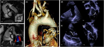
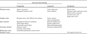
CHD affects 20–77% of patients with Turner syndrome, ranging in severity from incidental to potentially fatal (Table 1).Reference Mortensen, Andersen and Gravholt 1 CHD causes 8% of the threefold increased risk of early death, with left-sided anomalies being the most common.Reference Schoemaker, Swerdlow, Higgins, Wright and Jacobs 5 At the most severe end of the spectrum is hypoplastic left heart syndrome that affects 5%. Turner syndrome is diagnosed in 2% of hypoplastic left heart syndrome patients, and complication rates are, in the subset with Turner syndrome, increased at surgical palliation as well as with heart surgeries for other cardiovascular anomalies.Reference Cramer, Bartz, Simpson and Zangwill 6 – Reference Patel, Hickey and Mavroudis 8 Further left-sided obstructive anomalies include coarctation of the aorta, aortic stenosis, and, rarely, the Shone complex (Fig 1). Transthoracic echocardiography and cardiovascular MRI form the foundation for diagnosis and surveillance of these conditions (Table 2).
Timely detection of congenital cardiovascular anomalies in Turner syndrome requires systematic imaging in all patients and early targeted imaging when symptoms are present. A 17-year-old ( a ) has an occult aortic coarctation ( a , arrow) and bicuspid aortic valve ( a , inset), and a 55-year-old ( b ) has an “elongated transverse aortic arch” ( b , arrow) and mild regurgitation of a bicuspid aortic valve ( b , inset). A 44-year-old ( c ) incidentally has aberrant origins of left and right common carotid arteries (LCA, RCA) from a common arterial trunk and a retro-oesophageal right subclavian artery (RSA) that arises distal to the left subclavian artery (LSA). A 16-year-old ( d – g ) with Shone complex, comprised of aortic coarctation corrected surgically in infancy with no residual coarctation ( d ), a false tendon of the left ventricle ( e , arrow), subaortic obstruction ( f , arrow), and a supramitral ring ( g , arrow) above a parachute mitral valve. TTE=transthoracic echocardiography.

Cardiovascular abnormalities in Turner syndrome and their clinical significance.

Sensitivity of non-invasive imaging for the detection of cardiovascular disease in Turner syndrome, and suggested screening strategy for patients without concerning cardiac symptoms or urgent treatment-requiring anomaly.

ECHO: echocardiography
Scale: lowest sensitivity (+) increasing to highest sensitivity (++++); modalities with no score comprise those where secondary findings may be revealed but where the component in question cannot be directly assessed
* Patients with bicuspid aortic valve, aortic dilatation, and aortic coarctation that do not warrant surgery should be more frequently imaged than other patients
** Alternating strategy with transthoracic ECHO and cardiovascular MRI performed in separate years is recommended
Valvular disease
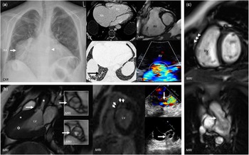
Bicuspid aortic valve morphology is, intriguingly, more frequent in Turner syndrome (15–30%) than in any other condition, and it renders these females prone to regurgitation and stenosis.Reference Mortensen, Andersen and Gravholt 1 , Reference Mortensen, Hjerrild and Andersen 9 Acquired aortic valve dysfunction is far more common than congenital, and may be seen both with the bicuspid aortic valve and the tricuspid aortic valve (Fig 2). Transthoracic echocardiography should be performed immediately at diagnosis of Turner syndrome. Poor echocardiographic windows can, however, hinder the differentiation of the tricuspid from the bicuspid valves in up to 6%.Reference Sachdev, Matura and Sidenko 10 Unresolved valve morphology at transthoracic echocardiography will necessitate either interrogation by high temporal and spatial resolution cine MRI in the valve plane or by transoesophageal echocardiography (Fig 2). Leaflet fusion mainly involves the right and left coronary cusps (82–95%) and tends to be complete, with partial fusions and unicuspid valve infrequently encountered.Reference Olivieri, Baba and Arai 11 , Reference Mortensen, Hjerrild and Stochholm 12 The bicuspid aortic valve severely increases the risk of aortic aneurysm formation and dissection (Figs 3 and 4), and warrants increased frequency of imaging surveillance for these complications. Less common aortic valve anomalies include congenital valvular stenosis (Fig 2), subaortic membrane and annular hypoplasia. The mitral valve is the second most common valve to be diseased in Turner syndrome with reported anomalies including leaflet prolapse, accessory leaflet, cleft leaflet, parachute mitral valve (Fig 1), supravalvar mitral stenosis, and myxomatous degeneration. Tricuspid and pulmonary valve disease is rare (Fig 2).Reference Cramer, Bartz, Simpson and Zangwill 6 Precise delineation of these valve anomalies and their impact on the left and right ventricles often necessitates a combination of echocardiography and MRI (Fig 2).
Bicuspid aortic valves, native or surgically replaced, render patients with Turner syndrome especially prone to aortic, valvular, and cardiomyopathic complications, but imaging surveillance is warranted irrespective of valve morphology because of an increased risk of complications also seen with tricuspid aortic valves. A 21-year-old ( a ) with valve replacement for a stenotic bicuspid aortic valve ( a , arrowhead) has biventricular cardiomyopathy with severe tricuspid regurgitation ( a , circle), right atrial (RA) and right ventricular (RV) dilatation, and smooth interlobular septal thickening consistent with pulmonary venous congestion ( a , black arrow). A 30-year-old ( b ) with a severely stenotic tricuspid aortic valve ( b , large arrows {aortic valve} and star {stenotic jet}) and concentric left ventricular (LV) hypertrophy ( b , asterisks) with preserved global systolic function with systolic mid and apical chamber obliteration ( b , circle), and patchy delayed enhancement in the subendocardial apical and mid-septal myocardium ( b , small arrows). An 18-year-old ( c ) with valvotomy for pulmonic stenosis has minimal flow acceleration ( c , arrow) across a trivially regurgitant valve, mild main pulmonary artery dilatation ( b , asterisk), and mild free-wall hypertrophy (6 mm) of the non-dilated RV ( c , arrowheads). CXR=chest X-ray; TOE=transoesophageal echocardiography; TTE=transthoracic echocardiography.

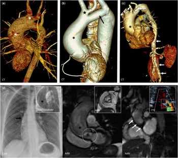
Aortic aneurysm is common in Turner syndrome and warrants imaging surveillance during the entire lifetime, which applies irrespective of cardiovascular phenotype, although some guidance can be taken by the presence of risk factors such as bicuspid aortic valves, elevated blood pressure, and aortic diameter. A 15-year-old ( a ), with childhood commissurotomy for a congenitally stenotic bicuspid aortic valve, has a 4.5-cm (2.7 cm/m2) ascending aorta ( a , asterisk) with a bovine arch ( a , star). A 50-year-old ( b ), with bicuspid aortic valve (normal function) and childhood end-to-end aortic coarctation repair, has a 5.0-cm (4.0 cm/m2) ascending aorta also with a bovine arch ( b , star) that is hypoplastic hypoplasia ( b , arrow). A 69-year-old ( c ), with mechanical aortic valve prosthesis (11 years before for a stenotic bicuspid aortic valve), had a 5.0-cm (4.0 cm/m2) ascending aorta ( c , asterisk) and extensive atherosclerosis of the major thoracic and abdominal arteries ( c , arrows). A 57-year-old ( d ) with severe stenosis of a thickened tricuspid aortic valve ( a , circles) and severe ascending aortic dilatation ( d , black arrow and stars) with a normally functioning aortic valve prosthesis and interposition graft in situ 12 months later ( d , white arrows and square box). CXR=chest X-ray; TTE = transeosophageal echocardiography.

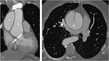
A low threshold for excluding aortic dissection should be adapted in Turner syndrome, and when reviewing imaging it is important to scrutinise for additional anomalies. A 44-year-old with acute Stanford type A dissection (asterisk [false lumen], star [true lumen], circle [entry point]) occurring years after mechanical aortic valve replacement (arrow) due to a stenotic bicuspid aortic valve. Note is made of an incidental, haemodynamically non-significant partial anomalous pulmonary venous return consisting of middle and right upper lobar pulmonary veins draining to the superior caval vein (arrowhead).

Aortic arch
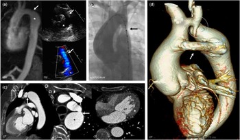
Coarctation of the aorta is common (17%) (Fig 5) and often seen in association with the bicuspid aortic valve.Reference Mortensen, Hjerrild and Andersen 9 The entire aorta should be considered abnormal even when coarctation is the only initial abnormal finding. In spite of 5–12% undergoing surgical or endovascular coarctation repair in childhood, an estimated 5–8% of adults have “occult” coarctation as defined by a shelf-like luminal narrowing on MRI (Fig 1).Reference Mortensen, Andersen and Gravholt 1 The overwhelming majority of these “occult” cases have no ancillary features to suggest significant flow restriction, such as arterial collateral formation, focal flow acceleration, or post-stenotic dilatation. Coarctation increases the risk of aortic aneurysm formation and this risk persists even after repair, necessitating continued imaging surveillance for both re-coarctation and aortic dilatation (Fig 5). Echocardiography can visualise the location of the narrowing with diastolic run-off being the most reliable sign of significant obstruction.Reference Baumgartner, Bonhoeffer and De Groot 13 Cardiac CT and MRI offer superior anatomical delineation compared with echocardiography and may demonstrate collateralisation. Cross-sectional imaging should be performed at coarctation diagnosis, with MRI generally being preferred in the non-acute setting due to a lack of radiation exposure plus additional information regarding left ventricular function, myocardial mass, and aortic flow patterns. Ultimately, invasive catheter angiography may be necessary, as it is the diagnostic gold standard for measuring pressure gradients and offers the opportunity for percutaneous intervention (Fig 5).Reference Baumgartner, Bonhoeffer and De Groot 13 The optimal coarctation treatment in Turner syndrome is not known. Complication rates at repair are generally comparable with the general population, although an internal wall frailty has been suggested to increase complication rates.Reference Mortensen, Andersen and Gravholt 1 A low threshold should be adapted for non-invasive imaging, often cardiac CT, in the immediate post-surgical state in order to assess complications such as haemorrhage, dissection, or pseudoaneurysms. Non-electrocardiogram-gated cardiac CT yields diagnostic images for coarctation site assessment, but if there are additional queries regarding the aortic root then electrocardiogram-gating may be preferred. In the ambulatory follow-up after coarctation repair, echocardiography and MRI are generally combined. The currently used stents are usually MRI safe, but stent artefacts may hamper accurate assessment of the repair site, although the diagnostic accuracy of MRI can be increased with optimised sequences.Reference Nordmeyer, Gaudin and Tann 14
Treatment of aortic anomalies in Turner syndrome does not abolish the need for continued imaging follow-up due to a high risk of recurrent coarctation and aortic aneurysm formation. A 17-year-old ( a ) has mild narrowing at a subclavian flap repair site ( a , arrows) with normal flow and only mild post-isthmic dilatation ( a , stars). A 35-year old ( b ) demonstrates re-coarctation ( b , arrow) of an end-to-end coarctation repair, and a 63-year-old ( c ) has severe narrowing (6 mm) of a subclavian flap coarctation repair ( c , arrows) with severe post-stenotic dilatation ( c , asterisk) and left ventricular (LV) hypertrophy ( c , star). A 47-year-old ( d ) has a patent 10 mm graft bypassing a hypoplastic transverse arch ( d , circle) with severe ascending and descending aortic dilatation. TTE=transthoracic echocardiography.

The common elongated transverse thoracic arch (47–49%) can be regarded as a forme fruste of coarctation (Fig 1), where the arch is elongated with a kink in the inferior curvature at the aortic isthmus.Reference Mortensen, Hjerrild and Andersen 9 , Reference Ho, Bakalov and Cooley 15 This anomaly forms a part of an aortic phenotype that includes dilatation and aneurysm of the head and neck arteries (44%).Reference Mortensen, Hjerrild and Andersen 9 Best appreciated on MRI and CT, the elongated transverse thoracic arch should always be noted as it increases the risk of hypertension and aortic aneurysm formation.Reference De Groote, Devos and Van Herck 16 Incidental arch anomalies are also often present, which mainly need consideration during surgical planning (Fig 1).Reference Mortensen, Hjerrild and Andersen 9
Cardiac shunts
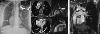
Partial anomalous pulmonary venous return is common (13–18%).Reference Mortensen, Andersen and Gravholt 1 The anomalous vein(s) forms a left-to-right shunt by draining into the right atrium, superior caval vein, or tributary veins such as the azygos and innominate veins (Fig 6). The shunt fraction is generally not clinically significant and will typically only warrant corrective surgery when the Qp:Qsratio>1.5 (pulmonary arterial flow relative to aortic flow) with concomitant right heart dilation (Fig 6). The more severe total anomalous pulmonary venous return is very rare in Turner syndrome. Owing to the risks associated with the presence of the shunt lesions, early screening for anomalous pulmonary venous return is warranted in all females with Turner syndrome.Reference Bondy 3 Cardiac CT provides exquisite high spatial resolution for precise anatomical delineation of the shunt anatomy. However, CT will unlike the radiation free MRI not assess the shunt fraction, which is an essential prognostic marker alongside right ventricular function and volumes that are also easily and reliably provided with MRI. Echocardiography lacks the ability to accurately locate a suspected shunt and quantify shunt fraction.Reference Baumgartner, Bonhoeffer and De Groot 13 MRI is thus the preferred modality for diagnosis and follow-up, and the surveillance frequency should be guided by symptoms, shunt fraction, and right ventricular parameters. Other cardiac shunts are infrequent in Turner syndrome, including atrial (1–2%) and ventricular septal (1–5%) defects.Reference Cramer, Bartz, Simpson and Zangwill 6 Partial anomalous pulmonary venous return frequently associates with a sinus venosus atrial septal defect in the general population, and the possibility of an atrial septal defect should always be considered also in Turner syndrome. Systemic venous anomalies are also frequent, such as a persistent left superior caval vein to the coronary sinus, but they are generally of limited relevance unless planning a major thoracic surgery, placing vascular lines, or inserting pacemaker leads.
The high prevalence of partial anomalous pulmonary venous return warrants screening early in life in Turner syndrome, although the shunt size often does not warrant surgical correction. A 26-year-old ( a ) has a haemodynamically significant partial anomalous pulmonary venous return, evident as right atrial (RA) and ventricular (RV) enlargement with pulmonary plethora ( a , circle) that result from anomalous middle and bilateral upper pulmonary veins ( a , arrows) that drain into a dilated superior caval vein (SVC) and innominate vein (INV). Incidentally, there is mild narrowing of the descending aorta, with a posterior shelf and an aneurysmal left subclavian artery ( a , square). A 51-year-old ( b ) with scimitar-type anomalous pulmonary venous drainage developed pulmonary vein thrombosis after surgical re-direction of right-sided pulmonary veins to the left atrium, which necessitated pneumonectomy for unilateral pulmonary hypertension with subsequent extreme cardiac dextroposition but normal cardiac function and volumes. CXR=chest X-ray; TTE=transthoracic echocardiography.

Acquired heart disease
Acquired heart disease accounts for 40% of excess deaths in Turner syndrome.Reference Schoemaker, Swerdlow, Higgins, Wright and Jacobs 5 CHD and acquired heart disease have significant overlap in Turner syndrome, and the distinction can be difficult, with cardiovascular diseases that are normally seen with ageing occurring even in very young girls.Reference Mortensen, Andersen and Gravholt 1
Thoracic aortic aneurysm and acute aortic syndrome
Aortic dissection is the leading cause of death in the young adult with Turner syndrome, occurring at a 100-fold increased incidence or in 2% of females with Turner syndrome at a median age of 35 years (Fig 4).Reference Gravholt, Landin-Wilhelmsen and Stochholm 17 , Reference Bondy 18 Dissection may occur at any location in the thoracic aorta, with Stanford type A seen in 63% (Fig 4) and type B in 37%.Reference Gravholt, Landin-Wilhelmsen and Stochholm 17 An apparent structural weakening involves the tunica media with a congenital defect likely present, possibly in the form of cystic media degeneration.Reference Gravholt, Landin-Wilhelmsen and Stochholm 17 Aortic diameter is currently the principal risk marker for aortic dissection, with the accuracy for predicting aortic events increased by indexation to body surface area. Average indexed diameters are enlarged in adults with Turner syndrome at all locations, except the classical coarctation site, with a prevalence of aortic dilation of 40% on MRI.Reference Mortensen, Hjerrild and Stochholm 12 Consistent with a propensity for type A and B dissection, both the ascending and descending aortae may be aneurysmal in Turner syndrome.Reference Mortensen, Hjerrild and Andersen 9 , Reference Mortensen, Hjerrild and Stochholm 12 Aortic wall stiffness and distensibility are also abnormal in Turner syndrome, but a lack of formal prognostic validation hampers the use of such markers in clinical practice.
Aortic dissection occurs at presumed normal aortic diameters in Turner syndrome, even following indexation to body surface area, which hinders any extrapolation of conventional size criteria onto this cohort.Reference Carlson and Silberbach 19 Risk factors for aortic dilation, and secondarily dissection, include bicuspid aortic valve, aortic valve dysfunction, aortic coarctation, hypertension, 45,X karyotype, and pregnancy (Fig 3).Reference Mortensen, Andersen and Gravholt 1 A critical size cut-off of >25 mm/m2 has been proposed for severe aortic disease in Turner syndrome, but this will fail to identify all at-risk patients because a significant proportion will progress to dissect even below this threshold.Reference Carlson and Silberbach 19 This cut-off was proposed by a relatively small study that demonstrated a 33% incidence of aortic dissection for females with diameters above this limit when following-up 126 females for nearly 3 years.Reference Matura, Ho, Rosing and Bondy 20 Owing to the complexity of aortic dilatation and dissection, balancing the risk of acute aortic syndrome for a given aortic diameter against that associated with prophylactic aortic surgery is difficult in daily clinical practice. This is compounded by higher mortality with complex heart surgery in Turner syndrome.Reference Cramer, Bartz, Simpson and Zangwill 6 Importantly, aortic size is also a better positive than negative predictor of aortic events, which even when used together with other risk factors will fail to identify at least 1 in 10 aortic dissections in Turner syndrome.Reference Carlson and Silberbach 19 Therefore, it remains difficult to identify the subset of females with Turner syndrome who require not just imaging surveillance but also lifestyle recommendations, medical treatment or surgical prophylaxis. Current risk assessment must therefore be highly individualised and be based on aortic size at any single time point, aortic growth over time, and other risk factors.Reference Erbel, Aboyans and Boileau 21 Measurement variability in the range of, at least, 1–3 mm must also be factored into the interpretation of serially measured aortic diameters.Reference Lu, Rizzo and Marques-Vidal 22 , Reference Groth, Henes and Mullerleile 23 For surveillance studies, it can be advocated for a single observer to re-measure each study at pre-defined positions in order to reduce measurement variability, and comparison of similar imaging techniques is a must.Reference Mortensen, Hjerrild and Stochholm 12 Electrocardiogram-gating or electrocardiogram-triggering is essential, especially for aortic root assessment, and diastolic measurements are possibly more reproducible than systolic measurements, although the relative value of measurements obtained over the cardiac cycle in risk stratification has not been determined.Reference Erbel, Aboyans and Boileau 21 Ascending aortic growth rates are 0.1–0.4 mm/year in Turner syndrome when assessed by MRI, which corresponds to other syndromic states with excessive risk of aortic dissection.Reference Mortensen, Hjerrild and Stochholm 12 Normative prospective MRI data for adults with Turner syndrome, incorporating the principal determinants of aortic diameter such as body size, age, and blood pressure, are available and can help determine if a female falls within her syndrome-specific, expected normative range.Reference Mortensen, Erlandsen, Andersen and Gravholt 24 Aggressive medical treatment is warranted in these often hypertensive females, typically combining angiotensin II receptor blockers with beta-blockers. Calcium channel blockers are often not favoured because of a propensity for peripheral oedema in Turner syndrome.
Coronary and systemic arterial disease
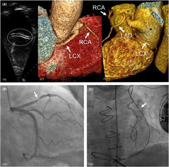
Congenital coronary arterial anomalies also occur, primarily in the form of aberrant origin (Fig 7) and coronary artery fistula.Reference Mortensen, Andersen and Gravholt 1 Systematic screening for these anomalies is not justified unless major cardiothoracic surgery is planned, when screening should be performed even in the young. Cardiac CT provides high spatial resolution anatomical information with a high negative predictive value for coronary atherosclerosis that is known to cause significant morbidity and mortality in Turner syndrome.Reference Schoemaker, Swerdlow, Higgins, Wright and Jacobs 5 No imaging study has assessed the true prevalence and extent of coronary atherosclerosis in these females, with the current evidence being mainly epidemiological. A plethora of aetiologies for myocardial ischaemia has been implicated.Reference Mortensen, Andersen and Gravholt 1 However, atherosclerosis is the most likely cause (Fig 7) with risk factors abundantly present from an early age such as hypertension, dyslipidaemia, insulin resistance, obesity, perturbed “growth hormone–insulin growth factor I” axis, and premature ovarian failure.Reference Mortensen, Andersen and Gravholt 1 Premature increased carotid artery intima-media thickness, endothelial dysfunction, and arterial stiffening further support atherosclerosis being a common phenomenon.Reference Mortensen, Andersen and Gravholt 1 , Reference Baguet, Douchin and Pierre 25
Congenital and acquired coronary artery anomalies in Turner syndrome should always be considered at baseline cardiovascular imaging with targeted coronary imaging warranted when congenital anomalies are suspected, in symptomatic patients, and before major cardiothoracic surgery. In a 27-year-old ( a ), a coronary artery was noted ( a , circle) in the left atrioventricular groove with the origin not seen but later confirmed to be a left circumflex coronary artery (LCX) with aberrant origin from the proximal right coronary artery (RCA) and a course between the left atrium (LA) and the aorta towards the left atrioventricular groove. A normal left anterior descending artery (LAD) was noted. In a 34-year-old ( b ), impaired left ventricular function (not shown) led to the diagnosis of a moderate atherosclerotic stenosis of the proximal LAD ( b , arrow). In a 61-year-old ( c ), with previous coronary artery bypass operation and implanted cardioverter defibrillator, an incidental persistent left superior caval vein was evident from an anomalous course of the pacemaker leads ( c , arrow). CCT=cardiac CT; TTE=transthoracic echocardiography; CAG = coronary angiography.

Cardiomyopathy
Left ventricular dysfunction and hypertrophy are common.Reference Mortensen, Gravholt, Hjerrild, Stochholm and Andersen 26 A plethora of metabolic and cardiovascular anomalies often seen in Turner syndrome can negatively impact the left ventricle, including insulin resistance, hypertension, aortic coarctation, and perturbed heart valve function.Reference Mortensen, Gravholt, Hjerrild, Stochholm and Andersen 26 Concentric left ventricular hypertrophy is common, associating with hypertension and bicuspid aortic valve, and myocardial metabolism has recently been found abnormal.Reference Trolle, Hjerrild and Mortensen 27 Even young females with no risk factors for myocardial disease have diastolic left ventricular dysfunction, and left atrial dilatation is common.Reference Andersen, Hjerrild and Sorensen 28 Conversely, overt systolic left ventricular dysfunction is uncommon.Reference Mortensen, Gravholt, Hjerrild, Stochholm and Andersen 26 Endocardial fibroelastosis may be seen in the very young girls with Turner syndrome, especially in the context of severe left ventricular outflow obstruction, but these girls rarely survive into adulthood. Other congenital myocardial anomalies such as non-compaction are rare. Transthoracic echocardiography is the principal clinical imaging modality for left ventricular cardiomyopathy because of the ability to delineate anatomy as well as systolic and diastolic function of the left ventricle. However, MRI is the reference standard for left ventricular mass, volume, and systolic function.
Best imaging strategy
Ambulatory patients
MRI should be performed at the earliest possible opportunity in all newly diagnosed females, with transthoracic echocardiography used as the sole imaging modality only in children where MRI either would necessitate general anaesthesia or is not indicated by clinical or echocardiographic findings.Reference Bondy 3 Focus on this is important as suboptimal cardiac investigations remain a major issue and systematic screening has repeatedly been shown to unveil clinically important but previously undetected disease. Owing to radiation concerns and lack of functional information, cardiac CT is a generally the second choice compared with MRI. Repeat imaging, even in asymptomatic patients with normal baseline MRI, will be necessary as many females will eventually need surgical or medicinal intervention as cardiovascular disease becomes apparent over time. The coronary arteries only need routine imaging when planning major cardiothoracic surgery or as justified by symptoms of angina.
MRI is the gold standard imaging test for diagnosis and surveillance of morphological anomalies of the thoracic aorta in Turner syndrome, because transthoracic echocardiography underestimates the size of both the ascending and the descending aortae.Reference Ostberg, Brookes, McCarthy, Halcox and Conway 29 , Reference Lanzarini, Larizza and Prete 30 A barrel-shaped chest often restricts echocardiographic windows.Reference Lanzarini, Larizza and Prete 30 , Reference Hjerrild, Mortensen and Sorensen 31 However, if a good echocardiographic window is obtained with adequate aortic views, then transthoracic echocardiography may be an appropriate surveillance method unless there is rapid growth or significant dilatation when confirmatory cross-sectional imaging must be deployed. MRI also more reliably diagnoses partial anomalous pulmonary venous return and other congenital cardiac anomalies. Of course, in complex and severe early-life anomalies such as hypoplastic left heart syndrome, transthoracic echocardiography remains the principal initial diagnostic modality of choice. Owing to the lack of a newborn screening programme, there is often a significant diagnostic delay for the diagnosis of Turner syndrome, especially in patients with less severe external phenotypes. A link is suggested between the externally visible neck webbing and congenital anomalies, but clinically significant cardiac disease is not exclusive to certain external features, to females with early diagnosis, or in the classical 45,X karyotype.Reference Ho, Bakalov and Cooley 15 , Reference Hjerrild, Mortensen and Sorensen 31 Therefore, a cardiovascular screening approach should be applied globally in Turner syndrome, focussing on the heart, heart valves, and great vessels. In contrast to this, routine investigation for ancillary features such as carotid atherosclerosis is not justified at present.
The frequency of follow-up and the imaging modality of choice should be determined by the disease spectrum encountered and the quality of acoustic windows at transthoracic echocardiography (Table 2).Reference Bondy 3 A reasonable approach to aortic disease in Turner syndrome would be to plan follow-up based on encountered aortic diameter compared with normative data for aortic diameter and growth rates in Turner syndrome, and according to the presence of cardiovascular co-morbidity such as hypertension and bicuspid aortic valves.Reference Mortensen, Hjerrild and Stochholm 12 , Reference Mortensen, Erlandsen, Andersen and Gravholt 24 Alternating transthoracic echocardiography and MRI may be helpful. Follow-up should be performed at 5- to 10-year intervals with normal baseline imaging, because disease may develop later in life, and clinicians should therefore also be careful to avoid loss to follow-up.Reference Bondy 3 Importantly, all current recommendations are based on clinical pragmatism and experience from other diseases, with an update of the expert consensus document needed.Reference Bondy 3
Acute patients
Sudden chest pain should in view of the high risk of acute aortic syndrome result in urgent imaging. The strategy chosen must not rely too heavily on the presence of risk factors for aortic dissection because at least 10% of aortic dissections in Turner syndrome occur with no apparent risk factors present.Reference Carlson and Silberbach 19 Cardiac CT outperforms echocardiography in suspected thoracic aortic dissection, with the latter having slightly inferior diagnostic accuracy. Echocardiography is, however, operator dependent and even the transoesophageal approach has limited acoustic windows with a blind angle in the distal ascending and proximal aortic arch. In the acute setting, MRI is limited by a relatively long examination time and difficulty in monitoring the unstable patient, and chest radiography has no real role. In comparison, cardiac CT rapidly images the entire aorta and the aortic branch vessels including coronary arteries, which makes it the preferred imaging modality. If acute aortic syndrome has been excluded in the female presenting with acute cardiological sounding symptoms, then other causes need to be considered such as myocardial ischaemia and valvular heart disease.
Conclusion
Cardiovascular disease is a major prognostic determinant in Turner syndrome. A heavy burden of congenital and acquired heart disease severely increases the risk of premature death and disability. All females with Turner syndrome must be screened at the earliest possible opportunity for cardiovascular disease at a specialist centre. Echocardiography is sensitive to many of the disorders encountered, but common shunt lesions may be missed and aortopathy may be suboptimally characterised. Owing to a higher sensitivity than echocardiography, cardiovascular MRI should be prescribed at the earliest possible opportunity when it can be performed without the use of general anaesthesia. Repeat assessments are warranted even in asymptomatic patients with normal baseline assessments because of a complex interplay between congenital and acquired cardiovascular disease as well as abundant endocrine factors. There is limited evidence to specific follow-up intervals even in females with established cardiovascular disease, and the timing of imaging follow-up should be determined on a patient-specific basis by the multidisciplinary team. A female with Turner syndrome who presents with symptoms potentially referable to acute aortic disease must be urgently assessed even if she is young, and cardiac CT together with echocardiography are cornerstone for exclusion of aortic events in this setting.
Acknowledgements
None.
Financial Support
Financial support was obtained from the Danish Ministry for Science and Technology and Innovation, the Danish Heart Foundation, and the Novo Nordisk Foundation. K.H.M. also has research support from the NIHR Cambridge Biomedical Research Centre.
Conflicts of Interest
None.