Introduction
The subclass Aspidogastrea Faust & Tang 1936, comprises a small group in the phylum Platyhelminthes, class Trematoda, which is composed of the families Rugogastridae Schell, 1973; Stichocotylidae Faust & Tang, 1936; Multicalycidae Gibson & Chinabut, Reference Gibson and Chinabut1984 and Aspidogastridae Poche, 1907 (Rohde, Reference Rohde, Littlewood and Bray2001, Reference Rohde, Jones, Gibson and Bray2005).
The family Aspidogastridae has three subfamilies: Aspidogastrinae Poche, 1907; Cotylaspinae Chauhan, 1954; and Rohdellinae Gibson & Chinabut, Reference Gibson and Chinabut1984 (Zamparo & Brooks, Reference Zamparo and Brooks2003). These three subfamilies are distinguishable by the number of longitudinal septa present in the adhesive disc (three septa are present in the first subfamily and two septa are present in the other two subfamilies). In particular, the subfamily Rohdellinae is differentiated from Cotylaspinae exclusively due to the presence of a hermaphroditic channel (Rohde, Reference Rohde, Jones, Gibson and Bray2005). Phylogenetic studies do not support the actual classification of the family Aspidogastridae, revealing that the subfamilies Aspidogastrinae and Cotylaspinae are monophyletic groups. However, according to Zamparo & Brooks (Reference Zamparo and Brooks2003), the subfamily Rohdellinae is a paraphyletic group.
Colomesus psittacus (Bloch & Schneider, 1801) is a demersal freshwater or estuarine fish that has been found in saltwater environments from the Gulf of Paria (Venezuela) to Pará State (Brazil) (Cervigon et al., Reference Cervigon, Cipriani, Fisher, Garibaldi, Hendrickx, Lemus, Márquez, Poutiers, Robaina and Rodriquez1993). In Brazil, this species is commonly found in estuarine regions of the Amazon Basin (Camargo & Maia, Reference Camargo and Maia2008).
During a helminthological survey of fish from the lower Tocantins River (Pará State), some Aspidogastridae species were found infecting the small intestines of C. psittacus. The present work, using light and scanning electron microscopy, describes a new species of the genus Rohdella, family Aspidogastridae, isolated from freshwater rivers of the Amazon Region. The description of the new taxon was supported by molecular data.
Materials and methods
Collection and examination of samples
A total of 112 specimens of C. psittacus were collected between January 2009 and April 2010 from the lower Tocantins River, Cametá Municipality, Pará State (02°14′S 49°49′W). Among these specimens, 85 were infected by trematodes of the subclass Aspidogastrea. The helminths were collected and rinsed in phosphate-buffered saline for in vivo analysis. Some specimens were compressed and fixed in AFA (2% glacial acetic acid, 3% formaldehyde and 95% ethanol 70°GL), stained with alcoholic carmine, dehydrated in an alcohol series, clarified in methyl salicylate and mounted on permanent slides for light microscopy analysis. Morphometric data and illustrations were made using an Olympus BX41 microscope (Olympus, Tokyo, Japan) with a drawing tube. Measurements, as means and with the range in parentheses, are given in millimetres unless otherwise noted.
For scanning electron microscopy (SEM), the helminths were fixed in 2.5% glutaraldehyde in 0.1 m sodium cacodylate buffer (pH 7.4), post-fixed in 1% osmium tetroxide, dehydrated with a graded alcohol series, critical-point dried, mounted on stubs, sputter-coated with gold and examined using an LEO 1450 VP scanning electron microscope (Carl Zeiss, Germany).
The excretory system was analysed in vivo after staining with neutral red and examined using an Olympus BX 41 microscope.
Molecular analysis
Total DNA was isolated according to a standard protocol (Sambrook et al., Reference Sambrook, Fritsch and Maniatis1989). Small subunit ribosomal DNA (SSU rDNA) and the internal transcribed spacers (ITS) (ITS1, 5.8S rDNA and ITS2) segments were amplified according to the original description with the primers WormA/WormB (Littlewood & Olson, Reference Littlewood, Olson, Littlewood and Bray2001) and NC5/NC2 (Zhu et al., Reference Zhu, Gasser, Podolska and Chilton1998), respectively. Amplicons were electrophoresed on a 1% agarose gel, excised and purified using GFX PCR DNA and a gel purification kit (GE Healthcare, Little Chalfont, Buckinghamshire, UK). An aliquot of each purified product was ligated into a pGEM-T vector (Promega, Madison, Wisconsin, USA) overnight and then desalted before electroporation into Escherichia coli TOP 10 cells (Invitrogen Life Technologies, Carlsbad, California, USA). The insert DNA of the white clones was obtained by polymerase chain reaction (PCR) directly from the colonies, using M13F/M13R primers, and sequenced automatically in a 3130 Genetic Analyzer (Applied Biosystems, Foster City, California, USA) according to the manufacturer's specifications. The 18S11 and PaceB (Littlewood & Olson, Reference Littlewood, Olson, Littlewood and Bray2001) primers were used as internal primers to obtain the SSU rDNA sequence. BioEdit software (Hall, Reference Hall1999) was used to align the forward and reverse sequences.
Results
Rohdella amazonica n. sp
Description from light microscopy and SEM
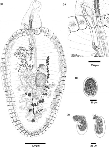
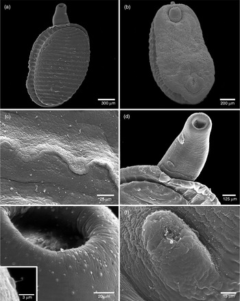
The following description is based on ten specimens. Body slightly longer than wide, narrow at the posterior region, measuring 5.92 (3.32–8.10) × 3.04 (1.85–4.44) (fig. 1a, fig. 2a, b). Tegument with small papillae distributed irregularly throughout the entire body. Adhesive disc occupying the entire ventral region composed of 29 (24–38) rows of transverse alveoli separated by two longitudinal septa (fig. 1a, fig. 2a, b). Sub-terminal oral aperture, the sucker absent and tegument covered by ciliated papillae randomly distributed (fig. 1, fig. 2d, e). Oral funnel 0.37 (0.14–0.54) × 0.3 (0.2–0.4) (fig. 2d). Pharynx oval and large 0.35 (0.2–0.53) × 0.21 (0.16–0.25), surrounded by pharyngeal glands. The excretory system forming long tubes situated laterally, starting close to the pharynx and discharging into a longitudinal, dorsal excretory pore with a longitudinal aperture close to the posterior extremity. Caecum blind and simple, terminated near the posterior extremity of the body 3.8 (2.44–5.06) in length. An eversible, medium-lateral, dexterous, hermaphroditic duct close to the pharynx, not measurable (fig. 1a, b; fig. 2d, f). A single, post-ovarian, medial, equatorially situated testis situated 2.97 (1.82–3.73) from the anterior region measuring 0.67 (0.4–0.86) × 0.47 (0.25–0.62) (fig. 1). Cirrus sac absent. A long and thick-walled seminal vesicle, with numerous prostatic glands, situated in the terminal portion of the vas deferens (fig. 1b). Ovary sub-equatorial, comma-shaped, situated at 2.48 (1.53–3.26) from anterior region and measuring 0.45 (0.29–0.54) × 0.32 (0.25–0.40). Ootype located between the testis and ovary. Uterus loops mainly in the dorsal field. Vitellarium follicular in lateral fields. Metraterm long and thick-walled, discharging at the hermaphroditic duct (fig. 1b). Ovoid eggs measuring 0.08 (0.06–0.09) × 0.05 (0.04–0.06), some eggs showing larvae emerging (fig. 1c, d).
Morphology of Rohdella amazonica n. sp.: (a) entire worm, ventral view; (b) genital pore; (c) egg; (d) emerging larva.

Ultrastructure of Rohdella amazonica n. sp.: (a) the ventral view; (b) dorsal view; (c) fringe between the transverse alveoli; (d) anterior end with the hermaphroditic channel; (e) oral aperture, inset shows ciliated papilla; (f) reverted hermaphroditic channel.

Taxonomic summary
Type host. Banded puffer, Colomesus psittacus (Bloch & Schneider, 1801) (Tetraodontiformes: Actinopterygii).
Type locality. Tocantins River, Cametá City, Pará State, Brazil. 2°14′S 49°49′W.
Site of infection. Intestine.
Prevalence. 66.3%.
Type data and depository. Specimens were deposited in an invertebrate collection at Museu Paranse Emílio Goeldi (MPEG): one holotype (MPEG 0030) and two paratypes (MPEG 0031 and MPEG 0032).
Etymology. The species is named after the biome where it was identified.
Molecular data
We obtained 1871 and 840 nucleotides for SSU rDNA (GenBank accession number, KC181852) and the ITS (accession number KC181853), respectively. These molecular markers present different phylogenetic signals, and they may contribute to future studies and understanding of R. amazonica n. sp. phylogenetic relationships.
Discussion
The genus Rohdella Gibson & Chinabut, Reference Gibson and Chinabut1984 has three valid species: R. siamensis Gibson & Chinabut, Reference Gibson and Chinabut1984 (a parasite of fish of the infraclass Teleostei), R. anodontiase Aboul-Dahab, Abdel-Salam & El-Damarany, Reference Aboul-Dahab, Abdel-Salam and El-Damarany1993 (a parasite of molluscs of the family Anodontidae) and R. clariasa Mohamadain, 1989, which is a parasite of a fish from the Nilo River.
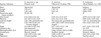
The new species is similar to R. siamensis regarding some morphological and morphometric characters (total length, width, number of transverse septa, oral funnel, pharynx, testis and ovary shape), but it has larger testis and ovary dimensions (testis: 0.4–0.86 × 0.25–0.62 versus 0.17–1.22 × 0.13–1.30; ovary: 0.29–0.54 × 0.25–0.4 versus 0.05–0.06 × 0.05–0.36).
The species R. amazonica n. sp. has a small, eversible hermaphroditic channel that is difficult to visualize using light microscopy, although it is easily observed using scanning electron microscopy (fig. 2d and f). The new species differs from R. anodontiase and R. clariasa, because those species do not have an eversible hermaphroditic channel, and it is similar to R. siamensis. However, R. siamensis has a longer hermaphroditic channel. Additionally, the hermaphroditic duct in R. siamensis and R. anodontiase is sinistral–lateral, whereas that in the new taxon is dextral–lateral, thus easily distinguishing the species and proposing R. amazonica as a new species for this genus.
The ultrastructure of the tegument of R. amazonica n. sp. displays numerous ciliated papillae that are concentrated at the cephalic region, primarily surrounding the oral aperture, and distributed randomly throughout the body. Although R. siamensis has been studied by SEM, Gibson & Chinabut (Reference Gibson and Chinabut1984) described the presence of pores in the margin of the lateral aveoli but did not observe the ciliated papillae covering the tegument surrounding the oral aperture, observed here for R. amazonica n. sp., thus enforcing the differences between the species.
Therefore, based on the morphological and morphometric characteristics presented in this study, the species could be considered a new taxon, as it has unique characteristics, including the presence of oesophageal glands, tegument covered by ciliated papillae and the position and shape of the hermaphroditic duct, which in this species is dextral, medial–lateral, short and eversible (table 1).
Morphometric characteristics (mm) of Rohdella amazonica n. sp. for comparison with R. siamensis and R. anodontiase.

Thus, R. amazonica n. sp. is the fourth species of this genus and an additional helminth parasite of C. psittacus. Additionally, we demonstrated new molecular data for an aspidogastrean species from the Amazon biome.
Acknowledgements
We are grateful to Dr Hilton Tulio Costi (Laboratory of Scanning Electron Microscopy/MPEG) for his technical support with the scanning electron microscopic analyses.
Financial support
This work was supported by the CAPES Foundation, Ministry of Education of Brazil (grants CAPES-PROCAD NF/2009, CAPES–PARASITOLOGIA BÁSICA/2010).
Conflict of interest
None.