Introduction
An aortic arch located on the right side of the trachea is referred to as a right aortic arch, occurring in approximately 0.01% to 0.1% of the population. In some cases, the right aortic arch is complicated by a vascular ring that encircles the esophagus and/or trachea with vascular structures. A right aortic arch with an aberrant left subclavian artery and left ductus arteriosus is among the most common forms of vascular rings, representing roughly 25% of cases. Coarctation of the aorta refers to the narrowing of the aorta, usually just distal to the left subclavian artery, affecting 0.06% to 0.08% of the population. While there are many reports on these anomalies individually, their combination is quite rare. Reference Law1–Reference Agarwal4 We found only seven published case reports describing these anomalies together, with a comprehensive literature review.
We present a rare case involving this combination of anomalies in a neonate. The patient was born with a right aortic arch with a unique branching pattern where the first branch was a bicarotid trunk, followed by the right subclavian artery, then an aberrant left subclavian artery. The aberrant left subclavian artery, which passed retro-oesophageally, formed a vascular ring with the ductus. Moreover, the patient had transverse arch coarctation. The condition was successfully treated with surgical repair.
Methods
A premature neonate, born via C-section at 33 weeks of gestation to a 31-year-old G1P2 female, received a fetal diagnosis of right-sided aortic arch and persistent left superior vena cava on obstetric ultrasound.
Postnatal echocardiogram revealed normal cardiac segmental anatomy but showed evidence of transverse arch coarctation with a peak gradient of 42 mmHg across the aortic arch, as well as continuous diastolic flow on spectral Doppler. The aortic branching pattern was not well-delineated on the echocardiogram, prompting further imaging.
A cardiac CT angiogram was performed to better define the aortic arch anatomy. The CT angiogram confirmed the transverse arch coarctation and identified a unique branching pattern: the first branch was a common carotid trunk that bifurcated into right and left common carotid arteries; the second branch was the right subclavian artery; and the third was a retro-oesophageal aberrant left subclavian artery originating from a diverticulum of Kommerell. The left ductal ligament connected to this aberrant vessel, completing a vascular ring. Importantly, there was no tracheal compression. In addition, the CT angiogram showed increased distance between the first and second arch branches, and a discrete narrowing of the transverse arch just proximal to the origin of the right subclavian artery, consistent with coarctation (z-score: –3.6; Figure 1).
Pre-operative three-dimensional reconstruction of cardiac computed tomography angiogram (CCTA). VR = vascular ring; KD = Kommerell diverticulum; ALSA = aberrant left subclavian artery; LPA = left pulmonary artery; BCT = bicarotid trunk; CoA = transverse aortic arch coarctation; DAA = distal aortic arch; DA = descending aorta.

Due to the patient’s low birth weight (1.4 kg), prostaglandin therapy was continued until definitive surgical repair was performed at two months of age, when the patient had reached 2.5 kg. The patient also had congenital ileal atresia and underwent an ileostomy.
Results
The neonate underwent surgical repair at two months of age. As informed by the cardiac CT angiogram findings, the surgical approach was via median sternotomy due to the contralateral location of the discrete transverse arch coarctation and aberrant left subclavian artery. Full aortic arch dissection was performed, and coarctation of the aorta was visualised between the common carotid trunk and right subclavian artery. The ductus ligamentum was identified and divided closer to the subclavian artery. The descending aorta and proximal aspect of an aberrant left subclavian artery were completely dissected. The aberrant left subclavian artery was notably larger than other branches of the aorta, contributing to a vascular ring that was compressing the oesophagus. The proximal and distal aspects of the aberrant left subclavian artery were clipped, and the artery was divided, after which the descending aorta was retracted, releasing the oesophagus from the vascular ring. The ductal tissue at the insertion of the ductus ligamentum was resected, and the distal aberrant left subclavian artery was translocated to the left common carotid artery. The coarctation was repaired with patch augmentation.
Post-operative echocardiogram and cardiac CT angiogram revealed progressive narrowing of the aortic arch between the first and second vessels, at the site of the repair (Figure 2). The transverse arch z-score was –2.8. On transthoracic echocardiogram, there was a worsening peak gradient across the aortic arch with an abnormal spectral Doppler profile, a mean gradient of 27 mmHg, indicative of recoarctation. The patient underwent cardiac catheterisation and balloon angioplasty of the stenotic area at four months of age with good results and no significant gradient.
Post-operative three-dimensional reconstruction of cardiac computed tomography angiogram (CCTA). TAA = transverse aortic arch; RSA = right subclavian artery; RPA = right pulmonary artery; LPA = left pulmonary artery.

Literature review and discussion
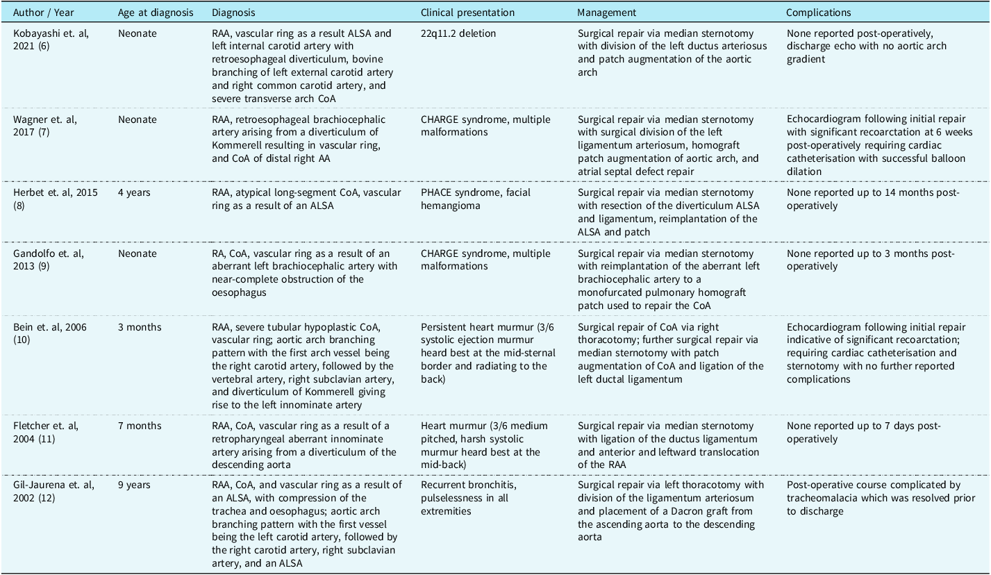
To our knowledge, the combination of right aortic arch, vascular ring, and coarctation of the aorta is rare, with only seven published case reports on patients with a combination of all three anomalies, and only three cases reported in a neonate. A summary of these reported cases, including clinical presentation, management, and outcomes, is provided in Table 1.
Reported cases with a combination of right aortic arch, vascular ring, and coarctation of the aorta. RAA: right aortic arch; coA: coarctation of the aorta; ALSA: aberrant left subclavian artery

The development of the aortic arch begins during the embryonic period along with the six primitive branchial arches and the dorsal and ventral aorta. Reference Agarwal4 Abnormal persistence, involution, or regression of these arches determines the type of aortic arch and gives rise to a variety of vascular rings. Attempts have been made to explain the embryology of the rare combination of right aortic arch, vascular ring, and coarctation by the principle of flow-related development of the central great vessels. Reference Ad and Vidne5 During fetal development, the descending aortic flow is supplied through both left and right ventricular outflow. Per this theory, coarctation can develop in situations with increased right-to-left ductal flow and decreased flow across the aortic arch. Increased blood flow from the ductus into the isthmus and descending aorta may result in aortic arch hypoplasia. Although this does not explain why all patients with a right aortic arch and vascular ring do not develop coarctation, the incidence is likely multifactorial, with flow theory being one contributing factor.
Surgical intervention for patients presenting with a combination of a right aortic arch, vascular ring, and coarctation of the aorta can be particularly difficult, and different techniques have been used to repair this rare combination. Reference Kobayashi, Harada and Shinkawa6–Reference Gil-Jaurena, Murtra, Gonçalves and Miró12 Though lateral thoracotomy allows easier access to the aortic arch, a complete repair would require bilateral incisions, increasing the risk of complications, making it an unfavourable option. As a result, median sternotomy is often preferred despite being more invasive and often leading to prolonged operative time, excessive bleeding, or injury to the surrounding structures. Reference Kim, Chee and Song13 Although the surgical approach itself was not novel, we believe this case offers educational value in emphasising the diagnostic challenges and surgical decision-making in the context of rare arch anomalies. In addition, it highlights the importance of close follow-up as coarctation has a recurrence rate ranging from less than 5% to more than 50%, especially in young infants and those who require prostaglandin infusion. Reference Allen, Shaddy, Penny, Feltes and Cetta14 Furthermore, in retrospect, this case underscored the significance of patch material selection during aortic arch repair. Recent studies have demonstrated that the choice of patch material is a crucial factor in determining the risk of restenosis and the need for reintervention. The patch material used in this case has since been associated with higher rates of restenosis, and, knowing this now, the surgical team would have opted for a different material. Reference van Beynum, Kurul and Krasemann15
Similarly, Kobayashi and colleagues reported a neonate with a right aortic arch, coarctation, aberrant left subclavian artery, and left internal carotid artery with retro-oesophageal diverticulum forming a vascular ring, bovine branch of the left external carotid artery, and right common carotid artery arising from the aorta. Reference Kobayashi, Harada and Shinkawa6 They underwent repair via median sternotomy with division of the left ductus arteriosus and patch augmentation of the aortic arch with an uncomplicated post-operative course.
Wagner and colleagues also identified a neonate with a right aortic arch, coarctation of the distal aortic arch, and a vascular ring formed by a retro-oesophageal brachiocephalic artery arising from a diverticulum of Kommerell. The patient underwent repair by midline sternotomy with homograft patch augmentation, with a residual gradient of 30 mmHg via direct measurement in the operating room. Follow-up echocardiogram 6 weeks later demonstrated a worsening peak gradient across the aortic arch, indicative of recoarctation. The patient underwent cardiac catheterisation and successful balloon angioplasty of the stenotic area with a decrease in gradient from 52 to 20 mmHg. Reference Wagner, Knowlton, Pastuszko and Shah7
Herbert and colleagues performed a similar repair to our case, of a right aortic arch with atypical, long-segment coarctation and vascular ring formed by an aberrant left subclavian artery. Reference Herbert, Guzmán-Pruneda, Sumner and McKenzie8 The patient underwent repair by median sternotomy with reimplantation of the aberrant left subclavian artery and translocation of the descending aorta to the proximal ascending aorta. Fourteen months post-operatively, the patient remained asymptomatic without activity limitations or need for cardiac medications, and there was no sign of hemodynamic compromise or re-coarctation. Gandolfo and associates reported a neonate with right aortic arch, coarctation of the transverse arch, and vascular ring (with near-complete obstruction of the oesophagus) formed by an aberrant left brachiocephalic artery. Reference Gandolfo, Albanese, Secinaro and Carotti9 The malformations were repaired via median sternotomy in one stage, including reimplantation of the aberrant left innominate artery to the branch of a monofurcated pulmonary homograft patch used to repair the coarctation. Chest CT performed three months post-operatively revealed optimal repair.
Fletcher and colleagues identified a right aortic arch coarctation, and a vascular ring formed by a retropharyngeal aberrant innominate artery arising from a diverticulum of the descending aorta. Reference Bein, Saba, Patel, Reinhartz and Hanley10 The patient’s defects were repaired successfully via median sternotomy with ligation of the ductus ligamentum and anterior and leftward translocation of the right arch. No complications were noted prior to discharge seven days post-operatively.
In contrast to the median sternotomy approach, Gil-Juarena and colleagues approached a right aortic arch, coarctation, and vascular ring (formed by an aberrant left subclavian artery and causing compression of the trachea and oesophagus) via a left thoracotomy with division of the ligamentum arteriosum and placement of a Dacron graft from the ascending aorta to the descending aorta. Reference Fletcher, Duncan, Schroeder and Danford11 The anomalies were repaired in one stage, though the authors recognised inadvertently creating a new vascular ring as a potential complication of this approach. The patient’s post-operative course was complicated by tracheomalacia, which was resolved before discharge.
Bein and colleagues initially identified a right aortic arch with an aberrant left subclavian artery arising from the diverticulum of Kommerell and severe tubular hypoplastic coarctation on echocardiogram. Reference Gil-Jaurena, Murtra, Gonçalves and Miró12 The patient underwent a right thoracotomy for coarctation repair, during which a long-segment coarctation extending from and involving the origin of the right subclavian artery to the origin of Kommerell’s diverticulum was noted along with a retro-oesophageal arch. Echocardiogram following repair indicated recoarctation. Further evaluation with cardiac catheterisation confirmed residual arch obstruction and demonstrated a right aortic arch with the first branch being the right carotid artery, followed by the right vertebral artery, the right subclavian artery, and the diverticulum of Kommerell (which gave rise to the left innominate artery). The patient underwent further surgical repair via a median sternotomy with patch augmentation of the coarctation and ligation of the left ductal ligamentum.
Conclusion
A right aortic arch in combination with coarctation and a vascular ring is a rare anomaly. This case is also unique due to the atypical aortic branching pattern, transverse arch coarctation, and neonatal presentation. While the surgical approach itself may not be novel, this case emphasised the diagnostic challenges and surgical decision-making in the context of rare arch anomalies, particularly in neonates. Cardiac CT angiogram can be helpful with diagnosis and surgical planning, with complete repair by way of a median sternotomy being the intervention of choice. Close follow-up is required to monitor for complications such as recoarctation and the need for reintervention.
Acknowledgements
None.
Financial support
This research received no specific grant from any funding agency, commercial, or not-for-profit sectors.
Competing interests
There are no conflicts of interest.