Introduction
The colonization of land by early vascular plants has been considered one of the most important evolutionary events in Earth history (Kenrick and Crane, Reference Kenrick and Crane1997; Taylor et al., Reference Taylor, Taylor and Krings2009), and the Silurian and Devonian fossil record is the major source of evidence for this transformative event. Nevertheless, previous studies have been focused mainly on the records from Europe, North America, and, more recently, South China, with contributions from Gondwana and northwestern China (e.g., Gensel and Andrews, Reference Gensel and Andrews1984; Anderson et al., Reference Anderson, Hiller and Gess1995; Kenrick and Crane, Reference Kenrick and Crane1997; Meyer-Berthaud et al., Reference Meyer-Berthaud, Scheckler and Wendt1999, Reference Meyer-Berthaud, Decombeix, Dunstone, Gerrienne, Momont and Young2016a, Reference Meyer-Berthaud, Servais, Vecoli and Gerrienneb; Berry et al., Reference Berry, Morel, Mojica and Villarroel2000; Hammond and Berry, Reference Hammond and Berry2005; Gensel, Reference Gensel2008; Taylor et al., Reference Taylor, Taylor and Krings2009; Hao and Xue, Reference Hao and Xue2013; H. Xu et al., Reference Xu, Zhao, Wang, Wen, Luo, Bao and Zhou2015; Xue et al., Reference Xue, Huang, Wang, Xiong, Liu and Basinger2018; Prestianni and Gess, Reference Prestianni and Gess2019), which constrains our understanding of early phytogeographic evolution of land floras.
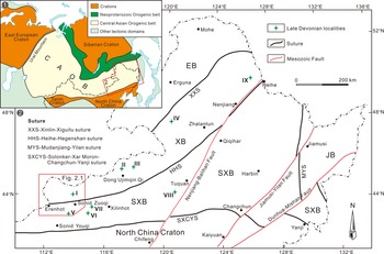
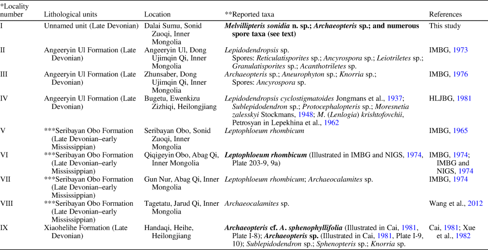
The vast area of northeastern China was formed through a complex tectonic amalgamation of crustal blocks, including the Erguna Block, Xing'an Block, Songliao-Xilinhot Block, and Jiamusi Block (Fig. 1; Liu et al., Reference Liu, Li, Feng, Wen, Neubauer and Liang2017). These constitute major components of the eastern part of the Central Asian Orogenic Belt (Sengör et al., Reference Şengör, Natal'in and Burtman1993). Studies of Devonian plants from these blocks have been limited, with only nine localities reported to contain Late Devonian plant fossils, including the new locality presented herein (Fig. 1.2, localities I–IX; see Discussion for a detailed review). However, only Leptophloeum rhombicum Dawson, Reference Dawson1862 from locality VI and two Archaeopteris species from locality IX have been illustrated and formally described (IMBG and NIGS, 1974; Cai, Reference Cai1981); all other reports listed only species names, and the reported fossils are apparently not available for re-investigation.
(1) Schematic map of Central Asian Orogenic Belt (CAOB) and adjacent tectonic units (modified from Sengör et al., Reference Şengör, Natal'in and Burtman1993); (2) location of Late Devonian plant fossil localities in northeastern China (locality I, this study; localities II-IX, other previous studies). For detailed information about the fossil localities, see Table 3 and the text in Discussion section. The eastern part of the CAOB is composed of several microcontinents and sutures, and here the division scheme of tectonic units proposed by Liu et al. (Reference Liu, Li, Feng, Wen, Neubauer and Liang2017) is followed. Abbreviations of microcontinents: EB, the Erguna Block; XB, the Xing'an Block; SXB, the Songliao-Xilinhot Block; and JB, the Jiamusi Block.

The recent discovery of a new flora of Late Devonian age from northern Sonid Zuoqi, Inner Mongolia (locality I in Fig. 1.2) presents an opportunity to describe a fossil assemblage, and to review the spatial and stratigraphic distribution of Late Devonian floras from NE China.
Geological setting and stratigraphy
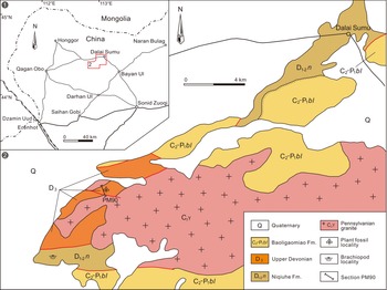
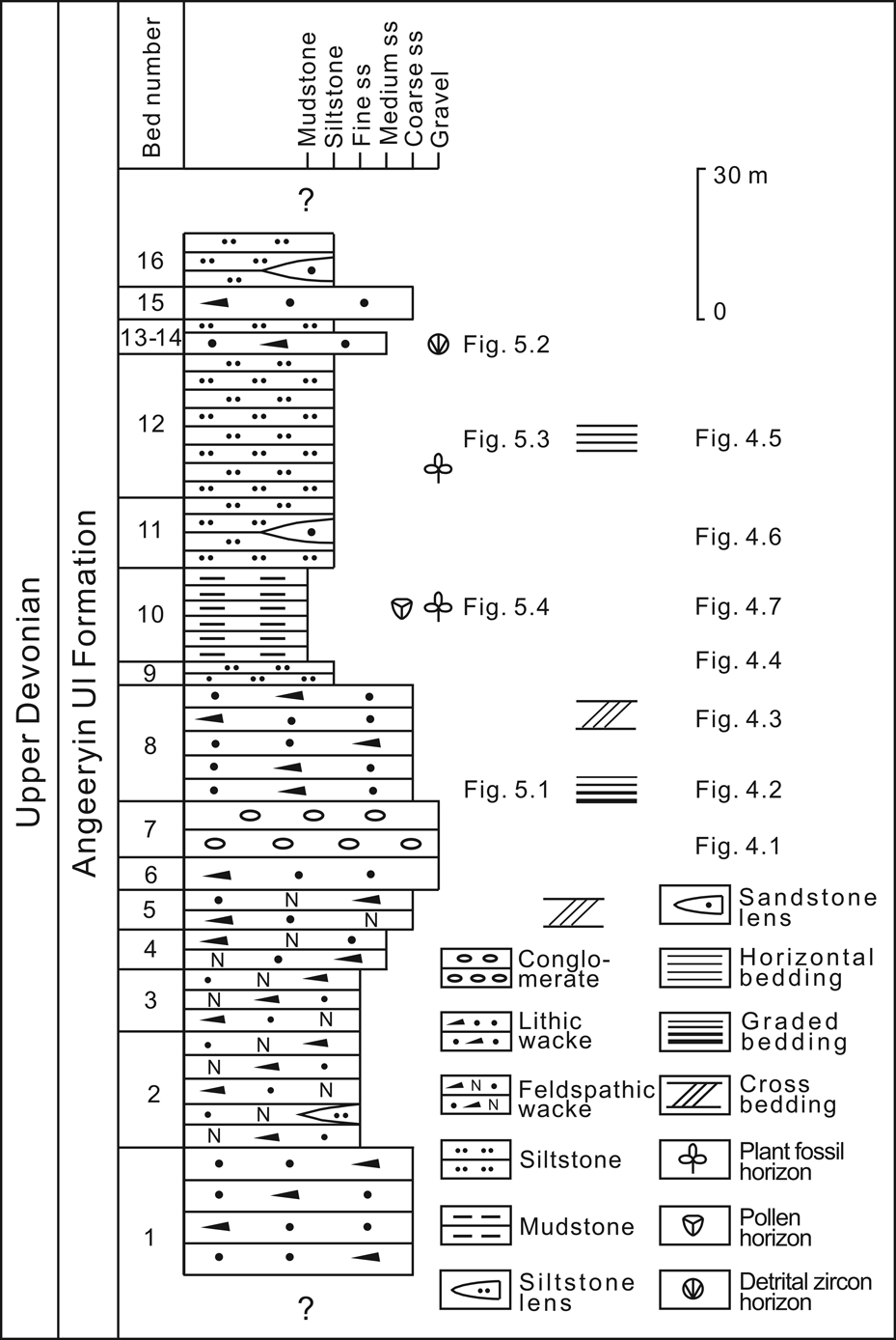
The fossil locality, here called section PM90, is located ~18 km southwest of Dalai Sumu Town, northern Sonid Zuoqi, Inner Mongolia, China, and the GPS location is 44°23′25″N, 112°53′13″E (Fig. 2). A 142-m-thick profile of the plant-bearing succession was measured and divided into 16 beds (PM90 succession for simplicity; Fig. 3). These strata were previously assigned to the upper Carboniferous Hong Obo Formation (IMBG, 1979), or to the upper Carboniferous–lower Permian Baoligaomiao Formation (IMGS, 2007). However, in light of the present discovery of megafossil plants and spores, we conclude that the PM90 succession belongs to the Upper Devonian, which was previously thought to be absent in the Dalai Sumu area. To the south of PM90, sequences with similar lithologies were intruded by Carboniferous-aged granites, or are in fault contact with the brachiopod-bearing Niqiuhe Formation (Early to Middle Devonian-aged marine clastic rocks), while to the north comparable sequences are unconformably overlain by the Baoligaomiao Formation (interbedded clastic and volcanic rocks) (Fig. 2).
(1) Map showing the location of the studied area. (2) Geological sketch map (modified from IMGS, 2007) showing the location of the studied section (PM90) and geologic setting near Dalai Sumu, Sonid Zuoqi, Inner Mongolia, NE China.


The PM90 succession at this time is considered to represent an unnamed lithological unit because it is difficult to compare this succession with any previously recognized unit in the area, where outcrops are usually not well exposed. Nevertheless, a potential candidate for the PM90 succession is the Angeeryin Ul Formation, the stratotype section of which is located near Angeeryin Ul, Dong Ujimqin Qi, Inner Mongolia (locality II in Fig. 1.2), where it is composed of conglomerate, sandstone, siltstone, and mudstone (IMBG, 1973; IMBGMR, 1991, 1996). Based on plant fossils and spores, a Late Devonian age was suggested for the Angeeryin Ul Formation; however, this formation in general is poorly understood. The Angeeryin Ul Formation has been suggested to reach over 4000 m in thickness, yet has been only roughly logged, with single beds varying from tens to hundreds of meters thick (IMBG, 1973; IMBGMR, 1996), and stratigraphic repetition due to tectonic activity has not been fully considered. Thus, at present, it is not possible to precisely compare the PM90 succession with the Angeeryin Ul Formation or indeed any part of it.
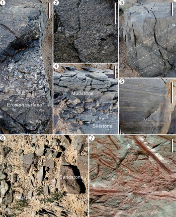
Four lithological facies are recognized in the PM90 succession. Facies Gm consists of pebble-supported, massive conglomerate, gray in color, and occurring only in bed-7 (Fig. 4.1); it typically occurs as units ranging from 1.5–2 m in thickness, with graded bedding. Sub-rounded cobbles up to 20 mm long are poorly sorted and are mainly lithic fragments (~50%) or quartz (~30%). Siltstone, tuff, andesite, granite porphyry, and mudstone constitute the lithic fragments, indicating low compositional maturity. An erosional surface and imbrications are evident at the base of bed-7 (Fig. 4.1), with dip angles of the imbrications ~15–30°. Gravels are irregularly distributed in bed-7, and lenses of sandstone are interbedded within the gravels of the Gm facies. The Gm facies are interpreted as representing channel lag deposits, in light of the massive coarse sediments, erosional surfaces, and imbricate structure (Allen, Reference Allen1964; Miall, Reference Miall1977; Makaske, Reference Makaske2001; Tucker, Reference Tucker2003; Nichols, Reference Nichols2009).
Sedimentary features at section PM90. (1) Conglomerate with erosion surface and imbrication at the base (bed-7); (2) gravelly coarse-grained sandstone with graded bedding (bed-8); (3) coarse-grained sandstone with planar cross-bedding (bed-8); (4) gradational contact between bed-9, siltstone with laminations (lower part of the photo), and bed-10, massive mudstone (upper part); (5) siltstone with horizontal bedding (bed-12); (6) siltstone, containing a lens of sandstone (bed-11); (7) mudstone with abundant plant fossils (bed-10). Scale bars = 20 mm.

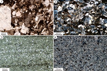
Facies Sp is a thinly to thickly bedded sandstone, with individual units ranging from 4 cm to 1 m in thickness, and cumulative thickness of >51 m (bed-1 to bed-6). Graded bedding and planar cross-bedding occur (Fig. 4.2, 4.3), with cross-bed dip angles ~20–30°. The grains are sub-rounded, poorly to moderately sorted, fine- to coarse-grained sand, and rare gravel (Fig. 5.1). The lithology is lithic wacke or feldspathic wacke (sensu Pettijohn, Reference Pettijohn1975), containing quartz (20–35%), feldspar (5–30%), and lithic fragments (30–70%) (Fig. 5.2). Facies Sp is interpreted as point-bar deposits in a fluvial setting, in that graded bedding and planar cross-bedding are generally found in such lateral-accretion deposits (Allen, Reference Allen1964; Miall, Reference Miall1977; Makaske, Reference Makaske2001; Tucker, Reference Tucker2003).
Photomicrographs of main rock types from section PM90. (1) Lithic wacke (bed-8), showing poorly sorted grains; (2) lithic wacke (bed-13); (3) siltstone in vertical profile (bed-12) showing horizontal lamination; (4) mudstone (bed-10). Q, quartz; F, feldspar; L, lithic fragments.

Facies Fl is composed of gray to dark-gray laminated (thinly bedded) siltstone, with rare sandstone lenses (Fig. 4.6). The grains are dominantly silt (70–75%), with some clay (25–30%) and rare fine-grained sand (5%). Thickness of beds ranges from 2 mm to 10 cm, and the cumulative thickness can reach up to 37 m. Horizontal bedding is common (Fig. 4.5), with laminae formed by variation in content of silt grains (Fig. 5.3), and the beds may be rich in plant fossils (bed-12). Facies Fl is interpreted as representing vertical-accretion deposits formed in a floodplain environment through overbank flooding (Allen, Reference Allen1964; Nanson and Croke, Reference Nanson and Croke1992; Makaske, Reference Makaske2001; Gradziński et al., Reference Gradziński, Baryła, Doktor, Gmur, Gradziński, Kędzior, Paszkowski, Soja, Zieliński and Żureke2003; Nichols, Reference Nichols2009).
Facies Fm is composed of massive mudstone (Fig. 5.4), devoid of visible bedding (Fig. 4.4), with a gray-green, but sometimes mottled red color. Plant fossils are found in abundance in some parts of the bed (Fig. 4.7). Like facies Fl, facies Fm is interpreted as representing vertical-accretion deposits in a floodplain environment, where plant remains were deposited near their growth site (Allen, Reference Allen1964; Nanson and Croke, Reference Nanson and Croke1992; Makaske, Reference Makaske2001; Gradziński et al., Reference Gradziński, Baryła, Doktor, Gmur, Gradziński, Kędzior, Paszkowski, Soja, Zieliński and Żureke2003).
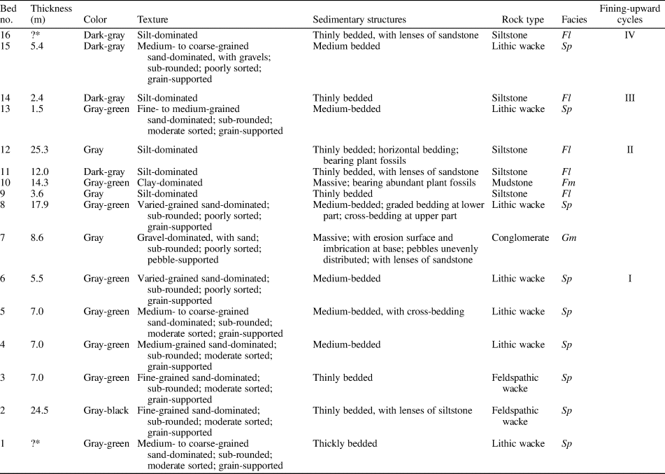
The PM90 succession includes sandstone at the base (bed-1 to bed-6; Cycle I), followed by three complete fining-upwards cycles (Cycles II–IV) (Table 1). The basal sandstone may represent an incomplete fining-upward cycle, but much has been lost due to erosion and deposition of the overlying conglomerate. Cycle II (bed-7 to bed-12) forms the main part of the section and is 81.7 m in overall thickness, including imbricated conglomerate with erosional surface at the base (facies Gm), sandstone with graded- and cross- bedding (facies Sp) in the lower part, and thick fines with abundant plant debris in the upper part (facies Fl and Fm). Cycles such as this typically develop in a fluvial environment: facies Gm is interpreted as a lag deposit; Sp, lateral point-bars; and Fl and Fm, overbank floodplain deposits (Allen, Reference Allen1964; Miall, Reference Miall1977; Makaske, Reference Makaske2001; Tucker, Reference Tucker2003; Bridge, Reference Bridge, Posamentier and Walker2006; Nichols, Reference Nichols2009). The third and fourth cycles are thinner, probably representing sediments of a lower energy environment, perhaps ephemeral rivers. Sediments throughout the section are interpreted as having been deposited close to their provenance, given the immature nature of the conglomerate and sandstone.
Lithological descriptions of the plant-bearing succession at section PM90 (see Fig. 3 for the logged column). *Bed thickness is uncertain because of heavy cover. Abbreviation of facies codes: Gm = massive conglomerate; Sp = sandstone with or without cross-bedding; Fl = laminated siltstone; Fm = massive mudstone.

Material and methods
Plant megafossils were obtained from bed-10 and bed-12 of the section PM90 (Fig. 3). Hundreds of specimens of a new euphyllophyte plant (Figs. 6–12) were recovered from both beds. A few specimens of Archaeopteris (Figs. 13, 14) were collected from bed-10, and a possible sphenophyllalean plant and some unidentified axes and roots (Fig. 15) were collected from bed-12. All specimens are preserved as adpressions in siltstone or mudstone.
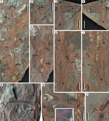
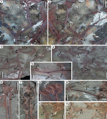
Melvillipteris sonidia n. sp. (1) Main axis; the upper node shows a pair of first-order branches, and immediately below this node is a bend of the main axis; in the middle, a node shows another pair of first-order branches; the axes show little contrast with the surrounding matrix, and thus a interpretative line drawing is presented in Figure 8; paratype, PKUB18861a; (2) counterpart of the upper node of the main axis in (1); PKUB18861b; (3, 4) part and counterpart; main axis with a pair of first-order branches, one of which is represented by a broken base; PKUB18859a, b; (5) two main axes with lateral branches; note that the nodal position is expanded (arrows n); PKUB18854b; (6, 7) part and counterpart; main axis with a pair of first-order branches; one branch is well preserved, and the other branch extends into the rock matrix; broken ultimate appendages occur at the base of a branch; paratype PKUB18860a, PKUB18860b; (8, 9) part and counterpart; main axis and two first-order branches; one branch shows a swollen base with a broken ultimate appendage (arrow U) and a second-order branch, distally; PKUB18873a, b; (10) enlargement of the broken ultimate appendage in (9, arrow U); (11) fertile structure attached to a probable main axis; sporangia can be confirmed on higher magnification; PKUB18865. M = main axis; a1 = first-order branch; a2 = second-order branch; U = sterile ultimate appendage; f = fertile structure. Scale bars: (1, 2) 20 mm; (3–5, 8–9) 10 mm; (6–7, 11) 5 mm; (10) 2 mm.

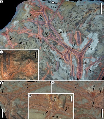
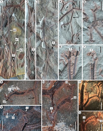
Melvillipteris sonidia n. sp. (1) Holotype; specimen showing main axes and two orders of lateral branches; arrows f1 and f2 indicate two fertile structures enlarged in (2) and Figure 12.2, respectively; the right main axis bears a pair of first-order branches; the distal part of this main axis is missing above the node; PKUB18801a; (2) fertile structure (f1 in 1) attached to a probable first-order branch with a long stalk; (3, 4) part and counterpart; PKUB18857a and paratype, 18857b; main axis and pair of first-order branches; distal part of the main axis above the node is missing in (3), but has been revealed through dégagement in (4); (5) main axis and pair of first-order branches; PKUB18876. M = main axis; a1 = first-order branch; a2 = second-order branch; f = fertile structure. Scale bars: (1) 20 mm; (2) 2 mm; (3, 4) 10 mm; (5) 5 mm.

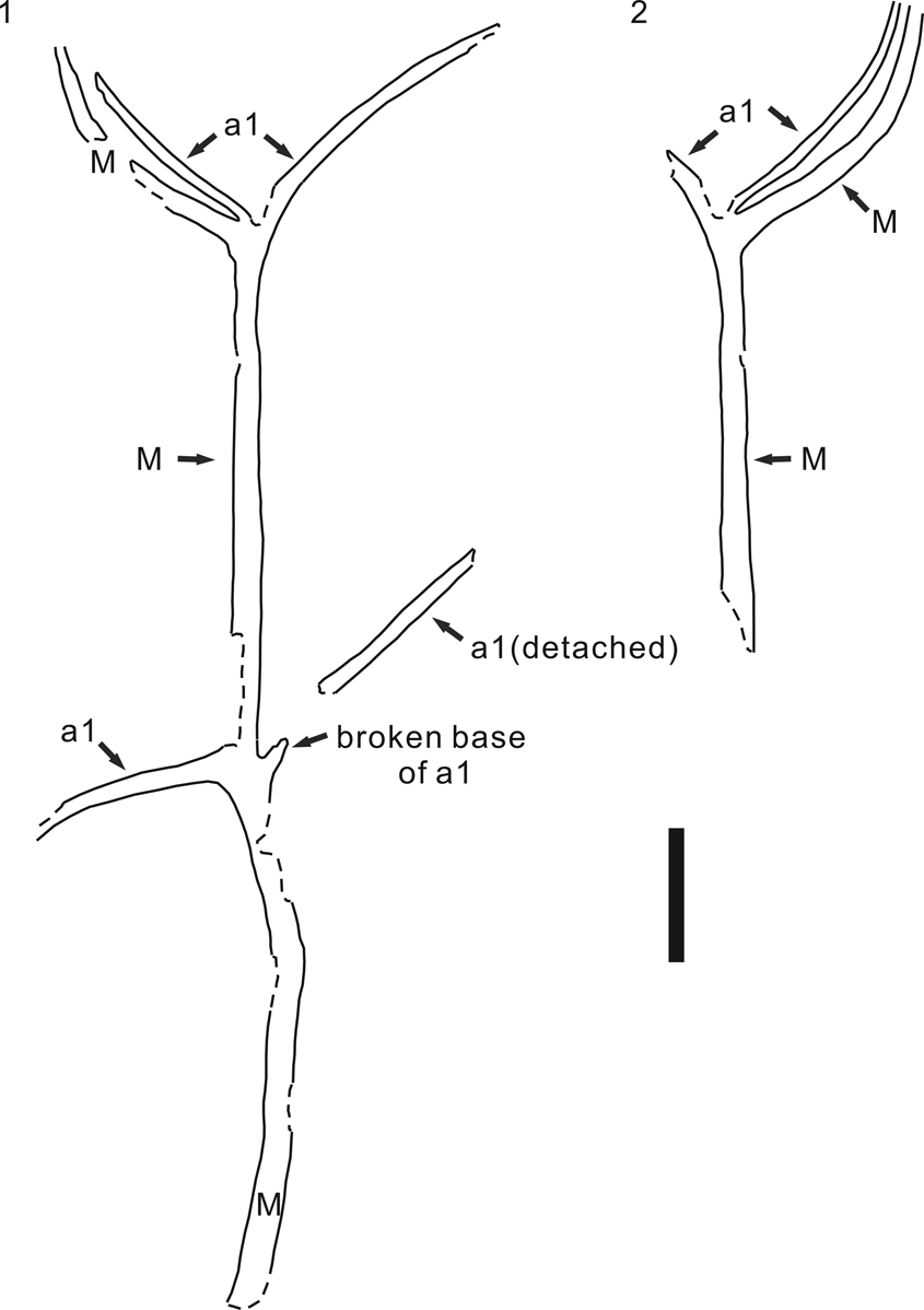
Interpretative drawings of a main axis and its first-order branches of Melvillipteris sonidia n. sp. (1, 2) Part and counterpart of the specimen shown in Figure 6.1 and 6.2. M = main axis; a1 = first-order branch. Scale bar: 20 mm.

Melvillipteris sonidia n. sp. (1, 2) Part and counterpart; PKUB18874a, 18874b, paratype; zigzag first-order branch with second-order branches; also showing main axis with expanded nodal position (arrow n); (3, 4) part and counterpart; PKUB18862a, c; first-order branch and second-order branches; arrow 5 points to the part enlarged in (5); (5) enlargement of second-order branch with sterile ultimate appendages; PKUB18862c; (6) first-order branch, and a fertile structure close to the branching point; PKUB18810b; (7) first-order branch, and a fertile structure attached at the branching point; a second-order branch demonstrated by its broken base; paratype PKUB18810c; (8) first-order branch with a second-order branch, the latter bearing a fertile structure; paratype PKUB18814; (9) enlargement of the fertile structure in (8); (10, 11) first-order branches with their second-order branches; (10) PKUB18858a; (11) PKUB18869b. M = main axis; a1 = first-order branch; a2 = second-order branch; U = sterile ultimate appendage; f = fertile structure. Scale bars: (1–4) 10 mm; (5) 2 mm; (6–8, 10, 11) 5 mm; (9) 1 mm.

Melvillipteris sonidia n. sp. (1) Second-order branches, one of which bears alternate laterals (white dots); paratype PKUB18816; (2) second-order branch, with alternate to sub-opposite laterals (white dots); PKUB18814-2; (3) second-order branch, with alternate laterals (white dots); PKUB18832; arrows f (1–3) indicate poorly preserved fertile structures, confirmed on higher magnification; (4) second-order branch and sterile ultimate appendages; PKUB18812b-1; (5) terminal part of a second-order branch; PKUB18821a-1; (6) second-order branch bearing ultimate appendages or fertile structures; PKUB18812b-2; (7) fertile structure attached to a second-order branch; paratype PKUB18821a-2; (8) enlargement of a sterile ultimate appendage (shown in 4, middle arrow U) composed of a twice dichotomously branching system (arrows I and II); (9, 10) incomplete sterile ultimate appendages, which are dichotomously branched three times (arrows I to III); (9) PKUB18875; (10) PKUB18812b-1; (11) two overlapped, incomplete ultimate appendages; the right one with terminal, recurved tips; PKUB18812a-2; (12) ultimate appendage, dichotomously branched twice (arrows I and II) and with terminal, recurved tips; PKUB18821a-3. M = main axis; a1 = first-order branch; a2 = second-order branch; U = sterile ultimate appendage; f = fertile structure. Scale bars: (1, 2) 10 mm; (3–5) 5 mm; (6–9, 11) 2 mm; (10, 12) 1 mm.

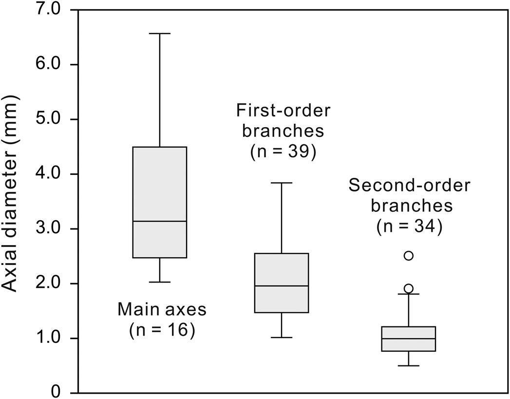
Box plot of the measured width of main axes, first-, and second-order branches. For each order of axis: the 25–75% quartiles of the measurements are drawn using a box; the median value, a horizontal line inside the box; and the minimal and maximum values, short horizontal line below and above the box. Circles represent some extreme values of the measured width of second-order branches.

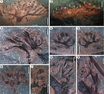
Melvillipteris sonidia n. sp. (1–9) Fertile structures: (1) fertile structure showing an initial dichotomy, two major sporangial clusters supported by branches S1 and S1′, and 13 sporangia (numerals 1–13); paratype PKUB18830; (2) fertile structure enlarged from Figure 7.1 (arrow f2), showing at least 12 sporangia (numerals 1–12); (3) incomplete fertile structure; PKUB18812c; (4, 5) incomplete fertile structure, showing paired sporangia on terminal divisions; part and counterpart, PKUB18821a-4, 18821b-4; (6) fertile structure with two sporangial clusters; PKUB18812a-4; (7) incomplete fertile structure, showing terminal, paired sporangia; PKUB18812a-2; (8) fertile structure with two sporangial clusters, the right cluster better preserved; enlarged from the counterpart of the specimen in Figure 9.6 (arrow f); PKUB18810a; (9) incomplete fertile structure, showing one of the sporangial clusters; enlarged from Figure 9.7 (arrow f). Scale bars: (1–9) 1 mm.

Some specimens were treated by acetic acid (2 mol/L) solution to remove surface carbonate film and to enhance contrast between plant axes and the surrounding rock matrix. The fossils were prepared by dégagement with steel needles to better expose morphology, and were photographed with a digital camera.
One palynological sample collected from bed-10 was treated using standard HCl-HF acid maceration at the Nanjing Institute of Geology and Palaeontology, Chinese Academy of Sciences (NIGPAS). The rock fragments were processed in 30% HCl, followed by decant washing in water to neutralize and then demineralization in 60% HF with repeated stirring. Organic matter from the residues was then mounted and the recognized palynomorphs photographed.
One sample of lithic wacke (PM90TW13) from bed-13 was collected for detrital zircon U-Pb dating, using Laser Ablation-Multicollector-Inductively Coupled Plasma-Mass Spectrometry (LA-MC-ICP-MS) at the Inner Mongolia Key Laboratory of Magmatic Mineralization and Ore-prospecting, China. The methodology and data of zircon U-Pb dating are available at Dryad (http://datadryad.org/; https://doi.org/10.5061/dryad.d51c5b042).
Repositories and institutional abbreviations
Types and other megafossil specimens examined in this study are deposited in the Geological Museum, School of Earth and Space Sciences, Peking University (PKU), Beijing, China. All slides of palynomorphs are deposited at NIGPAS.
Systematic paleobotany
Phylum Tracheophyta Sinnott, Reference Sinnott1935 ex Cavalier-Smith, Reference Cavalier-Smith1998
Subphylum Euphyllophytina Kenrick and Crane, Reference Kenrick and Crane1997
Order Rhacophytales Taylor et al., Reference Taylor, Taylor and Krings2009
Family Incertae sedis
Genus Melvillipteris Xue and Basinger, Reference Xue and Basinger2016
Type species
Melvillipteris quadriseriata Xue and Basinger, Reference Xue and Basinger2016, by original designation, from the Parry Islands Formation (Famennian), Upper Devonian, eastern Melville Island, Canadian Arctic Archipelago.
Diagnosis
See Xue and Basinger (Reference Xue and Basinger2016, p. 603).
Melvillipteris sonidia Bai, Huang, Basinger, and Xue, new species
Figures 6–12
Holotype
PKUB18801a (Fig. 7.1), from the Late Devonian of northern Sonid Zuoqi, Inner Mongolia, northeastern China.
Paratypes
PKUB18810c, 18814, 18816, 18821a, 18830, 18857b, 18860a, 18861a, and 18874b (Figs. 6.1, 6.6, 7.4, 9.2, 9.7, 9.8, 10.1, 10.7, 12.1).
Diagnosis
Main axes bent slightly or moderately, ~3.5 (2.0–6.6) mm wide and at least 215 mm long, with internode length ~80 mm. First-order branches with a swollen base, inserted on main axes in pairs. First-order branches with a zigzag appearance, ~2.1 (1.0–3.8) mm wide and up to at least 108 mm long, bending at each node at angles of ~139° (125–147°). Second-order branches ~1.0 (0.5–2.5) mm wide and up to at least 95 mm long, borne helically or alternately, departing from first-order branches at angles of ~102° (82–122°), with internode length 14–46 mm, and bearing alternate to sub-opposite ultimate appendages that may be fertile or sterile. Sterile ultimate appendages inserted alternately on second-order branches, with internode length 4.0–39 mm, with two or three successive dichotomies and paired recurved tips, ~0.5 mm wide at the proximal portion, up to ~6.8 mm in overall length. Each fertile structure with a short stalk ~2.3 (1.4–3.8) mm long and an initial dichotomy forming two sister branches, each of which supports a sporangial cluster. Each sporangial cluster branched dichotomously two or three times, with six or more terminal sporangia. Sporangia fusiform, borne singly or in pairs on ultimate divisions, ~1.3 (0.8–1.9) mm in length and ~0.4 (0.2–0.7) mm in maximum width.
Occurrence
Upper Devonian, an unnamed lithological unit, 18 km southwest of Dalai Sumu Town, northern Sonid Zuoqi, Inner Mongolia, China (GPS location: 44°23′25″N, 112°53′13″E).
Description
Main axes.—Orders of branching are distinguished by size, morphology, arrangement of lateral organs, and organic connections. The most robust axes are interpreted as main axes, although this interpretation is tentative because no roots have been found. Main axes show different preservation status. (1) They clearly show a pair of first-order branches at the nodes of some main axes (Figs. 6.1 upper node, 6.2, 8). (2) In some cases, one of the first-order branches is usually well preserved or exposed, while the other one is represented by a broken base (Fig. 6.3, 6.4) or is demonstrated to extend into the rock matrix (Fig. 6.6, 6.7). (3) Some main axes appear to bear one only lateral branch at the node (Fig. 6.5), but we consider that, in these cases, the second of a pair of branches is either unexposed or has been broken off during preservation. (4) In some specimens, a main axis bears a pair of first-order branches, but above the node the distal part of main axis may appear to be missing (Figs. 6.8, 6.9, 7.1, 7.3), although distal remains of the axis may be recovered through careful dégagement (Fig. 7.4, 7.5). In all cases, the first-order branches show a swollen base, a criterion for distinguishing them from second-order branches.
Main axes are bent slightly to moderately at the nodes where first-order branches depart, and are straight or nearly so between the nodes (Fig. 6.1–6.7). The diameter of main axes ranges from 2.0–6.6 mm, average 3.5 mm (N = 16; Fig. 11), with the width decreasing when approaching the branching point. The longest preserved main axis is 21.5 cm long, with two nodes bearing first-order branches, and shows an interval of ~8.0 cm between the nodes (Fig. 6.1). A fertile structure has been found attached to a probable main axis (Fig. 6.11).
First- and second-order branches.—First-order branches have been found attached to their main axes (Figs. 6–8), while many others are detached (Fig. 9). First-order branches show a consistent morphology: they have a swollen base and are bent at each node where a second-order axis departs, such that the bending angles of 125–147°, with an average of 139° (N = 14), creates a characteristic zigzag appearance (Figs. 7.1, 9.1–9.4, 9.10, 9.11). First-order branches range from 1.0–3.8 mm in width, average 2.1 mm (N = 39; Fig. 11), but the swollen base is much wider, reaching up to ~9.1 mm wide, and the most proximal portion of a first-order branch may exceed 4.0 mm in diameter, as for example in Figure 7.1. The longest preserved first-order branch is 10.8 cm in length, without showing taper. A fertile structure has been found on a probable first-order branch (Fig. 7.1 arrow f1, 7.2).
Second-order branches found attached to first-order branches appear to be arranged alternately, although this also could be attributed to a helical pattern; presently there is insufficient evidence to distinguish between these two patterns. Second-order branches depart from first-order branches at an average angle of 102° (range = 82–122°, N = 13), and the interval between two successive second-order branches ranges from 14.2–45.7 mm (average = 27.4 mm, N = 6).
Second-order branches are slender and straight to slightly flexed in appearance (Figs. 9.5, 9.8, 10.1–10.4), with a width of 0.5–2.5 mm (average = 1.0 mm, N = 34; Fig. 11). The longest preserved second-order axis is 95 mm long (Fig. 10.1). Second-order branches appear to terminate in a series of anisotomies in quick succession (Fig. 10.5). Lateral organs are attached to the second-order branches in an alternate to sub-opposite pattern (Fig. 10.1–10.3, white dots), and may be sterile ultimate appendages (Fig. 10.4 arrows U) or fertile structures (Fig. 10.1–10.3, 10.6, 10.7 arrows f). The interval between appendages along the second-order branches ranges from 4.0–38.6 mm (average = 20.2 mm, N = 7).
Sterile ultimate appendages (SUAs).—In only two specimens, ultimate appendages that are probably sterile are found attached to the base of first-order branches, but they are very poorly preserved, with only a short, undivided fragment left (Fig. 6.6–6.10). Numerous other first-order branches show no evidence of SUAs.
SUAs regularly occur on second-order branches, but most are incomplete. In the specimen shown in Figure 10.8, a SUA departs from its parent axis at a right angle, extends ~4.3 mm, and then divides once (dichotomy I); one of the daughter branches extends into rock matrix, while the other daughter branch extends ~1.8 mm and then further divides (dichotomy II); the dichotomy II produces a sharply recurved tip ~1.3 mm long, while its sister tip runs into matrix. SUAs shown in Figure 10.9 and 10.10 are similar, but show a third dichotomy: their initial dichotomy (dichotomy I) produces two daughter branches, the first branch is partially buried in the matrix or has been broken off, and the second is visible and shows further divisions (dichotomies II and III); of the two daughter branches produced by dichotomy II, one is either incomplete or lies buried within the matrix, and a second isotomously divides once (dichotomy III) and produces two terminal divisions with oppositely recurved tips. It is clear that the branching of the SUAs is three-dimensional. The specimen in Figure 10.11 shows two overlapped, incomplete SUAs; compared with the SUA in Figure 10.10, the left one may represent dichotomy II of a SUA, while the right one represents an ultimate dichotomy (dichotomy III) with two recurved tips. Other SUAs show consistent branching patterns (e.g., Fig. 10.12). Thus, the features of SUAs of second-order branches can be summarized as follows: they divide two or three times, with a branching angle of 70–140°, forming a three-dimensional, dichotomous branching system, and are terminated in oppositely recurved tips. In overall length, these SUAs reach up to 6.8 mm and at the base are ~0.5 mm wide. The tips (ultimate divisions) are short, 0.5–1.3 mm long, sharply recurved, and tapering.
Fertile structures.—Most fertile structures are detached from their axes. One fertile structure has been found attached to what may be a main axis (Fig. 6.11, based on its larger width), and another one to a first-order branch (Fig. 7.1 arrow f1, 7.2), but these are rare occurrences, and it may be that the interpretation of their parent axes as main axis or first-order branch is incorrect. Nevertheless, one specimen shows a fertile structure at the branching point of a first-order branch (Fig. 9.7), and a second specimen shows a fertile structure very close to the branching point of a first-order branch (Fig. 9.6). We interpret these as fertile structures borne at the base of second-order branches. More commonly, however, fertile structures are found attached along the length of second-order branches at an acute to right angle (Figs. 9.8, 9.9, 10.1–10.3, 10.6, 10.7).
Fertile structures are dichotomously branched. Each fertile structure extends 1.4–3.8 mm (average = 2.3 mm, N = 7) to form a short, straight stalk (Figs. 9.9, 10.6, 10.7), with a width of 0.6–0.9 mm (average = 0.7 mm, N = 8). This stalk then divides dichotomously at an angle of 70–120° to form daughter branches 0.3–0.6 mm in width (average = 0.4 mm, N = 15) and 0.8–1.5 mm in length (average = 1.1 mm, N = 6). Subsequent dichotomies occur at shorter intervals, making this initial dichotomy more conspicuous and forming a pair of sporangial clusters (Fig. 12.1, 12.9), which themselves consist of two or more trusses of sporangia.
The fertile specimen shown in Figure 12.1 is the best preserved. The stalk of this fertile structure initially dichotomizes to form the branches S1 and S1′ that support the two sporangial clusters. The branch S1 then divides to support two fertile trusses, each of which consists of at least three sporangia. The branch S1′ also supports two fertile trusses: one composed of at least four sporangia (sporangia 7–10 in Fig. 12.1), the other with at least three sporangia (sporangia 11–13 in Fig. 12.1). In some specimens, numerous sporangia are densely compressed together (Fig. 12.2), and in some others, fertile structures are only partially preserved (Fig. 12.3–12.7). In the specimen shown in Figure 12.8, the right sporangial cluster shows at least seven sporangia. In the specimen shown in Figure 12.9, the left sporangial cluster, similarly, contains two fertile trusses, each with at least four sporangia. Many other specimens show only part of a complete fertile structure, such as one of the two sporangial clusters (Fig. 12.3), or simply a fertile truss with several sporangia (Fig. 12.4, 12.5, 12.7). The entire fertile structure consists of two sporangial clusters, collectively with 12–16 sporangia. The sporangia are borne singly (e.g., Fig. 12.1 sporangia 3 and 11), or in pairs (Fig. 12.1, 12.4, 12.5, 12.7), on terminal divisions.
The sporangia are fusiform in shape, 0.8–1.9 mm long (average = 1.3 mm, N = 61) and 0.2–0.7 mm in maximum width (average = 0.4 mm, N = 61), and with a pointed tip. A dehiscence line was not observed due to poor preservation. No in situ spores were found.
Etymology
From Sonid Zuoqi, where the plant was collected.
Remarks
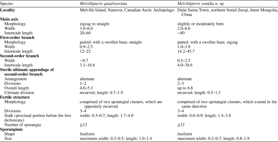
Our material is assigned to the genus Melvillipteris as a new species, M. sonidia n. sp. This genus was established from the Late Devonian (Famennian) of Arctic Canada (Xue and Basinger, Reference Xue and Basinger2016). The similarities between M. sonidia n. sp. and the type species, M. quadriseriata, are striking (Table 2): both plants show main axes with paired first-order branches; their first-order branches show a swollen base, and in rare cases bear ultimate appendages on the base, although in both plants this structure has been poorly preserved; in both, sterile ultimate appendages and fertile structures are alternately arranged along the second-order branches; their sterile ultimate appendages are terminated in oppositely recurved tips; their fertile structures are similar in showing a two-clustered appearance, and each sporangial cluster dichotomizes two to three times, with sporangia borne singly or in pairs on terminal divisions; in addition, quantitatively, the measurements of main axes, first- and second-order branches, sterile ultimate appendages, and fertile structures are quite similar between the Inner Mongolian plant and M. quadriseriata (Table 2). In the new plant, we are not able to demonstrate how the paired first-order branches are arranged on main axes because most main axes are preserved in fragments. Given the above numerous similarities, however, a quadriseriate arrangement, as in M. quadriseriata, can be inferred in M. sonidia n. sp. (i.e., pairs of first-order branches are alternately arranged along the main axes).
Morphological comparisons between Melvillipteris quadriseriata and M. sonidia n. sp. Data for M. quadriseriata based on Xue and Basinger (Reference Xue and Basinger2016). All measurements are in mm.

The differences between M. sonidia n. sp. and M. quadriseriata are also notable: first-order branches of M. quadriseriata are straight and bear sterile ultimate appendages in the proximal portions, but those of M. sonidia n. sp. are typically zigzag and bear no appendages in the proximal portions; sterile ultimate appendages of second-order branches dichotomize once or twice in M. quadriseriata, but dichotomize two or three times in M. sonidia n. sp.; in M. quadriseriata, each sporangial cluster of a fertile structure is supported by a long, recurved branch, while in M. sonidia n. sp., the two sporangial clusters extend in the same direction and are not recurved.
Xue and Basinger (Reference Xue and Basinger2016) provided detailed comparisons between Melvillipteris and other related plants, particularly those with quadriseriate branching, and tentatively assigned this genus to the Rhacophytales (sensu Taylor et al., Reference Taylor, Taylor and Krings2009). Their comparisons remain valid and applicable to M. sonidia n. sp. Besides Melvillipteris, many other plant genera (e.g., Rhacophyton Crépin; Cephalopteris Nathorst; Protocephalopteris Ananiev; Ellesmeris Hill, Scheckler, and Basinger; Chlidanophyton Gensel; Eocladoxylon Koidzumi; Protopteridophyton Li and Hsü) were collectively included in the Rhacophytales (Hilton, Reference Hilton1999; Berry and Wang, Reference Berry and Wang2006; Taylor et al., Reference Taylor, Taylor and Krings2009), and the quadriseriate branching in these taxa may be a shared derived character (synapomorphy) (Xue and Basinger, Reference Xue and Basinger2016). However, taxonomic assignment of Melvillipteris to the Rhacophytales remains uncertain and requires further phylogenetic analyses. First, the lack of anatomy for Melvillipteris hinders a comparison with the well-studied genus Rhacophyton, which shows a clepsydroid-shaped strand of primary xylem surrounded by secondary xylem (Leclercq, Reference Leclercq1951; Andrews and Phillips, Reference Andrews and Phillips1968). Second, the arrangement of fertile structures in Melvillipteris along second-order branches differs from the fertile structures of Rhacophyton, which are borne at the base of lateral branches, occupying a catadromic position (Leclercq, Reference Leclercq1951; Andrews and Phillips, Reference Andrews and Phillips1968), and fertile structures of Cephalopteris and Protocephalopteris are borne at a similar position (Ananiev, Reference Ananiev1960; Schweitzer, Reference Schweitzer1968). Nevertheless, some specimens in M. sonidia n. sp. appear to show fertile structures at the base of second-order branches (e.g., Fig. 9.6, 9.7), which could be considered a possible equivalent of the catadromic fertile structures in Rhacophyton and other members of the Rhacophytales. Studies based on additional materials are needed to resolve the phylogenetic position of Melvillipteris and plants collectively assigned to the Rhacophytales.
Class Progymnospermopsida Beck, Reference Beck1960
Order Archaeopteridales Zimmermann, Reference Zimmermann1930
Family Archaeopteridaceae Schmalhausen, Reference Schmalhausen1894
Genus Archaeopteris (Dawson, Reference Dawson1871) Stur, Reference Stur1875
Type species
Archaeopteris hibernica (Forbes, Reference Forbes1853) Stur, Reference Stur1875, by original designation.
Archaeopteris sp.
Figures 13, 14
Description
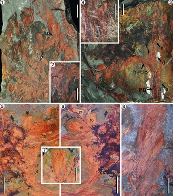
One slab shows ultimate branches and scattered leaves (Fig. 13.1). Five complete leaves are obovate in shape, 14.6–17.1 mm in length and 4.4–5.8 mm in maximum width, and possess a crenulate to entire distal margin (Fig. 13.2, 13.8). One of the ultimate branches, ~0.6 mm wide and ~19 mm long, bears multiple leaves along one only side, while other leaves are not preserved (Fig. 13.8).
Archaeopteris sp. (1) Slab with ultimate branches and scattered leaves; arrow 2 points to the leaf enlarged in (2); arrow 8 points to the part enlarged in (8); PKUB18888a; (2) enlargement of a leaf showing serrate margin; (3) penultimate branch with an ultimate branch (arrow b = UB); other laterals (arrows a, c–e) can be recognized by their broken bases; see line drawing in Figure 14.1; PKUB18802a; (4) enlargement of lateral a in (3), a partially preserved leaf; (5, 6) part and counterpart, enlargement of the ultimate branch shown in (3), with at least 17 leaves (numerals 1–17); leaf 15 in (6) is enlarged in (7); see line drawing in Figure 14.2; PKUB18802a, b; (7) enlargement of a leaf; dissection of its distal margin may be a preservational artifact; (8) leaves attached on ultimate branch. Scale bars: (1, 3, 5, 6) 10 mm; (2, 4, 7, 8) 5 mm.

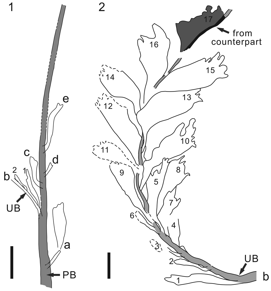
Interpretative line drawings of Archaeopteris sp. (1) Penultimate branch (arrow PB), based on the specimen shown in Figure 13.3, with at least five lateral organs (a–e); only very proximal portions of lateral b, with two leaves, shown (numerals 1, 2) (complete drawing of this lateral is shown in 2); (2) ultimate branch and leaves, based on the specimen shown in Figure 13.5, with leaf 17 from the counterpart shown in Figure 13.6. Labels for the laterals and leaves same as in Figure 13.3–13.6. Leaf venation is poorly preserved in most leaves, and thus is not shown. Scale bars: (1) 10 mm; (2) 5 mm.

Specimen PKUB18802a and its counterpart show a penultimate branch and laterals (Figs. 13.3–13.7, 14). The penultimate branch is incomplete, ~2.4 mm wide and ~89 mm long, with broken ends, and bears at least five lateral organs that depart at an acute angle of 30–40° (Figs. 13.3, 14 letters a–e). While most laterals are poorly preserved, one clearly represents a leafy ultimate branch (Figs. 13.3 arrow b = UB, 14 lateral b = UB). The ultimate branch is curved downward, ~0.8 mm wide and ~66 mm in preserved length, with distal parts absent. At least 17 leaves can be observed along this ultimate branch (Figs. 13.5, 13.6, 14.2 numerals 1–17). These leaves are densely packed, making it difficult to determine the arrangement. Some leaves appear to lie beneath the branch (Fig. 14.2 leaves 4, 7, 8, 12, 17), while others lie on the upper surface of the branch (Fig. 14.2 leaves 1, 2, 5, 9, 13). It is neither possible to distinguish between adaxial and abaxial surfaces of leaves, nor possible to demonstrate whether there is leaf dimorphism. Distal margins of most leaves are incomplete, but one leaf shows that the distal margin is shallowly dissected (Fig. 13.7), although this is considered to be a preservational artifact (compare Fig. 13.2, 13.7, 13.8). More complete leaves are obovate in shape, depart at an angle of 35–48°, appear sessile with no distinct petiole, and are 12.5–15.8 mm long and 4.8–5.4 mm in maximum width. Venation appears to be open dichotomous.
Remarks
Specimens are assignable to the genus Archaeopteris based on gross leaf and branch morphology. Archaeopteris was an important component of global Late Devonian floras (Fairon-Demaret et al., Reference Fairon-Demaret, Leponce and Streel2001; Guo and Wang, Reference Guo and Wang2009; Taylor et al., Reference Taylor, Taylor and Krings2009), and has been reconstructed as a tall tree (Meyer-Berthaud et al., Reference Meyer-Berthaud, Scheckler and Wendt1999). Leaves in this genus show great variation in morphology. Our specimens are distinct from those with deeply dissected leaves (e.g., A. fissilis Schmalhausen, Reference Schmalhausen1894, A. macilenta Lesquereux, Reference Lesquereux1884, and A. notosaria Anderson, Hiller, and Gess, Reference Anderson, Hiller and Gess1995), but similar with those with crenulate to entire margins (e.g., A. halliana [Göppert, Reference Göppert1859] Stur, Reference Stur1875 = A. roemeriana [Göppert, Reference Göppert1859] sensu Stockmans, Reference Stockmans1948) (Kenrick and Fairon-Demaret, Reference Kenrick and Fairon-Demaret.1991; Anderson et al., Reference Anderson, Hiller and Gess1995; Fairon-Demaret et al., Reference Fairon-Demaret, Leponce and Streel2001; Taylor et al., Reference Taylor, Taylor and Krings2009). The shallow dissection in some of our leaves appears to be a result of poor preservation.
Among the species of Archaeopteris, our plant is most similar to A. halliana. As in our specimens, the leaves of A. halliana usually have been described as entire, while some show crenulate margins (e.g., Fairon-Demaret and Leponce, Reference Fairon-Demaret and Leponce2001, figs. 3, 12). Leaves of our specimens, ~15 mm long and ~5 mm in maximum width, fit well within the range of leaves of A. halliana from Belgium (Fairon-Demaret and Leponce, Reference Fairon-Demaret and Leponce2001, based on >350 leaves). Anisophylly as an essential character is demonstrated in A. halliana (i.e., an ultimate branch bears both smaller and larger leaves) (Fairon-Demaret and Leponce, Reference Fairon-Demaret and Leponce2001), although it is not possible to demonstrate this character in our specimens due to poor preservation. Thus, it is appropriate to treat our specimens as Archaeopteris sp.
Archaeopteris has been previously reported from two other localities of the Xing'an Block, or more broadly, NE China. The first record is from the Angeeryin Ul Formation near Zhunsaber, Dong Ujimqin Qi, Inner Mongolia (Fig. 1.2, Table 3, locality III; IMBG, 1976); however, there is no description or illustration available for a comparison with our specimens. Cai (Reference Cai1981) described two species of Archaeopteris (A. cf. A. sphenophyllifolia Lesquereux, Reference Lesquereux1884, and Archaeopteris sp.) from the Xiaohelihe Formation at a locality near Handaqi, Heihe, Heilongjiang Province (Fig. 1.2, Table 3, locality IX). Cai's first species shows deeply dissected leaves, which is quite different from our specimens. His second species is poorly preserved, with leaves of similar shape and size as in our specimens, but distal margins of the leaves are unclear.
List of macrofossil plants and spores from the Late Devonian deposits of northeastern China. *Locality numbers, same as in Figure 1.2. **Taxa with published illustrations are indicated in bold; all other taxa have been neither illustrated nor described in previous reports. ***Plant fossils occur only in the lower part of the Seribayan Obo Formation, while marine fossils of early Mississippian age occur in the upper part (IMBGMR, 1991, 1996).

A possible sphenophyllalean plant
Figure 15.1–15.4
Description
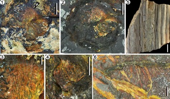
This plant is poorly preserved and the fossils show very low contrast with the surrounding rock matrix. Leaves that can be recognized in the first specimen (Fig. 15.1, 15.2) apparently are arranged in a whorl, but are highly overlapped; one leaf is wedge-shaped, at least 16 mm long, and shows open dichotomous venation (Fig. 15.3). Dégagement of this specimen shows that the leaf remains occupy a circular area that is ~37 mm in diameter and can be clearly distinguished from the surrounding matrix (Fig. 15.2), indicating the outline of a leaf whorl. The second specimen also shows a wedge-shaped leaf ~16 mm long (Fig. 15.4). Leaf veins indicate that the leaves are flabellate.
Other plants. (1–4) A possible sphenophyllalean plant: (1) a whorl of many leaves, which are highly overlapped, and are partially covered and surrounded by white carbonate materials; arrow points to the best preserved leaf enlarged in (3); PKUB18803-2; (2) dégagement stage of the specimen in (1), after removal of carbonate materials; a circular outline is shown, indicating the trace of a leaf whorl; (3) enlargement of distal part of a leaf shown in (1), with vestige of leaf veins shown as ridges on the surface; (4) a wedge-shaped leaf; PKUB18803-1; (5, 6) indeterminate plants; (5) two axes with longitudinal grooves on surface; PKUB18804; (6) axes, with the upper one bearing adventitious roots on one side; PKUB18803-3. Scale bars: (1, 2, 4, 6) 10 mm; (3) 2 mm; (5) 20 mm.

Remarks
Plants in the order Sphenophyllales constitute an important component in the Paleozoic floras, spanning the Late Devonian to late Permian (Batenburg, Reference Batenburg1982; Wang et al., Reference Wang, Hao and Wang2005, Reference Wang, Hao, Tian and Xue2006; Bashforth and Zodrow, Reference Bashforth and Zodrow2007; Xiong and Wang, Reference Xiong and Wang2008; Taylor et al., Reference Taylor, Taylor and Krings2009; Prestianni and Gess, Reference Prestianni and Gess2019). Slender axes with nodes and internodes, and linear, spatulate, or wedge-shaped leaves borne in whorls, are key shared characters (Galtier and Daviero, Reference Galtier and Daviero1999; Bashforth and Zodrow, Reference Bashforth and Zodrow2007; P. Huang et al., Reference Huang, Liu, Qin, Liu, Deng, Wang, Basinger and Xue2018). Our material is potentially assignable to the Sphenophyllales, in the presence of wedge-shaped leaves that apparently are arranged in a whorl. Our plant differs from species with linear, or deeply dissected, or lobed leaves, such as Hamatophyton verticillatum Gu and Zhi, Reference Gu1974; Rinistachya hilleri Prestianni and Gess, Reference Prestianni and Gess2019; Rotafolia songziensis (Feng, Reference Feng1984) Wang et al., Reference Wang, Hao and Wang2005; Sphenophyllum oblongifolium (Germar and Kaulfuss, Reference Germar and Kaulfuss1831) Unger, Reference Unger1850; S. pseudotenerrimum Sze, Reference Sze1936; S. changxingense Huang et al., Reference Huang, Yan, Piper, Zhang, Yi, Yu and Zhou2018; and others (Galtier and Daviero, Reference Galtier and Daviero1999; Wang et al., Reference Wang, Hao and Wang2005, Reference Wang, Hao, Tian and Xue2006; Deng et al., Reference Deng, Huang, Liu, Wang and Xue2016; P. Huang et al., Reference Huang, Liu, Qin, Liu, Deng, Wang, Basinger and Xue2018; Prestianni and Gess, Reference Prestianni and Gess2019), while similar wedge-shaped, unlobed leaves occur in the Late Devonian species S. lungtanense Gothan and Sze, Reference Gothan and Sze1933 (Wang et al., Reference Wang, Wan and Cui2008).
Axes and roots of indeterminate plants
Figure 15.5, 15.6
Description
Axes of unknown plants are found within the beds (Fig. 15.5, 15.6). One of the axes reaches ~40 mm in width, indicating it is a part of a large-bodied plant (Fig. 15.5). Several adventitious roots, ~2.2 mm wide, are attached as a cluster to one side of an axis (Fig. 15.6, upper axis).
Remarks
These axes and roots cannot be assigned to any known taxon, and they are shown here only for the purpose of documentation.
Spores
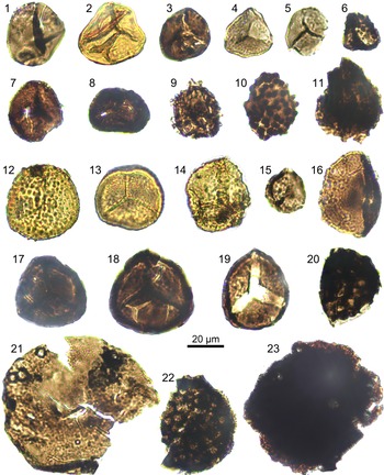
The sample from bed-10 mainly yielded miospores (Fig. 16), which are in low abundance and in poor to moderate preservation. The recognized taxa include: Ambitisporites dilutes (Hoffmeister, Reference Hoffmeister1959) Richardson and Lister, Reference Richardson and Lister1969; Ambitisporites sp.; Aneurospora sp.; Apiculiretusispora spp.; Cymbosporites conatus Bharadwaj et al., Reference Bharadwaj, Tiwari and Venkatachala1971; Cymbosporites cyathus Allen, Reference Allen1965; Densosporites sp.; Dibolisporites sp.; Gulisporites hiatus Lu, Reference Lu1997; Gulisporites intropunctatus Lu, Reference Lu1981; Gulisporites spp.; Lophotriletes sp.; Lycospora sp.; Reticulatisporites sp.; and Retusotriletes sp. Most cannot be identified at the species level with certainty. Additionally, there are some indeterminable spores and rare acritarchs.
Palynomorphs from bed-10 at section PM90. (1) Gulisporites intropunctatus Lu, Reference Lu1981; (2) Gulisporites sp. 1; (3) Gulisporites hiatus Lu, Reference Lu1997; (4) Apiculiretusispora sp. 1; (5) Aneurospora sp.; (6) Lophotriletes sp.; (7) Apiculiretusispora sp. 2; (8) Densosporites sp.; (9) Acritarch; (10) Dibolisporites sp.; (11, 23) indeterminable spores; (12) Cymbosporites cyathus Allen, Reference Allen1965; (13) Cymbosporites conatus Bharadwaj et al., Reference Bharadwaj, Tiwari and Venkatachala1971; (14) Gulisporites sp. 2; (15) Lycospora sp.; (16) Retusotriletes sp.; (17, 18) Ambitisporites sp.; (19) Ambitisporites dilutes (Hoffmeister) Richardson and Lister, Reference Richardson and Lister1969; (20, 22) Reticulatisporites sp.; (21) Apiculiretusispora sp. 3.

Discussion
Age of the flora
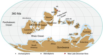
Megafossil plants from the PM90 succession include Melvillipteris sonidia n. sp., Archaeopteris sp., a possible sphenophyllalean plant, and fragments of other plants. The type species of Melvillipteris has been reported from the Late Devonian (Famennian) Parry Islands Formation of Melville Island, Canadian Arctic Archipelago (Fig. 17; Xue and Basinger, Reference Xue and Basinger2016). The genus Archaeopteris has a worldwide distribution, mostly reported from the Upper Devonian of South China, North America, Europe, Morocco, Australia, South Africa, and Colombia (Fig. 17; Beck, Reference Beck1960; Hao and Mei, Reference Hao and Mei1987; Beck and Wight, Reference Beck, Wight and Beck1988; Anderson et al., Reference Anderson, Hiller and Gess1995; Cai and Wang, Reference Cai, Wang, Li, Zhou, Cai, Sun, Ouyang and Deng1995; Meyer-Berthaud et al., Reference Meyer-Berthaud, Scheckler and Wendt1999; Berry et al., Reference Berry, Morel, Mojica and Villarroel2000; Guo and Wang, Reference Guo and Wang2009, Reference Berry, Morel, Mojica and Villarroel2011; Orlova et al., Reference Orlova, Jurina and Snigirevsky2016), although there are also some Middle Devonian occurrences (Berry and Fairon-Demaret, Reference Berry, Fairon-Demaret, Gensel and Edwards2001). Archaeopteris apparently became extinct at about the Devonian-Carboniferous boundary, and rare reports from the earliest Carboniferous remain to be verified (Fairon-Demaret, Reference Fairon-Demaret1986; Scheckler, Reference Scheckler1986). Based on data essentially from Laurasia, seven floral assemblage zones were established by Banks (Reference Banks, Dilcher and Taylor1980) for the interval from the late Silurian to earliest Carboniferous. The Archaeopteris Zone, marked by the appearance of Archaeopteris, was said to be Late Devonian (Frasnian and Famennian) in age, above which is the Rhacophyton Zone with an age of latest Devonian to earliest Carboniferous (Banks, Reference Banks, Dilcher and Taylor1980; Fairon-Demaret, Reference Fairon-Demaret1986; Scheckler, Reference Scheckler1986). However, the development of provincialism may hinder the application of such biozonations outside of Laurasia (Edwards and Berry, Reference Edwards, Berry and Cleal1991). Nevertheless, by combining the evidence of megafossil plants (the co-occurrence of Melvillipteris and Archaeopteris), spores, and U-Pb ages of detrital zircons, the PM90 succession is suggested to be Late Devonian, and most probably Famennian, in age.
Paleogeographic base map modified from B. Huang et al. (Reference Huang, Yan, Piper, Zhang, Yi, Yu and Zhou2018), showing the localities from which Late Devonian Archaeopteris (black dots) and Melvillipteris (black squares) are known. Tectonic blocks: IC = Indochina; MOB = Mongolia; NCB = North China; NQ = North Qiangtang; Qm = Qaidam; SCB = South China; SXB = Songliao-Xilinhot. XB, the Xing'an Block, is considered to be a part of the MOB in this reconstruction. JB, the Jiamusi Block, is considered to be a part of the SXB. Localities of Archaeopteris: 1, Heihe, Heilongjiang, China (Cai, Reference Cai1981; locality IX in Table 3); 2, Sonid Zuoqi, Inner Mongolia, China (flag, this study); 3, Southeastern Altay, Russia (Gutak et al., Reference Gutak, Antonova and Ruban2011); 4, North Timan, Russia (Orlova et al., Reference Orlova, Jurina and Snigirevsky2016); 5, Bear Island, Norway (Nathorst, Reference Nathorst1902; Edwards and Berry, Reference Edwards, Berry and Cleal1991); 6, Namur, Belgium (Fairon-Demaret and Leponce, Reference Fairon-Demaret and Leponce2001); 7, Kiltorcan Hill, Ireland (Dawson, Reference Dawson1871); 8, Ellesmere Island, Canada (Andrews et al., Reference Andrews, Phillips and Radforth1965); 9, New York, USA (Carluccio et al., Reference Carluccio, Hueber and Banks1966); 10, Pennsylvania, USA (Beck, Reference Beck1971); 11, West Virginia, USA (Phillips et al., Reference Phillips, Andrews and Gensel1972); 12, Eastern Cordillera, Colombia (Berry et al., Reference Berry, Morel, Mojica and Villarroel2000); 13, Sierra de Perijá, Venezuela (Berry et al., Reference Berry, Morel, Mojica and Villarroel2000); 14, Eastern Anti-Atlas, Morocco (Meyer-Berthaud et al., Reference Meyer-Berthaud, Scheckler and Wendt1999); 15, Eastern Cape, South Africa (Anderson et al., Reference Anderson, Hiller and Gess1995); 16, Nanjing, Jiangsu, China (Wu et al., Reference Wu, Zhao and Deng1982); 17, Changyang, Hubei, China (Hao and Mei, Reference Hao and Mei1987; Guo and Wang, Reference Guo and Wang2011); 18, Chizhou, Anhui, China (Guo and Wang, Reference Guo and Wang2009); 19, Xinhui, Guangdong, China (Cai et al., Reference Cai, Wen and Chen1987); 20, New South Wales, Australia (Young, Reference Young2007; Meyer-Berthaud et al., Reference Meyer-Berthaud, Decombeix, Dunstone, Gerrienne, Momont and Young2016a). Localities of Melvillipteris: 2 (this study) and Melville Island, Canadian Arctic Archipelago (Xue and Basinger, Reference Xue and Basinger2016).

Spores from the PM90 succession, although only preliminarily reported herein, also suggest a Late Devonian age. While Cymbosporites cyathus was first reported from the Middle Devonian (Givetian) of Vestspitsbergen (Allen, Reference Allen1965), this species commonly occurs in the Late Devonian of South China (Ouyang et al., Reference Ouyang, Lu, Zhu and Liu2017), and in the basal part of the Heishantou Formation (around the Devonian-Carboniferous boundary) in Xinjiang, northwestern China (Lu, Reference Lu1999). Cymbosporites conatus shows a similar temporal distribution as the type species, also with a record from the basal Heishantou Formation of Xinjiang (Lu, Reference Lu1999; Ouyang et al., Reference Ouyang, Lu, Zhu and Liu2017). Gulisporites intropunctatus was described from the lower Upper Devonian of Sichuan, and Gulisporites hiatus, from the Famennian Shaodong Formation of Hunan, South China (Lu, Reference Lu1981, Reference Lu1997). Regionally, previous studies have reported spores from the Angeeryin Ul Formation (Fig. 1.2, Table 3, localities II, III), but the lack of illustrations for these reports prevents a comparison with our results. Spores assigned to 22 genera and 34 species were described from the Heitai Formation of Mishan, Heilongjiang Province, NE China, indicating a Middle Devonian age (Ouyang, Reference Ouyang1984), although no taxa at the species level are shared by the Heitai assemblage and that of the PM90 succession.
Another line of evidence for the age of the flora is based upon 206Pb/238U ages of detrital zircons from bed-13 (Supplementary Data). The youngest age populations of the measured detrital zircons show a peak value at ca. 365 Ma (Supplementary Data, Fig. 2), within the range of the Famennian Stage (Becker et al., Reference Becker, Marshall, da Silva, Gradstein, Ogg, Schmitz and Ogg2020).
Late Devonian plants from blocks of NE China
Previous records of Late Devonian vascular plants from NE China are sparse, having been described or briefly mentioned from only eight localities, mainly during regional geological surveys during the 1970s and 1980s (Fig. 1.2, Table 3, localities II-IX) (IMBG, 1965, 1973, 1974, 1976; IMBG and NIGS, 1974; HLJBG, 1977, 1981; Cai, Reference Cai1981; Xue et al., Reference Xue, Zhao and Zhang1982; Wang et al., Reference Wang, Lu, Zhang and Zhi2012). Our locality is a new addition to the list. These localities are found on two different crustal blocks: the Xing'an (XB) and Songliao-Xilinhot (SXB) blocks (Fig. 1.2). Among the previously reported plants from XB, two species of Archaeopteris from the Xiaohelihe Formation were illustrated and described by Cai (Reference Cai1981) (Fig. 1.2, Table 3, locality IX), while all others have yet to be identified.
The XB is almost, but not precisely, equivalent to the Uliastai active continental margin (Xiao et al., Reference Xiao, Windley, Hao and Zhai2003) or the Nuhetdavaa terrane (Badarch et al., Reference Badarch, Dickson Cunningham and Windley2002), and it may have been a microcontinent since the early Paleozoic (Liu et al., Reference Liu, Li, Feng, Wen, Neubauer and Liang2017), or even the Neoproterozoic (B. Xu et al., Reference Xu, Jiang, Zhang, Wang, Fu and Feng2015; Zhou et al., Reference Zhou, Wilde, Zhao and Han2018). It has been suggested that the XB had become part of the Mongolian Block (MOB) by the Late Devonian, when the MOB occupied a position at 30–60°N (e.g., Fig. 17; based on B. Huang et al., Reference Huang, Yan, Piper, Zhang, Yi, Yu and Zhou2018). Thus, the Late Devonian floras of the XB provide data for mid-latitude vegetation of the Northern Hemisphere, which is less well known than lower latitudes.
Four localities on the western segment of the SXB were reported to yield Late Devonian plants from the Seribayan Obo Formation (Fig. 1.2, Table 3, localities V-VIII) (IMBG 1965, 1974; IMBG and NIGS, 1974; Wang et al., Reference Wang, Lu, Zhang and Zhi2012). However, only two genera, Leptophloeum and Archaeocalamites, were recorded, and only the former was illustrated and described.
Geographically, it seems that during the Late Devonian, the XB was in mid-latitude and the SXB in low-latitude positions (e.g., Fig. 17; B. Huang et al., Reference Huang, Yan, Piper, Zhang, Yi, Yu and Zhou2018). A comparison among the Late Devonian floras from the SXB and XB yields the interesting observation that Leptophloeum and Archaeopteris, two index plants of Late Devonian age, were found in the SXB and XB, respectively, but not in both. Nevertheless, this may well be a result of very limited sampling from both blocks, and such an interpretation would be somewhat premature.
Conclusions
A Late Devonian flora, which is described from a newly discovered locality near Dalai Sumu, northern Sonid Zuoqi, Inner Mongolia, NE China, includes Melvillipteris sonidia n. sp., Archaeopteris sp., and a possible sphenophyllalean plant. Melvillipteris sonidia n. sp. represents the second species of Melvillipteris, a genus previously only known from the Upper Devonian (Famennian) of Arctic Canada. Melvillipteris sonidia n. sp. is characterized by paired first-order branches that are typically zigzag in appearance; its second-order branches bear three-dimensional sterile ultimate appendages, as well as fertile structures that terminate in fusiform sporangia. An associated palynological assemblage, as well as U-Pb ages of detrital zircon grains from adjacent horizons, are also reported, supporting a Late Devonian age, which in accord with the megafossil plants. Vascular plants have been rarely recorded from Late Devonian deposits of tectonic blocks that constitute present-day NE China. This new flora, from the Xing'an Block, permits comparison with coeval floras from Europe, North America, and South China, and aids our understanding of Late Devonian mid-latitude vegetation of the Northern Hemisphere.
Acknowledgments
The authors thank X.C. Jin for his helpful discussions, and Y.C. Su, Z. Xu, G.D. Qiu, J.X. Du, J.C. Guo, and J.S. Wang for their help with fieldwork and fossil photography. This research was supported by the National Natural Science Foundation of China (Nos. 41722201, 41802003), Geological Survey projects of the China Geological Survey (Nos. DD2016041-15, DD20190008), Strategic Priority Research Program of Chinese Academy of Sciences (No. XDB26000000), and the Natural Science Foundation of Jiangsu Province (No. SBK2018042780). The anonymous reviewers and Associate Editor (J. Nathan) are thanked for their constructive comments that greatly improved the quality of this paper.
Data availability statement
Data available from the Dryad Digital Repository: https://doi.org/10.5061/dryad.d51c5b042