Introduction
Since the introduction of antibiotics, facial paresis secondary to acute otitis media in adults has become a rare but nevertheless relevant complication. The reported incidence is approximately 0.05 per cent.Reference Ellefsen and Bonding1,Reference Ren, Sethi and Stankovic2 However, the pathogenesis is still unclear and different hypotheses have been discussed. Possible explanations include: (1) direct involvement of the facial nerve by infection through pre-existing, small bony dehiscences; (2) osteitis of the Fallopian canal, with bone erosion and facial nerve involvement; (3) reactivation of a latent neurotropic virus (e.g. herpes virus); and (4) inflammatory oedema with compression of the vasa nervorum and nerve damage.Reference Redaelli de Zinis, Gamba and Balzanelli3 Previous studies have also indicated that dehiscences of the facial canal are found in up to 48–55 per cent of all temporal bones, and in the vast majority of all cases the tympanic segment is affected.Reference Baxter4–Reference Yu, Wang, Yang, Han and Zhang6
From a diagnostic perspective, high-resolution temporal bone computed tomography (CT) supplements clinical and audiological investigations, whenever intracranial or extracranial complications are suspected. Additionally, high-resolution temporal bone CT provides a bone anatomy ‘roadmap’ for potential surgical procedures, and allows detailed assessment of the facial nerve course within the labyrinthine, tympanic and mastoid segments.Reference Yu, Wang, Yang, Han and Zhang6,Reference Juliano, Ginat and Moonis7 However, to our knowledge, no previous study has systematically investigated the correlation between acute otitis media with secondary facial paresis and the presence of bony dehiscence in the tympanic facial canal.
This retrospective case–control study aimed to investigate the prevalence and distribution pattern of bony dehiscences in the tympanic facial canal in adult acute otitis media patients with or without secondary facial paresis.
Materials and methods
Study design
This retrospective study received ethical approval from the relevant authorities (identification number: KEK 2018-00196). We reviewed the data for all adult patients with available high-resolution temporal bone CT scans, who presented with acute otitis media and secondary facial paresis at the Department of Otorhinolaryngology, Head and Neck Surgery, at the University Hospital Zurich (Switzerland), between January 2000 and May 2019. In total, 24 patients were identified and formed the index group.
For every index group patient, we included an age- and sex-matched control patient with acute otitis media and available high-resolution temporal bone CT scans, but without facial paresis, who presented during the same study period (n = 24). All control group patients had undergone high-resolution temporal bone CT to rule out intracranial or extracranial complications of acute otitis media, such as subperiosteal abscess, mastoiditis, sigmoid sinus thrombosis or intracranial abscess.Reference Leskinen and Jero8
Patients with a prior history of ear surgery, no available high-resolution temporal bone CT scans or an unwillingness to contribute their data were excluded.
The following patient and disease-associated data were collected: age at diagnosis, sex, affected ear (right, left, or both), conservative treatment regimen (antibiotics, steroids), microbiological examination findings of the middle-ear fluid, surgical treatment modalities (paracentesis or ventilation tube insertion, antrotomy), course of facial paresis as documented by House–Brackmann score (baseline score, peak score (highest documented score), nadir, follow-up time), and baseline bone conduction and air conduction pure tone audiometry data.
Diagnostic investigation and treatment characteristics
Routine diagnostic investigation of the following was conducted: past medical history, otoscopy, tuning fork tests, vestibular assessment and pure tone audiometry (following standard procedures in accordance with International Organization for Standardization audiometric test methods (ISO 8253-1: air conduction, bone conduction)). As mentioned above, contrast-enhanced, high-resolution temporal bone CT (bone and soft tissue kernel) was performed in all included patients to rule out intracranial or extracranial complications of acute otitis media, and as part of a bone anatomy roadmap for subsequent potential surgery.Reference Mather, Yates, Powell and Zammit-Maempel9
Treatment cornerstones consisted of antibiotic treatment (first choice of amoxicillin/clavulanic acid, or third generation cephalosporin), corticosteroids (systemic or intratympanic administration), paracentesis or ventilation tube insertion, and antrotomy.Reference Smith, Gutteridge, Elliott and Cronin10 In one particular patient, an additional mastoidectomy and decompression of the facial nerve in its mastoid segment was performed. Eye protection was prescribed in cases of facial paresis, to prevent lagophthalmos-associated morbidity. The individual treatment algorithm was determined based on clinical, audiological and radiological findings, and with regard to the patient's will.
Imaging procedure
Bilateral temporal bone CT imaging was performed using a single-source CT scanner (Somatom Definition; Siemens Healthcare, Erlangen, Germany) in helical mode and with intravenous contrast-enhancement. A clinical protocol was used with the following parameters: acquisition = 16 × 0.6 mm, pitch of 0.85, reference kV = 120, effective mAS = 160, gantry rotation time of 1 second, slice thickness and increment of 0.6 mm, using automated attenuation-based tube current modulation (Care Dose 4D; Siemens Healthcare). For each side, reconstructions in coronal and axial planes, and in bone and soft tissue kernel, were performed.
Imaging analysis
The high-resolution temporal bone CT scans for each patient were independently reviewed by two radiologists, one with six years (SP; board-certificated neuroradiologist) and another with one year (NN) of experience in neuroradiology. The osseous roofing of the fallopian canal was evaluated by each radiologist in the axial and coronal plane reconstructions using the digital radiology imaging system in our hospital (picture archiving and communication system; Agfa Impax™ version 6.7.0.1071). The two readers were blinded to patient information and clinical symptoms.
A dehiscence of the bony tympanic facial canal was assumed if there was no bony shelter over the tympanic segment of the fallopian canal. The contralateral side was used for comparison. The presence of a dehiscence was rated as: obvious dehiscence, no dehiscence or unclear. Additional findings, such as sigmoid sinus thrombosis, subperiosteal abscess and intracranial abscess, were assessed. In cases of inter-reader disagreement, a final consensus decision was achieved by bilateral re-evaluation of the images, which occurred for 9 out of 48 subjects (18.8 per cent).
Variables and statistical analysis
Ordinal non-dichotomous variables are expressed as median (with range), nominal non-dichotomous variables are expressed as mode (percentage), and ratio variables are expressed as geometric mean ± standard deviation. For comparison of the index group (acute otitis media with secondary facial paresis) versus the control group (acute otitis media without facial paresis), a 2 × 2 contingency table and Fisher's exact test were used. A post-hoc power analysis was performed, assuming a power of 80 per cent and an alpha of 0.05, to determine whether the group size was sufficient. The sensitivity, specificity, positive predictive value, negative predictive value and accuracy for the variable ‘dehiscence of bony tympanic facial canal’ in predicting facial paresis were calculated, including asymptotic 95 per cent confidence interval (CI). Differences in age between the index group and control group were compared using a Mann–Whitney U test. A p-value of less than 0.05 was regarded as statistically significant. Statistics were calculated using SPSS for Windows software, version 25.0 (SPSS, Chicago, Illinois, USA).
Results
Patient and treatment characteristics
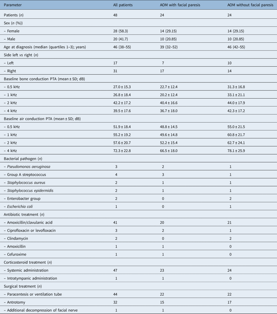
In total, 48 patients were included in the study: 24 had acute otitis media with facial paresis, and 24 had acute otitis media without facial paresis. Table 1 provides detailed information on patient demographics, diagnostic investigation findings and treatment characteristics. Both groups revealed a comparable age distribution (p = 0.11).
Patient demographics, diagnostic investigation findings and treatment characteristics

AOM = acute otitis media; PTA = pure tone average; SD = standard deviation
Microbiological examination of the middle-ear fluid was performed in 44 of the 48 patients, who underwent paracentesis or ventilation tubes insertion. A bacterial pathogen was identified in 14 of these 44 patients (31.8 per cent), with a similar distribution in both groups (7 patients in the acute otitis media with facial paresis group and 7 patients in the acute otitis media without facial paresis group) (Table 1).Reference Mofatteh, Shahabian Moghaddam, Yousefi and Namaei11
The mean bone conduction baseline pure tone average threshold, used as a measure of initial cochlear damage, was similar in both groups (30 ± 10 dB in the acute otitis media with facial paresis group, and 37.2 ± 6.4 dB in the acute otitis media without facial paresis group), as was the mean bone conduction pure tone average threshold after six months (20.9 ± 6.7 dB vs 24.9 ± 2.2 dB, respectively).
The acute otitis media with facial paresis patients had a mean initial House–Brackmann score of 3.1 ± 1.0 (House–Brackmann grade II in 6 patients, grade III in 13 patients, grade IV in 1 patient, grade V in 4 patients) and a mean peak score of 3.2 ± 0.9 (grade II in 5 patients, grade III in 12 patients, grade IV in 1 patient, grade V in 3 patients). The mean peak score was reached after 6.7 days (3–14 days). The final documented House–Brackmann score corresponded to grade I in 20 patients (normal facial function) and grade II in 4 patients (with very mild, persistent asymmetry). The time for recovery varied from two weeks to six months (in a case of an initial House–Brackmann grade V score). The mean follow-up time after initial documentation of facial paresis was 8.4 ± 4.6 months (range, 1–12 months). The mean follow-up time in the four patients with uncomplete recovery was 6.3 ± 3, 9 months (range, 1–12 months), with initial House–Brackmann scores corresponding with grade III in three patients and grade V in one patient.
Radiological assessment
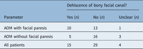
As presented in Table 2, we systematically evaluated high-resolution temporal bone CT scans for the presence of a bony dehiscence in the tympanic segment of the facial canal. In 44 out of 48 patients (91.7 per cent), the neuroradiological review allowed definitive determination of the absence or presence of dehiscence. The prevalence of bony dehiscence did not significantly differ between the index group and control group (p = 0.21), although absolute numbers showed a trend towards a higher prevalence in the acute otitis media with facial paresis group (Table 2). However, a post-hoc power analysis revealed that the study is underpowered; 33 patients are required in the index group to show significant differences.
Systematic evaluation of high-resolution temporal bone CT for determining bony dehiscence*

*In the tympanic segment of the facial canal. CT = computed tomography; AOM = acute otitis media
A bony dehiscence of the tympanic facial canal was associated with a sensitivity of 43.5 per cent (95 per cent CI = 23.2–65.5), specificity of 76.2 per cent (95 per cent CI = 52.8–91.8 per cent), positive predictive value of 66.7 per cent (95 per cent CI = 45–83.1), negative predictive value of 55.2 per cent (95 per cent CI = 44.4–65.4 per cent) and accuracy of 59.1 per cent (95 per cent CI = 43.3–73.7 per cent) in predicting acute otitis media with secondary facial paresis. No other complications, such as sigmoid sinus thrombosis, subperiosteal abscess or intracerebral abscess, were seen on the high-resolution temporal bone CT images.
Discussion
Main findings
This retrospective case–control study on adult acute otitis media patients who underwent high-resolution temporal bone CT indicates that patients with or without suspected bony dehiscence of the tympanic facial canal can develop secondary facial paresis. An intact bony facial canal in the tympanic segment does not seem to protect from the development of a secondary facial paresis.
Results in context of existing literature
In line with previous studies, we confirmed secondary facial paresis in adult acute otitis media patients, thus revealing a favourable prognosis, leading to restitutio ad integrum in the vast majority of all cases.Reference Ellefsen and Bonding1,Reference Redaelli de Zinis, Gamba and Balzanelli3 Treatment algorithms should rely on clinical, audiological and radiological findings, and must incorporate the presence or absence of concomitant serous labyrinthitis due to bacterial toxin and inflammatory mediators in the middle ear.Reference Kitsko and Dohar12,Reference Choi, Han, Nahm, Shin and Kim13 In addition to conservative treatment cornerstones, there are surgical options to maximally evanish ototoxic agents due to concomitant labyrinthitis, including paracentesis or ventilation tube insertion and antrotomy.Reference Smith, Gutteridge, Elliott and Cronin10 In our cohort, antrotomy, the more extreme treatment, was performed in 32 out of 48 patients (66.7 per cent; 15 in the acute otitis media with facial paresis group, 17 in the acute otitis media without facial paresis group).
Clinical and audiological examinations should be accompanied by high-resolution temporal bone CT in acute otitis media cases, whenever intracranial or extracranial complications are suspected. These complications usually form per continuitatem or via haematological spread, and include sinus venous thrombosis, meningitis, labyrinthitis, epidural or intracranial abscess, or mastoiditis with development of osteitis and subperiosteal abscess.Reference Mather, Yates, Powell and Zammit-Maempel9 Radiological key findings of ‘uncomplicated’ acute otitis media include fluid within the middle ear and mastoid, resulting in an opacification, with intact bony architecture of the ossicular chain and mastoid septae.
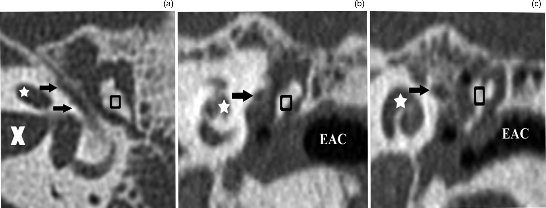
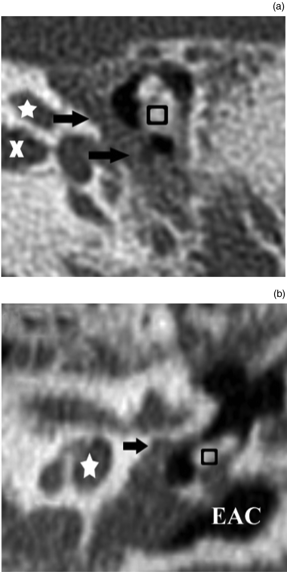
The course of the tympanic facial nerve with its bony shelter can usually be best visualised on axial and particularly coronal sections in high-resolution temporal bone CT, whereby the labyrinthine leads to the tympanic segment and forms the so called ‘snake eyes’ sign.Reference Juliano, Ginat and Moonis7 The tympanic segment of the facial nerve runs inferior to the lateral semicircular canal and superolateral to the oval window.Reference Juliano, Ginat and Moonis7 In line with previous studies, we showed that assessment of the bony tympanic facial nerve, in terms of presence or absence of bony dehiscence, can reliably be achieved in the vast majority of cases (91.7 per cent) (Figures 1–3).Reference Yu, Wang, Yang, Han and Zhang6 However, it is unclear whether radiological dehiscence dependably correlates with intra-operative findings or post-mortem studies.
Axial (a) and coronal (b) high-resolution temporal bone computed tomography images of a 76-year-old patient with otomastoiditis and secondary facial paresis on the left site. The images show an intact bony canal of the left facial nerve in its tympanic segment (black arrows), cochlea (white asterisk), internal auditory canal (white ‘X’), ossicle (black rectangle), superior semicircular canal (black arrowhead) and external auditory canal (EAC). There is opacification of Prussak's space, mesotympanum and mastoid air cells due to inflammation.

Axial (a) and coronal (b & c) high-resolution temporal bone computed tomography images of a 41-year-old patient with acute otomastoiditis and secondary facial paresis on the left site. The images show an intact bony canal of the facial nerve in its tympanic segment (black arrows), cochlea (white asterisk), external auditory canal (EAC), internal auditory canal (white ‘X’) and ossicle (black rectangle).

Axial (a) and coronal (b) high-resolution temporal bone computed tomography images of a 38-year-old patient demonstrating a large dehiscence (black arrows) in the tympanic segment of the bony facial canal. Note opacification in the hypotympanum, Prussak's space and mastoid air cells due to acute inflammation. The images also show the cochlea (white asterisk), internal auditory canal (white ‘X’), external auditory canal (EAC) and ossicle (black rectangle).

Two previous studies investigating the correlation of high-resolution temporal bone CT information and surgical findings in terms of the facial canal indicated that radiological dehiscence can usually be confirmed intra-operatively.Reference Fuse, Tada, Aoyagi and Sugai5,Reference Yu, Wang, Yang, Han and Zhang6 However, other authors have reported that high-resolution temporal bone CT can overestimate the prevalence of superior canal dehiscence, when compared to intra-operative or post-mortem findings.Reference Mittmann, Ernst, Seidl, Skulj, Mutze and Windgassen14,Reference Tavassolie, Penninger, Zuniga, Minor and Carey15
When comparing the index group (acute otitis media with facial paresis) with the control group (acute otitis media without facial paresis), the prevalence of bony dehiscence did not significantly differ. Although the presence of a bony dehiscence was associated with a positive predictive value of 66.7 per cent in terms of predicting acute otitis media with secondary facial paresis, 33.3 per cent of all patients did not develop facial paresis despite the presence of dehiscence (Figure 3). On the other hand, an intact bony tympanic facial canal did not prevent secondary facial paresis in 44.8 per cent of cases (95 per cent CI = 34.6–55.6) (Figures 1 and 2). However, a post-hoc power analysis of our data revealed a power of 68 per cent (assuming an alpha of 0.05); a total of 33 patients would have been needed to achieve a power of 80 per cent. Thus, given the lack of power of our study, a conclusive statistical statement is not possible.
A review of the absolute number of cases revealed a trend towards a higher prevalence in the index group, but this must be interpreted with caution. Based on our findings, a radiologically apparent direct contact between the tympanic facial nerve and inflammatory agents in the middle ear does not seem to be mandatory for the development of secondary facial paresis, although such paresis may be less likely in cases without this contact. Based on these findings, we consider multifactorial pathogenesis to be most likely. Firstly, pre-existing, radiologically inapparent bony micro-dehiscences could cause inflammatory agents in the middle ear to affect the facial nerve and its vasa nervorum. Secondly, one could hypothesise that an inflammatory effect of the facial nerve along with acute otitis media is common, but is often clinically inapparent and possibly more likely in the presence of a pronounced bony dehiscence. Thus, the pathogenic potential of adjacent inflammatory agents might influence the course of disease.
Strengths and limitations
To the best of our knowledge, this is the first study to address the role of a dehiscent tympanic facial canal and its association with the development of a secondary facial paresis in adult acute otitis media patients. Besides its retrospective design, we acknowledge that our study has some noteworthy limitations. Although all acute otitis media patients with facial paresis and available high-resolution temporal bone CT scans at our institution were included, our study was underpowered. Furthermore, it remains unclear whether bony dehiscences are pre-existing or sequelae of middle-ear infection and consecutive bony erosion.
Radiological assessment by high-resolution temporal bone CT was considered the ‘gold standard’, and no surgical exploration of the middle ear or cadaveric studies were performed. However, tympanoscopy is not part of routine surgical treatment in complicated acute otitis media cases and therefore is not justified. The accuracy of high-resolution temporal bone CT to detect a tympanic facial canal dehiscence has been debated previously. Some studies have shown that image-based assessment by high-resolution temporal bone CT correlated with surgical findings in 75 per cent of all cases;Reference Fuse, Tada, Aoyagi and Sugai5,Reference Yu, Wang, Yang, Han and Zhang6 other studies showed that thin bony structures can be missed in high-resolution temporal bone CT.Reference Mittmann, Ernst, Seidl, Skulj, Mutze and Windgassen14,Reference Tavassolie, Penninger, Zuniga, Minor and Carey15 Four patients received radiological assessment outside of our hospital on different scanners; thus, the slice thickness of high-resolution temporal bone CT in these cases varied between 0.75 mm and 0.8 mm, while it was 0.6 mm as part of the standardised protocol in our hospital (44 out of 48 patients).
• Facial paresis secondary to acute otitis media in adults is a rare but relevant complication (incidence of 0.05 per cent)
• Neuroradiological review of high-resolution temporal bone computed tomography scans allowed definitive determination of dehiscence in 44 out of 48 patients
• An intact tympanic bony facial canal does not protect from development of secondary facial paresis
• Radiologically apparent contact between tympanic facial nerve and inflammatory agents in the middle ear is not mandatory for secondary facial paresis
• Pathogenesis of facial paresis secondary to acute otitis media is most likely multifactorial
Conclusion
An intact tympanic bony facial canal does not protect from the development of secondary facial paresis. Vice versa, a radiologically apparent direct contact between the tympanic facial nerve and inflammatory agents in the middle ear does not seem to be mandatory for the development of secondary facial paresis. Additional, ideally prospective studies, conducted on larger numbers of patients, are warranted to determine the pathogenesis of secondary facial paresis in acute otitis media, which is most likely multifactorial.
Competing interests
None declared