Introduction
Freshwater snails and bivalves have been used frequently as bioindicator organisms and in several studies of chemical contaminants. Due to the large size and limited mobility of freshwater snails and bivalves, they have two important advantages over most other freshwater organisms for biomonitoring research. In addition, they are commonly found in freshwater environments and are relatively easy to collect and identify (Elder & Collins, Reference Elder and Collins1991). Ansaldo et al. (Reference Ansaldo, Nahabedian, Di Fonzo and Wider2009) reported that the availability and the relatively easy aquaculture of Biomphalaria glabrata snails made them an accessible tool for contamination bioassays. Moreover, B. glabrata was used as a bioindicator for assessing lead (Pb) contamination in aquatic ecosystems, especially at low levels of metal exposure (Aisemberg et al., Reference Aisemberg, Nahabedian, Wider and Guerrero2005). The closely related species B. alexandrina has historically been confined to the Nile Delta, Egypt (Vidigal et al., Reference Vidigal, Spatz, Nunes, Simpson, Carvalho and Neto1998), but recently it appears to have been expanding its range upstream in Egypt. Changes in the hydrology of the Nile basin, controlled water flow, and new irrigation networks following construction of the Low and High Dams at Aswan, in 1902 and 1968, respectively, have been implicated in increasing appropriate habitats for B. alexandrina (Spatz et al., Reference Spatz, Vidigal, Silva, Cappa and Carvalho2000; Vidigal et al., Reference Vidigal, Caldeira, Simpson and Carvalho2000). This species has been associated with transmission of Schistosoma mansoni and Echinostoma liei in Egypt (Mostafa et al., Reference Mostafa, Bakry, Sleem and El-Dafrawy2001).
In Egypt, the Nile and its waterways used to receive about 85% of industrial wastewater. Also, agricultural wastewater, untreated domestic wastewater, discharged oil and grease have been dumped into the Nile (Abdel-Shafy et al., Reference Abdel-Shafy, Abo-El-Wafa and Azzam1987). Marcogliese & Pietrock (Reference Marcogliese and Pietrock2011) reviewed the combined effects of parasites and contaminants on animal health and reported that these combined stressors might reduce either resistance or tolerance to infection.
Infection of freshwater molluscs with their natural parasites alters the concentrations of metallic ions in their bodies. This was demonstrated in a variety of snail–trematode models using a number of different analytical methods (Gabrashanska et al., Reference Gabrashanska, Damyanova and Kanev1991; Layman et al., Reference Layman, Dory, Koehnlein, Fried and Sherma1996; Ong et al., Reference Ong, Chejlava, Fried, Koehnlein, Bosavage and Sherma2004; Hassan, Reference Hassan2008; Mostafa, Reference Mostafa2008; Ma et al., Reference Ma, Zhu, Wang, Zhou, Peng and Liu2010; Mostafa & Dajem, Reference Mostafa and Dajem2010). Furthermore, interactions between the effects of heavy metals and trematode infections on the snails' biological and physiological systems have been investigated (Abd Allah et al., Reference Abd Allah, Wanas and Thompson1997; Morley et al., Reference Morley, Crane and Lewis2002, Reference Morley, Crane and Lewis2004; Salice & Roesijadi, Reference Salice and Roesijadi2002; Soliman, Reference Soliman2009). In the field of ecotoxicology, the effects of parasitic infections on pollutant uptake and accumulation in different intermediate and final hosts are very important (Sures, Reference Sures2006).
Due to the importance of snails in the field of environmental monitoring, the present study aimed to compare the heavy metal concentrations in trematode-infected and uninfected snails (B. alexandrina) collected from Kafer Alsheikh and Menofia provinces, Egypt. The results will increase our understanding of the use of these snails as bioindicators for heavy metal pollution.
Materials and methods
Collection and examination of snails
Using a net, 15 cm deep and a mesh size of 3 mm, two samples of 100 B. alexandrina were collected from the River Nile in provinces of Kafer Alsheikh and Menofia, Egypt during September and October 2011. Snails were transferred to the Medical Malacology laboratory at Theodor Bilharz Research Institute (TBRI) and examined immediately for any evidence of trematode infections of E. liei or S. mansoni.
Shedding snails were isolated and kept at − 20°C for later analysis. Snails from Kafer Alsheikh were infected with cercariae of either S. mansoni or E. liei, whereas snails from Menofia were only infected with E. liei. The remaining snails were crushed and examined under a binocular microscope and any uninfected snails were isolated and kept at − 20°C for use as controls. Uninfected, lab-bred snails, obtained from the Theodor Bilharz Research Institute (TBRI), were used for comparison with uninfected snails collected from the field. The range in diameter of shells of snails used in the present study was 5–8 mm.
Heavy metal and data analyses
The entire shell and soft tissues of four snail groups with three individuals per group were prepared for analysis. These groups included those shedding S. mansoni or E. liei cercariae in addition to uninfected field and lab-bred snails. Samples of known wet weight were digested in 10 ml of concentrated nitric acid by boiling to dryness. The residue from each digested sample was diluted to 25 ml with deionized water in a volumetric flask.
Aluminium (Al), cadmium (Cd), copper (Cu), iron (Fe), manganese (Mn), lead (Pb) and zinc (Zn) were measured by flame atomic absorption spectrometry (model 3100; Perkin Elmer Beaconsfield, Bucks, UK) in both the soft parts and shells of the snails. The flame wavelength and sample aspiration rate were optimized according to the manufacturer's recommendations. Four aqueous acidified dilutions of the standards were prepared at concentrations within the linear response range of the instrument. Each sample, standard and blank was analysed using three 10-s integrations. The reagent blank value was subtracted from the sample values to give the final concentration. Elemental concentration (C) was calculated according to the following equation:
 $$\begin{eqnarray} \end{eqnarray}$$
where F is the standard factor calculated from the standard curve, V is the volume of sample and W is the sample weight. Data were expressed in micrograms of element per gram of tissue.
Data analysis
Results were expressed as means ± standard error (SE). Data analysis with the Statistical Package for Social Sciences (SPSS 8.0 for Windows; SPSS Inc., Chicago, Illinois, USA), using one-way analysis of variance (ANOVA) followed by Duncan's test for comparison between different treatment groups. Statistical significances were set at P< 0.05.
Results
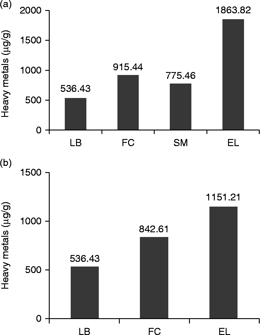
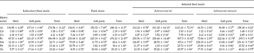
The total concentrations (the sum of each mineral element concentration in shell and soft parts) of heavy metals were compared for field collected, lab-bred and uninfected snails (fig. 1a and b). Results showed that levels of Al, Cd, Cu, Fe, Mn, Pb and Zn were significantly higher in uninfected, field-collected snails compared with uninfected, lab-bred snails in both samples collected from Kafer Alsheikh and those from Menofia province (tables 1 and 2). An exception was at Kafer Alsheik, where Al and Cu concentrations in the uninfected snails collected from the field did not differ significantly from lab-bred, uninfected snails.
Concentration (μg/g) of the heavy metals Al, Cd, Cu, Fe, Mn, Pb and Zn in uninfected laboratory-bred (LB) and field-collected Biomphalaria alexandrina (FC) for comparison with B. alexandrina infected with cercariae of Schistosoma mansoni (SM) and/or Echinostoma liei (EL) from (a) Kafer Alsheik and (b) Menofia.

Mean heavy metal concentrations (μg/g± SE) in uninfected laboratory-bred and field snails (Biomphalaria alexandrina) and field snails infected with cercariae of Echinostoma liei or Schistosoma mansoni from the province of Kafer Alsheik, Egypt; values not showing common superscripts (a, b, c, d) differ significantly at P<0.05.
Mean heavy metal concentrations (μg/g± SE) in uninfected laboratory-bred and field snails (Biomphalaria alexandrina) and field snails infected with cercariae of Echinostoma liei from the province of Menofia, Egypt; values not showing common superscripts (a, b, c, d) differ significantly at P<0.05.
Soft body parts had higher Al, Fe and Zn content compared with the shells (infected, uninfected, field-collected or lab-bred). However, the opposite was true for the rest of the metals, that is, the levels of Cd, Cu, Mn and Pb were higher in shells than in soft body parts of all snails.
In addition, levels of heavy metals in field collected E. liei-infected and S. mansoni-infected snails were compared with their uninfected counterparts (table 1). Type of infection, either by E. liei or S. mansoni, affected the concentrations of heavy metals in the host snail. In general, total concentrations of heavy metals in E. liei-infected snails were greater compared to uninfected snails but were less in S. mansoni-infected snails when compared with uninfected snails (fig. 1). The metal contents of E. liei-infected snails collected from both sites (Kafer Alsheikh and Menofia provinces) were compared with uninfected snails collected from same sites. Results showed significant increase in total concentrations of Al, Cd, Cu, Fe, Mn and Zn. However, Pb concentration was less in E. liei-infected snails. Field-collected S. mansoni-infected snails were compared with uninfected snails at the same site. Results showed a significant decrease in the concentrations of Al, Cd, Cu, Fe and Pb. However, there was significant increase in the total concentrations of Mn and Zn in S. mansoni-infected snails. Interestingly, Cd concentration was the lowest in all snail samples (tables 1 and 2). The results and degrees of statistical significance are displayed in tables 1 and 2.
Discussion
The present study showed that total concentrations of heavy metals of uninfected field-collected snails were higher than uninfected lab-bred snails, demonstrating the bioaccumulation of heavy metals in B. alexandrina. This was in agreement with the findings of Ma et al. (Reference Ma, Zhu, Wang, Zhou, Peng and Liu2010). They demonstrated the bioaccumulation of heavy metals of the river sediment in Bellamya aeruginosa, specifically in the hepatopancreas of snails. Furthermore, heavy metals didn't distribute evenly in the snails' body parts, as concentrations of Al, Fe and Zn were higher in soft parts compared with shells and the converse was true for Cd, Cu, Mn and Pb. This pattern was observed in both uninfected and trematode-infected B. alexandrina snails. This led to the suggestion that these snails might have the ability to detoxify accumulated Al, Fe and Zn. This suggestion paralleled that reported by Hoang & Rand (Reference Hoang and Rand2009) who stated that Cu levels were higher in live apple snails (Pomacea paludosa) compared to the dead ones, suggesting that apple snails have the ability to detoxify (survive?) accumulated Cu. Siwela et al. (Reference Siwela, Nyathi and Naik2010) showed that Lymnaea natalensis snails can survive elevated levels of heavy metal accumulation through some physiological modifications. The same authors reported that L. natalensis responded to pollution by increasing the activity of catalase and selenium-dependent glutathione peroxidase specific activity in an effort to detoxify peroxides produced as a result of metal-induced oxidative stress.
The elevated concentration of Zn in the soft parts of both E. liei-infected and S. mansoni-infected snails was in agreement with the findings of previous studies, Layman et al. (Reference Layman, Dory, Koehnlein, Fried and Sherma1996) in Echinostoma trivolvis on metallic ions of Helisoma trivolvis snails, Mostafa (Reference Mostafa2008) on Fasciola gigantica-infected L. natalensis, Hassan (Reference Hassan2008) in Lanistes carinatus infected with gymnocephalus cercariae or xiphidiocercariae, and Pokora et al. (Reference Pokora, Szilman and Pokora1993) in digestive gland tubules of Lymnaea stagnalis snails. However, this result disagrees with the findings of Gabrashanska et al. (Reference Gabrashanska, Damyanova and Kanev1991) from their study on L. stagnalis-infected Echinostoma revolutum and also with the study by Ong et al. (Reference Ong, Chejlava, Fried, Koehnlein, Bosavage and Sherma2004) on S. mansoni-infected B. glabrata.
Furthermore, the current study reported that the Mn concentration was higher in all infected snails compared to uninfected ones. This corresponded with studies carried out by Ong et al. (Reference Ong, Chejlava, Fried, Koehnlein, Bosavage and Sherma2004) on experimentally infected snails and Hassan (Reference Hassan2008) on naturally infected snails, but is in disagreement with the findings of Layman et al. (Reference Layman, Dory, Koehnlein, Fried and Sherma1996) in experimentally infected snails. The significant increase in Cu concentration in E. liei-infected snails compared with uninfected snails was supported by several studies on experimentally infected snails (Layman et al., Reference Layman, Dory, Koehnlein, Fried and Sherma1996; Ong et al., Reference Ong, Chejlava, Fried, Koehnlein, Bosavage and Sherma2004; Mostafa, Reference Mostafa2008). The reduced Cu levels found in S. mansoni-infected snails was different from that reported by the three studies mentioned above, but it was in agreement with the findings of Gabrashanska et al. (Reference Gabrashanska, Damyanova and Kanev1991) on experimentally infected snails. There was a positive correlation between miracidia and Cu accumulation, where increasing the experimental dose of miracidia, increased the Cu accumulation. Sullivan & Palmieri (Reference Sullivan and Palmieri1979) found similar results in Lymnaea rubiginosa snails infected with miracidia of Echinostoma audyi.
The concentrations of Pb decreased in both E. liei- and S. mansoni-infected snails compared to uninfected field-collected snails. This was in agreement with the findings of Ong et al. (Reference Ong, Chejlava, Fried, Koehnlein, Bosavage and Sherma2004) and Mostafa (Reference Mostafa2008). In contrast, however, Hassan (Reference Hassan2008) reported an increase in Pb concentration of infected L. carinatus. The lowest concentration of all heavy metals recorded in the present investigation in all snails examined, regardless of infection status, was Cd. This finding was supported by the study of Ma et al. (Reference Ma, Zhu, Wang, Zhou, Peng and Liu2010) who found less bioaccumulation of Cd compared with the bioaccumulation of other heavy metals in B. aeruginosa. Cd present in snails infected with E. liei or S. mansoni was not evenly distributed, as Cd concentrations were significantly higher in the soft parts compared to shells, and this was supported by the finding recorded by Ong et al. (Reference Ong, Chejlava, Fried, Koehnlein, Bosavage and Sherma2004).
Several mechanisms have been proposed to explain the effects of parasitic infection on the levels of heavy metal bioaccumuled in snails. Kaufer et al. (Reference Kaufer, Chejlava, Fried and Sherma2002) and Ong et al. (Reference Ong, Chejlava, Fried, Koehnlein, Bosavage and Sherma2004) reported that it is not yet known why the concentrations of some metals are significantly increased with infection, while others are significantly decreased. In contrast, Layman et al. (Reference Layman, Dory, Koehnlein, Fried and Sherma1996) reported that the changes in metallic ions associated with parasitism in the snails reflect an influx of certain ions and an efflux of other ions from the larval trematodes to the snail hosts. In addition, Evans et al. (Reference Evans, Irwin and Fitzpatrick2001) attributed the concentrations of certain elements in trematode-infected snails to the pathology of the digestive gland due to the presence of parasitic larvae. Evans et al. (Reference Evans, Irwin and Fitzpatrick2001) stated that an important function of the digestive gland cells is the storage of various elements, therefore destruction of the digestive gland cells by larval trematodes probably reduces the storage and holding capacity of elements in the infected snails. Sures (Reference Sures2006) summarized the various reasons proposed to explain the levels of metallic ions in parasite-infected hosts, and stated that parasites: (1) may influence the metabolism of pollutants in infected hosts; (2) interact with pollution in synergistic or antagonistic ways; and (3) may induce physiological reactions in hosts which were thought to be pollutant-induced.
The present study has important implications for the use of B. alexandrina and other snails as bioindicators in freshwater habitats. Such snails are usually heavily parasitized with larval trematodes, which could alter baseline levels of heavy metals within such snails. Environmental scientists who use these snails as bioindicators should be aware of the effects of parasitic infections and should make efforts to distinguish between infected and uninfected snails prior to using such snails as bioindicators.
Acknowledgements
Grateful thanks are due to Professor Judith Humphries, Biology Department, Lawrence University, USA for valuable suggestions and for critically reading the manuscript.