Introduction
Hemimandibulectomy involves the removal of up to half the mandible, as treatment for locally advanced malignant or aggressive benign disease which has invaded the mandible. At the author's centre, complex gingivobuccal cancers infiltrating the alveolar part are the commonest indication for hemimandibulectomy. Hemimandibulectomy should be avoided whenever possible, as it results in significant deterioration of quality of life.Reference Wilson, Rizk, Armstrong and Gluckman1 When hemimandibulectomy is unavoidable, attempts should be made to reconstruct the bone and soft tissue defect by free tissue transfer.
However, bony reconstruction may be challenging when oncological safety mandates removal of the vertical ramus or condyle.Reference Chepeha, Teknos, Fung, Shargorodsky, Sacco and Nussenbaum2 The few publications on the usage of metallic condylar prostheses have reported high complication rates.Reference Nahabedian, Tufaro and Manson3 Extrusion of non-vascularised prostheses (e.g. metal, Silastic® or irradiated bone) used for the reconstruction of such defects is a common clinical problem, especially in patients receiving adjuvant radiation therapy.Reference Patel and Maisel4 Certain patients, such as the elderly and those with significant co-morbidity, are not suitable for prolonged microvascular reconstructive bone surgery.
The pectoralis major myocutaneous flap is the workhorse for reconstruction of such defects. However, this flap has certain limitations in terms of patient acceptability, bulk, limited arc of rotation, shoulder dysfunction etc.
In some patients, segmental resection is feasible but the inability to offer microvascular reconstruction may warrant hemimandibulectomy. Such inability may be due to: advanced age, significant co-morbidity, lack of surgical expertise, lack of infrastructure, demineralised remnant vertical ramus etc.
Oral cancer is a disease of impoverished nations, where most institutions may not have the infrastructure to offer microvascular reconstruction. Therefore, hemimandibulectomy and primary mucosal closure continues to be a common procedure at several centres, especially in developing nations.
Hemimandibulectomy has previously been reported as a simple, safe, reliable and cost-effective option which produces acceptable post-operative function. Objective cosmesis scores for reconstruction of lateral segment defects have been reported to be significantly better than those for hemimandibulectomy. However, many hemimandibulectomy patients accept the resultant deformity and do not let it affect their routine activities.Reference Patel, Deshmukh, Savant and Bhathena5
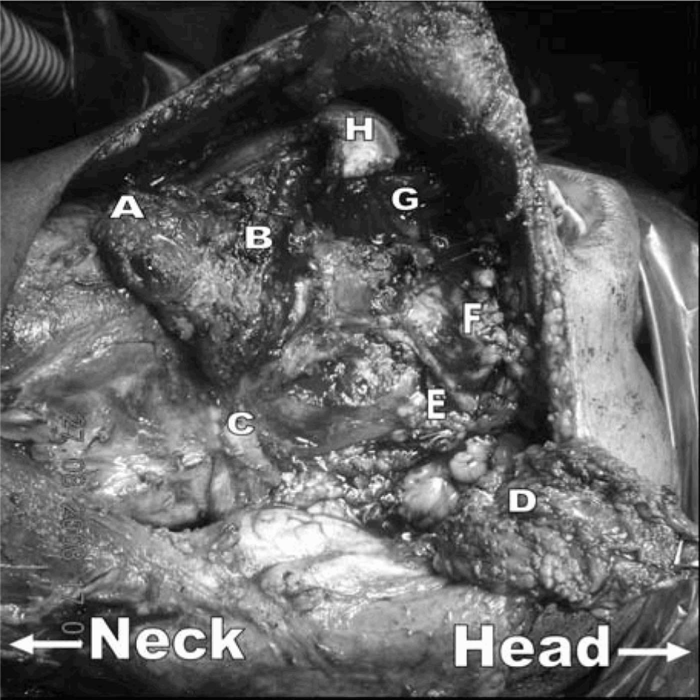
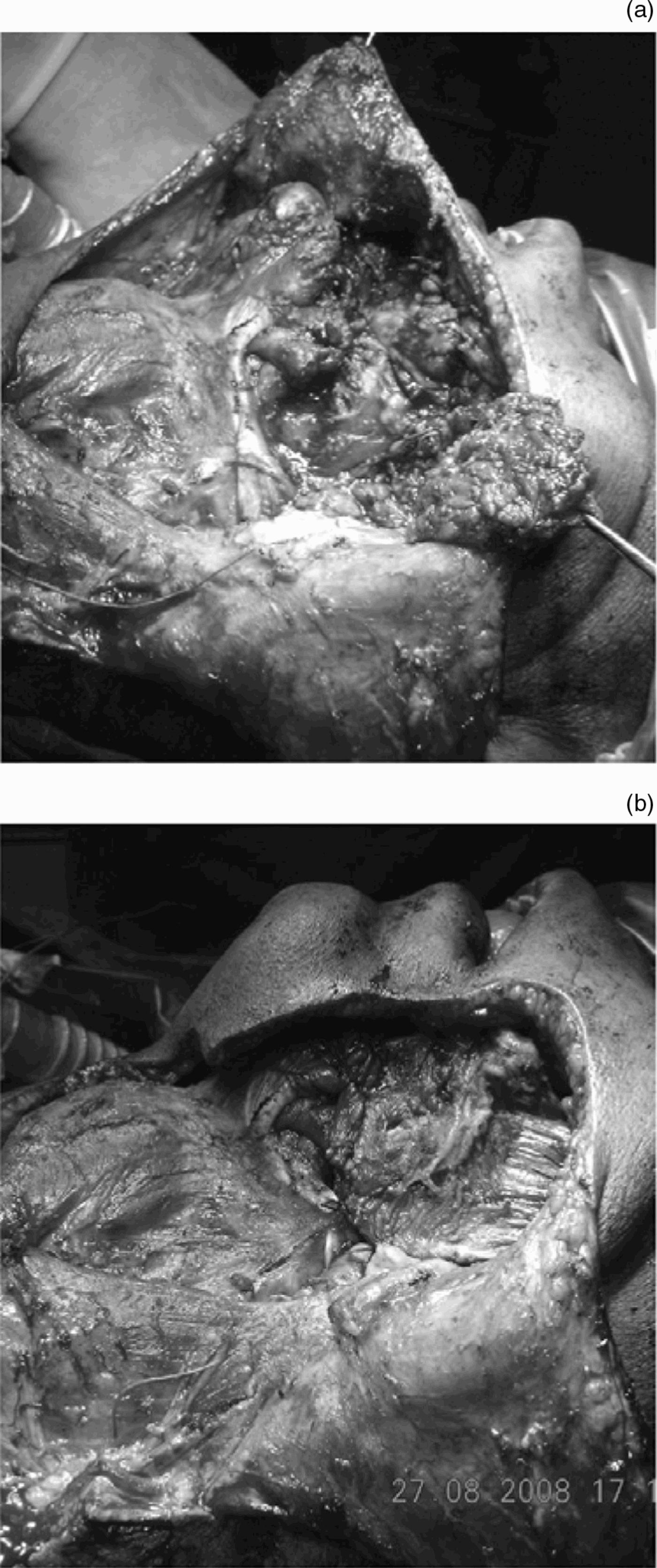
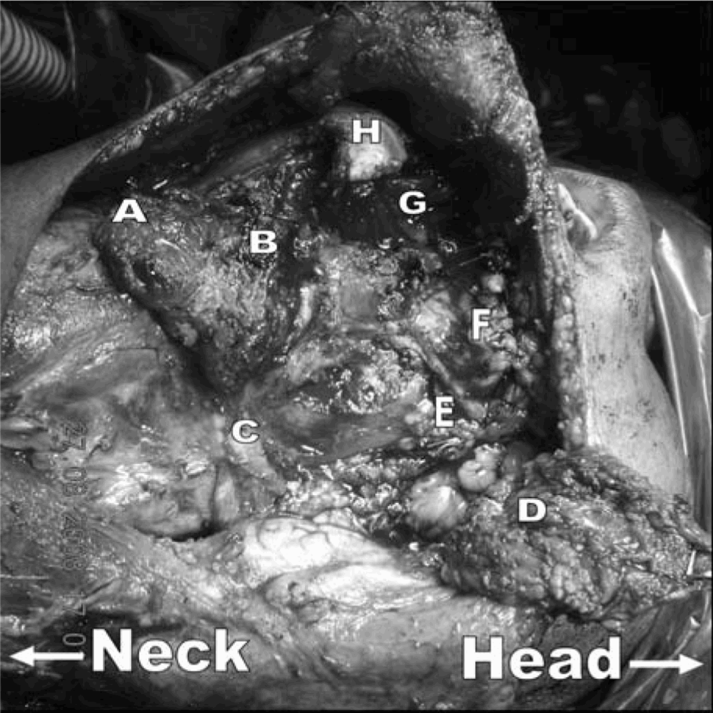
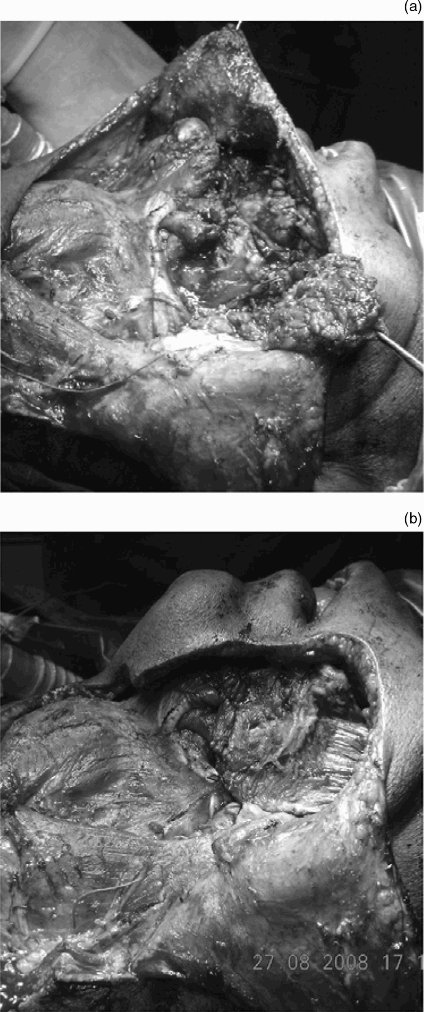
This report proposes a simple technique to improve cosmesis following hemimandibulectomy in a highly selected group of patients in whom the masseter can be preserved. It suggests that, whenever possible and without compromising oncological safely, the masseter muscle, mylohyoid muscle and anterior belly of the digastric muscle should be saved. This is applicable to selected lesions of the tonsil and posterior third of the tongue and to primary jaw tumours, for which hemimandibulectomy does not necessitate removal of the masseter muscle. The remaining muscles can be approximated as shown in the Figures 1 and 2 to prevent the lateral hollow that is a common sequel of hemimandibulectomy. The masseter, mylohyoid and digastric muscles can be easily preserved in the case of primary jaw tumours such as ameloblastoma, giant cell tumour and osteosarcoma. The masseter muscle can also be preserved when the lesion is situated away from the muscle belly. Similarly, attempts should be made to preserve the digastric and mylohyoid muscles, without compromising oncological safely.
Intra-operative photograph showing digastric, masseter, pterygoid and mylohyoid muscle remnants following a typical hemimandibulectomy for an oral cancer involving the alveolus. A = digastric muscle; B = mylohyoid muscle; C = digastric tendon; D = masseter muscle; E = pterygoid muscle; F = sublingual gland; G = genioglossus muscle; H = mandibular remnant
Intra-operative photographs showing the mylohyoid muscle approximated to the masseter muscle. Distal fibres of the masseter are stitched to the digastric tendon, and some fibres are approximated to the substance of the parotid. The digastric muscle is stitched to the outer periosteum to cover the cut end of the mandible and the genioglossus muscle. This procedure covers the disfiguring hollow created by hemimandibulectomy with primary closure.
Procedure
Firstly, the uninvolved masseter muscle is dissected away from the lower border of the mandible, and from the parotid gland. The muscle is released from the posterior border of the mandible and held with Babcock forceps. The dissection is continued on the lateral surface of the mandible up until the tip of the coronoid process. The anterior belly of the uninvolved digastric muscle can then be dissected off the lower border of the mandible.
After dividing the mandible, while incising the mucosa in the floor of the mouth, the mylohyoid muscle is divided close to its attachment on the mylohyoid line. The hemimandibulectomy is then undertaken, with primary closure of the floor of the mouth with remnant buccal mucosa.
The digastric muscle is stitched to the outer periosteum of the mandible near the divided bony edge with 3-0 vicryl sutures, so as to cover the cut edge entirely. This prevents the development of a hollow near the cut end of the mandible. Some fibres of the mylohyoid can also be stitched to that area to cover the defect.
The posterior fibres of the mylohyoid muscle are then approximated to the lower-most edge of the masseter muscle, using 3-0 vicryl sutures. In some cases, the length of the mylohyoid may not be sufficient for approximation. In such cases, the masseter may be stitched to the hyoglossus muscle. If the length of the masseter is insufficient, the mylohyoid may be stitched to the pterygoid muscles or directly to the hyoglossus. The aim is to prevent the lateral hollow which is a common sequel of hemimandibulectomy.
After achieving haemostasis, a suction drain is placed and the skin closed.
The patient should be instructed to undertake active jaw exercises from the first post-operative day. Oral intake may be allowed from the fifth post-operative day. Provision of a bite guide prosthesis will assist in preventing malocclusion.
As mentioned previously, this modified myopexy procedure is only applicable to a limited number of patients; in a centre undertaking nearly 1000 oral surgery procedures annually, this myopexy procedure was able to be performed in only 13 patients in one year. Most of these patients had a satisfactory appearance following surgery.
Objective parameters are currently being developed to enable assessment of the outcomes of this modified procedure, compared with those of routine primary closure.
Conclusion
In patients requiring hemimandibulectomy, attempts should be made whenever possible to preserve the condyle and to reconstruct the bony defect with microvascular free tissue transfer. In cases in which microvascular surgery is not possible, the use of a pedicled flap (such as a pectoralis major myocutaneous flap) should be kept in mind. In highly selected patients in whom neither of the above are possible, a simple myopexy technique is suggested to prevent the hollow that is a common sequel of hemimandibulectomy and primary mucosal closure. The suggested technique involves approximation of the remaining masseter, mylohyoid, digastric, pterygoid and hyoglossus muscles, following excision of the mandible. This simple technique has the potential to improve the patient's appearance and thus their quality of life.