Introduction
The health risks associated with electromagnetic radiation generated by mobile phones and base stations are becoming more widespread. The use of mobile phones has increased tremendously in recent years because of the ease of device accessibility and communication.Reference Kayabasoglu, Sezen, Eraslan, Aydın, Coskuner and Unver1 Energy, carried by radiofrequency waves, is absorbed by the body and transmitted to the tissues. The effects of radiofrequency waves are classified as thermal and non-thermal. The thermal effects caused by this energy are generated by the movement (oscillation) of ions and water molecules.2
Mobile phone systems use 800–950 MHz and 1800–1900 MHz frequencies for the transmission and reception of signals, with unique repetition rates. These frequencies penetrate the human body to a depth of a few centimetres, and the majority of the energy is absorbed relatively superficially.2 The maximum output power of the mobile phone, which determines the amount of absorbed radiofrequency energy, is limited in modern digital Global System for Mobile Communications (‘GSM’) systems to 1–2 W.Reference Stanislaw and Sobiczewska3, Reference Otto and von Muhlendahl4 The detrimental effects of high-power and low-power electromagnetic radiation are created by heat production and chemical changes respectively.Reference Otto and von Muhlendahl4 International guidelines on electrical field limits of exposure to non-ionising radiation for high-frequency electromagnetic fields (100 kHz to 300 GHz) were introduced by the International Commission on Non-Ionizing Radiation Protection.5
Dosimetry plays an important role in risk evaluation of human exposure to radiofrequency fields.Reference Vecchia, Matthes, Ziegelberger, Lin, Saunders and Swerdlow6 Specific absorption rate is used to evaluate the rate of radiation energy absorption per unit weight of tissue. This rate is expressed as W/kg in different biological tissues.5–Reference Esmekaya, Seyhan and Omeroglu7
Several studies have investigated the effects of mobile phone use on the auditory system.Reference Sievert, Eggert, Goltz and Pau8–Reference Monnery, Srouji and Bartlett11 These studies, which frequently evaluated outer hair cell functions using distortion product otoacoustic emission (DPOAE) testing, showed no hearing changes associated with mobile phone use.Reference Sievert, Eggert, Goltz and Pau8, Reference Monnery, Srouji and Bartlett11–Reference Galloni, Parazzini, Piscitelli, Pinto, Lovisolo and Tognolo13 To our knowledge, there has been no histopathological study investigating the effect of radiofrequency energy caused by electromagnetic radiation on cochlear development.
This study aimed to assess the changes in hearing in rats exposed to electromagnetic radiation emitted by a radiofrequency source in intrauterine life, with exposure lasting until the end of the cochlear development period. Electron microscopic evaluation and DPOAE testing were used to show changes in cellular structure and functional hearing respectively.
Materials and methods
Animals and groups
The study protocol was approved by the Local Institutional Animal Care and Use Committee (2009/54), and the animals were treated in accordance with protocols approved by this committee. Eight pregnant (weight, 320–400 g) and 81 newborn (weight, 55–60 g) albino Wistar rats were included in this study. All rats had free access to commercial food and water, and were maintained in an environment with a controlled temperature (21°C) and 12-hour light/dark cycles.
Initially, female rats were mated with males in different cages. Eight female rats, identified as pregnant by daily examination of vaginal dilatation and mucous plug formation, were separated from the males and the first day of pregnancy was determined.
The 8 pregnant rats and subsequent newborns were divided into 3 groups: a control group (2 pregnant rats and 24 newborn rats); a 900 MHz electromagnetic radiation group (3 pregnant rats and 31 newborn rats); and a 1800 MHz electromagnetic radiation group (3 pregnant rats and 26 newborn rats).
The pregnant rats in the radiation groups were exposed to electromagnetic radiation for 1 hour per day starting on the 12th day of pregnancy until delivery. The newborn rats in these two groups were exposed to electromagnetic radiation for 1 hour per day for 21 days after delivery.
Newborn rats were monitored for 21 days after delivery. Distortion product otoacoustic emission (DPOAE) testing was carried out on day 22. Eight rats in each group were randomly selected for electron microscopic evaluation.
Electromagnetic radiation
The electromagnetic radiation exposure system was designed by the Department of Biophysics, and quality control assessments were made by the Department of Electric and Electronic Engineering. Everest Global System for Mobile Communications simulators (models 900 CW4 and 1800 CW2; Adapazari, Turkey) were used for electromagnetic radiation exposure. The 900 MHz simulator was set, equivalent to mobile phone antenna, with the following parameters: 850–950 MHz band interval, 4 W maximum output power, two radiofrequency output channels, and circular polarisation with high-gain specification. Similarly, the parameters of the 1800 MHz simulator were: 1750–1850 MHz band interval, 2 W maximum output power, two radiofrequency output channels, and circular polarisation with high-gain specification. A spectrum analyser and satellite receiver (MC-877C model; Promax, Barcelona, Spain), and an electrical field meter (PMM 8053 Portable Field Meter; ELA Medical Sorin, Milan, Italy), were used to detect the background effects of other radiofrequency sources in the environment; this was recorded as zero. The electrical field probe model used was the EP-330 (Costruzioni Elettroniche Centro Misure Radioelettriche Srl, Savona, Italy).
The monopole antenna of the exposure system was placed in the centre of the pie cage restrainer in order to sustain an equal distribution of the electrical field. Each rat in the two electromagnetic radiation groups was put into different rooms of the pie cage restrainer. During exposure, the output power of the generator was fixed at 2 W for the 900 MHz group and 1 W for 1800 MHz group.2, Reference Stanislaw and Sobiczewska3 The electrical field exposure values radiated to the pregnant rats in the 900 MHz and 1800 MHz groups were 9–25 V/m and 9–18 V/m respectively. The electrical field exposure values radiated to the newborn rats in the 900 MHz and 1800 MHz groups were 19–25 V/m and 10–18 V/m respectively. The electrical field exposure values were measured weekly on the back of each newborn rat.
Distortion product otoacoustic emission test
After electromagnetic radiation exposure, all newborn rats in the three groups underwent otoscopic examination followed by DPOAE testing. The rats that had cerumen in the ear canal, and/or problems in the tympanic membrane and/or middle ear were excluded from the study. In order to carry out DPOAE testing, the rats were anaesthetised with ketamine hydrochloride (50 mg/kg, intraperitoneal) and xylazine (10 mg/kg, intraperitoneal).
The DPOAE measurements were carried out with the ILO V6 instrument (Otodynamics, Hatfield, UK) using newborn probe tips. Calibration and probe placement controls were automatically performed by the otoacoustic emissions (OAEs) device. The intensity of stimulus presentation was recorded as L1 for the f1 frequency (80 dB SPL) and L2 for the f2 frequency (80 dB SPL); the f2/f1 ratio was 1.22. The DPOAEs were recorded for 30 seconds at 1, 1.5, 2, 3, 4, 6 and 8 kHz. Frequencies lower than 1 kHz were not recorded because of the internal noise of rats. The DPOAE results were evaluated as signal-to-noise ratio values; these values were recorded for each frequency and compared among the groups.
Electron microscopic evaluation
The middle segment of each cochlear turn was preferred for the cell count in order to obtain the largest possible number of cells. The specimens were immersed in a fixative solution of 2.5 per cent glutaraldehyde in 0.1 M phosphate buffer for 12 hours, and decalcified in 10 per cent ethylene diamine tetra-acetic acid in phosphate buffer (pH 7.4) for 2 months. Decalcified specimens were post-fixed in 1 per cent osmium tetroxide in 0.1 M phosphate buffer for 2 hours, dehydrated in a graded series of ethanol (30–100 per cent) and embedded in Epon 812 resin. Ultra-thin sections (70 nm) were stained with uranyl acetate and lead citrate for electron microscopic examination (JEM 1400; Jeol, Tokyo, Japan), and photographed using a SC1000 Orius CCD camera (Gatan, Pleasanton, California, USA).
The numbers of cells were counted using electron microscopic evaluation. Cells were divided into three types: normal cells, apoptotic cells and necrotic cells. For each cochlea, 20 cells were counted from the middle segment sections of the same animal. Stereocilia shapes and arrangement were used to define hair cell integrity. Three cochleas in the control group were not evaluated because of technical problems and therefore were not included in the study.
Statistical analysis
Statistical evaluations were carried out with the Statistical Package for the Social Sciences software, version 15 (SPSS, Chicago, Illinois, USA). Statistical analysis was performed using a Kruskal–Wallis analysis of variance, and a Mann–Whitney U test with Bonferroni correction.
Results
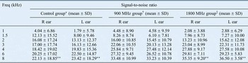
The distortion product otoacoustic emission results for the newborn rats, according to group and ear, are shown in Table I. No statistically significant differences were found among the groups and ears for the following frequencies: 1, 1.5, 2, 3, 4 and 6 kHz. The 1800 MHz electromagnetic radiation group had statistically significant higher signal-to-noise ratio values compared with the control group for 8 kHz.
Distortion product otoacoustic emission test results

Kruskal–Wallis analysis of variance, and Mann–Whitney U test with Bonferroni correction data. *n = 24; †n = 31; ‡n = 26. **p < 0.01. Freq = frequency; SD = standard deviation; R = right; L = left
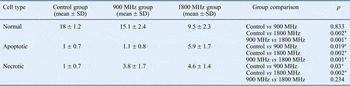
The distributions of mean cell numbers for each group are shown in Table II. There was a statistically significant difference between the groups in terms of normal cell distribution. With regard to apoptotic cells, there was a significant difference between the 1800 MHz electromagnetic radiation group and the other groups. The number of necrotic cells was greater in the electromagnetic radiation groups compared with the control group; the differences were statistically significant.
Distribution of cell types

Kruskal–Wallis analysis of variance, and Mann–Whitney U test with Bonferroni correction data. *p < 0.05. SD = standard deviation
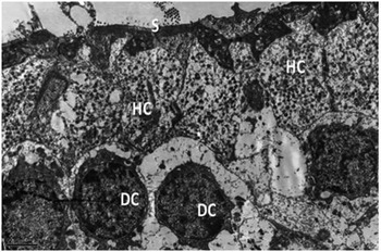
Electron microscopy images for each group are shown in Figures 1–3. In the control group (Figure 1), the organ of Corti showed a normal appearance: a single inner hair cell and three outer hair cells separated by the tunnel of Corti. The stereocilia of outer hair cells were embedded in the tectorial membrane. Inner and outer pillar cells, inner and outer phalangeal cells (Deiters' cells), and basilar membrane appeared to be normal.
Electron microscopy image of control group cells (uranyl acetate and lead citrate; ×8000). S = stereocilia; HC = hair cell; DC = Deiters' cell

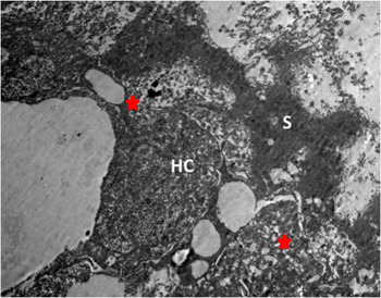
Electron microscopy image of 900 MHz group cells (uranyl acetate and lead citrate; ×5000). The red stars indicate damaged intercellular junctions. S = (degenerated) stereocilia; HC = hair cell

Electron microscopy image of 1800 MHz group cells (uranyl acetate and lead citrate; ×2000), showing loss of stereocilia in apical part of hair cells and swollen Deiters' cells.

In the 900 MHz electromagnetic radiation group (Figure 2), hair cell nuclei and cytoplasm were normal, but the junctional complexes between cells were damaged. The stereocilia of the hair cells were degenerated. The swollen Deiters' cells displayed signs of necrosis. The Deiters' cells were slightly damaged.
In the 1800 MHz electromagnetic radiation group (Figure 3), there was a higher degree of damage to hair cells and stereocilia. Chromatin condensation and compaction were found in some of the hair cells. Intercellular junctions were degenerated. The swollen Deiters' cells had large vacuoles in the cytoplasm. Condensation of chromatin and cytoplasm was observed in the pillar cells.
Discussion
Wireless communication technologies transmit electromagnetic waves at a given frequency using air as the transmission environment. Non-ionised electromagnetic waves are eventually scattered, resulting in exposure to both users and non-users.
Electromagnetic waves are known to have short and long-term effects. Short-term effects include heart disease, narrowing of field of vision, temporary and permanent hearing disorders, tinnitus, dizziness, headaches, increased risk of brain tumour, memory loss, intense stress and fatigue, impairment of attention and concentration, weakening of the immune system, and increased risk to the unborn fetus. Long-term effects include high blood pressure, genetic degradation, reduced sperm count, lymphoma, skin cancer formation and injury to the blood brain barrier.2
Mobile phones are one of the most important radiofrequency radiation sources, and they produce both radiofrequency radiation and a low-frequency electromagnetic field.Reference Sage, Johansson and Sage14 The radiofrequency radiation emitted from mobile phones may have thermal and non-thermal effects. Human and animal studies indicate that radiofrequency fields can be harmful because of the excessive heating of internal tissues. The sensitivity of different tissues and cells to thermal damage varies; absolute increases in temperature, the duration of heating and the thermoregulatory capacity of the body are important determinants of harm caused by tissue heating. All thermal effects are dependent on the electrical parameters (especially the conductivity) of the tissue exposed to radiofrequency energy.Reference Gabriel15 As tissues with a high fluid content have increased dielectric properties, they are subjected to higher thermal effects and absorb more radiofrequency energy.Reference Peyman, Rezazadeh and Gabriel16 Radiofrequency exposure leads to changes in cell membrane function, metabolism and cellular signal communication, and causes nerve cell death because of reactive oxygen species generation.Reference Sage and Carpenter17 Radiofrequency energy exposure also affects biological tissues via electrical and magnetic field mechanisms.Reference Challis18
Mobile phones are frequently used in the vicinity of ears. For this reason, the auditory system is thought to be affected by the electromagnetic waves. However, experimental studies investigating hearing changes caused by electromagnetic field exposure generated by short and long-term mobile phone use have revealed no significant differences.Reference Sievert, Eggert, Goltz and Pau8–Reference Monnery, Srouji and Bartlett11, Reference Peyman, Rezazadeh and Gabriel16, Reference Moussa19, Reference Oktay and Dasdag20
Otoacoustic emissions are low-intensity acoustic signals emitted by the healthy cochlea. The OAE is a reliable method to assess in vivo cochlear functionality and is a subject of choice in experimental studies.Reference Monnery, Srouji and Bartlett11–Reference Galloni, Parazzini, Piscitelli, Pinto, Lovisolo and Tognolo13, Reference Kizilay, Ozturan, Erdem, Kalcioglu and Miman21 In this study too, distortion product otoacoustic emission (DPOAE) testing was utilised for functional hearing assessment. The DPOAE test results showed no functional hearing changes and this is consistent with the literature.
• Electromagnetic radiation generated by mobile phones and base stations is becoming a more widespread health risk
• Studies have investigated effects of mobile phone use on the auditory system using distortion product otoacoustic emission testing
• Few studies have investigated electromagnetic radiation radiofrequency effects on cochlear development using electron microscopic evaluation
• This study exposed rats to electromagnetic radiation emitted by a radiofrequency source, from intrauterine life until the end of the cochlear development period
• Radiofrequency radiation exposure during cochlear development led to cochlear cellular structural damage in the rat model
Although several studies have investigated the effects of radiofrequency energy exposure on functional hearing, no study has previously investigated the cellular changes or cochlear development process following radiofrequency energy exposure. To the best of our knowledge, this is the first study to investigate cellular changes and cochlear development anomalies caused by radiofrequency energy exposure, detected by electron microscopy. The rat cochlea starts to develop on intrauterine day 12 and is formed on intrauterine day 16.Reference Angula, Merchan and Merchan22 Cochlear development proceeds until post-partum day 21.Reference Roth and Bruns23 Based on this knowledge, the rats in the current study were exposed to electromagnetic radiation for 28 days, and received radiofrequency energy throughout the cochlear development period.
Histopathological evaluation revealed adverse cellular changes in the cochlea caused by electromagnetic radiation exposure. Both of the electromagnetic radiation groups showed structural damage, apoptosis and necrosis at the cellular level. Our results clearly show cochlear cellular damage, despite the presence of normal functional hearing.
In this study, the DPOAE findings revealed no functional hearing loss in the groups. However, ultrastructural evaluation of the organ of Corti revealed significant cellular destruction in the electromagnetic radiation groups. The cellular destruction can be explained by the increased thermal effect of radiofrequency energy on tissues with high fluid content.
Our findings indicate cellular structural damage in the cochlea caused by radiofrequency radiation exposure during cochlear development in the rat model. Further animal and human studies will be needed to validate the findings and determine the effects of radiofrequency radiation exposure on cochlear development in humans.
Acknowledgement
The authors would like to thank Assistant Prof Mesut Kahraman from Suleyman Demirel University, Electronic and Communication Engineering Department, for supplying us with the software used in the calculation of specific absorption rate.