I. INTRODUCTION
The bismuth phosphate BiPO4-family has been the subject of intensive research due to its potential applications. It is known as catalyst (Chang et al., Reference Chang, Li, Shin, Lee and Yun2000; Pan et al., Reference Pan, Li, Ma, Chen and Zhu2011), orthophosphate ion sensor (Alekseev et al., Reference Alekseev, Gorelov and Kornilov2000), microwave dielectric (Cho et al., Reference Cho, Kim, Kim and Hong2006), and in separation of radioactive elements (Holgye and Poliak, Reference Holgye and Poliak1991). Recent focus on BiPO4 is due to its application as a host lattice for rare earth luminescent centers and as a photocatalyst (Roming and Feldmann, Reference Roming and Feldmann2009; Guan et al., Reference Guan, Sun, Tao and Xu2008; Pan and Zhu, Reference Pan and Zhu2010, Reference Pan and Zhu2011). X-ray diffraction studies showed that BiPO4 crystallizes in three different structures with trigonal and monoclinic (low and high temperature modifications) symmetries (Mooney, Reference Mooney-Slater1962; Masse and Durif, Reference Masse and Durif1982; Romero et al., Reference Romero, Bruque, Aranda and Iglesias1994). In many cases, the structure-property relations were studied to derive its importance from both fundamental and technological aspects (Arunkumar et al., Reference Arunkumar, Jayajothi, Jeyakumar and Lakshminarasimhan2012). The trigonal BiPO4 crystallizes with the P3121 space group (PDF#15-766). The monoclinic low temperature variety has a monazite-type structure (space group P21/n; PDF#15-767). Note that the standard space group P21 /c should be reported as the corresponding space group for the room temperature modification of BiPO4. This last variety can be also converted to high monoclinic (P21/m space group, PDF#43-637) form when it is heated at 700 °C and above. The high-temperature form is a slight distortion of the monazite form. Note that the transition from the hexagonal to the monazite type is not reversible. Structure of all the BiPO4 phases consist of three dimensional networks made up of PO4 tetrahedra and BiO8 polyhedra.
Generally, in order to obtain a variety of application for A IIIPO4 phases, various cationic substitutions were realized in A sites leading thus to a series of A (1− x ) A′ x PO4 compositions. Note that the A (1− x ) A′ x PO4 phases can include a mixing of A II/A′IV ions leading to (AII 0 . 5A′IV 0 . 5)PO4 materials (Orlova et al., Reference Orlova, Kitaev, Kazantsev, Samoilov, Kurazhkovskaya and Vopilina2002; Kitaev et al., Reference Kitaev, Volkov and Orlova2004; Clavier et al., Reference Clavier, Podor and Dacheux2011) or a mixture of A II, A′III and A″IV ions as in the case of A II 1/3 A′III 1/3 A″IV 1/3PO4 compounds (A II = Mg, Ca, Cd, Sr; A′III = Sm, Gd, Nd; A″IV = Zr, Ce, Th) (Orlova et al., Reference Orlova, Kitaev, Kemenov, Orlova, Kazantsev, Samoilov and Kurazhkovskaya2003). Depending on the ionic radii and the nature of the A, A′ and A″ cations, a wide variety of structural arrangements were obtained. In fact, the larger A cation can cause structural distortions, specifically a rotation of the PO4 tetrahedra. Thus a series of new family of oxides crystallizing in different structural types as monazite, zircon (xenotime), scheelite, cheralite, nasicon and yavapaiite type compounds are characterized (Clavier et al., Reference Clavier, Podor and Dacheux2011). The monazite and zircon structures can be viewed as being composed of alternating edge-sharing AO8 or AO9 polyhedra and PO4 tetrahedra while scheelite has tetrahedral sharing corners with adjacent dodecahedra. On heating, polymorphs in both scheelite and monazite structures, as well as in the zircon structure are observed as in the case of the orthovanadate Pb0.5Th0.5VO4 (Calestani and Andretti, Reference Calestani and Andretti1984). Cheralite-type structure is a monazite-related compound (Rose, Reference Rose1980). For smaller AIV ions, a monoclinic structure isotypic of the K0 . 5Fe0 . 5SO4 yavapaïte is stabilized as evidenced for the BaII 0 . 5 A IV 0 . 5PO4 (A IV = Ti, Zr, Hf, Ge, Sn, Mo) series (Fukuda et al., Reference Fukuda, Moriyama and Iwata2005; Popa et al., Reference Popa, Bregiroux, Konnings, Gouder, Popa Aurelian, Geisler and Raison2007, Reference Popa, Wallez, Bregiroux and Loiseau2011). The yavapaiite BaII 0 . 5 A IV 0 . 5PO4 has a structure which is made of layers of BaII cations in eight- or tenfold coordination, alternating with dense slabs built up of corner-connected of AIVO6 octahedra and PO4 tetrahedra.
In our recently reported works we have isolated some phosphates including a mixture of A II, A′III and A″V ions and crystallizing with the nasicon type-phases [i.e., A II 0 . 5SbVFeIII(PO4)3 (A III = Ca, Sr)] (Aatiq et al., Reference Aatiq, Tigha, Hassine and Saadoune2006, Reference Aatiq, Tigha and Benmokhtar2012). In a continuation of our research, herein the synthesis and structural characterization of a new CaII 1/3SbV 1/6BiIII 1/2PO4 phase has been undertaken.
II. EXPERIMENTAL
Syntheses of Ca1/3Sb1/6Bi1/2PO4 was carried out using conventional solid-state reaction techniques. Powder crystalline samples were prepared from mixtures of carbonates CaCO3 (Riedel-de Haën, 99%), oxides Sb2O3 (Riedel-de Haën, 99.9%), Bi2O3 (Prolabo, 99%) and NH4H2PO4 (Riedel-de Haën, 99%) in stoichiometric proportions. The mixture was heated progressively with intermittent grinding at 200 °C (12 h), 400 °C (6 h), 600 °C (12 h), 800 °C (24 h) and 900 °C (24 h) in air atmosphere. The products of reaction were characterised by XRPD data at room temperature with a Panalytical X′Pert-PRO (θ-2θ) diffractometer; (CuKα) radiation (45 kV, 40 mA); divergence slit of 1° and antiscatter slit of 1°. The data were collected from 10 to 90°2θ, in steps of 0.008°, with a counting time of 30 s per step. The Rietveld refinement of the structure was performed using the Fullprof program (Rodriguez-Carvajal, Reference Rodríguez-Carvajal1993). The infrared (IR) spectra were recorded using a Bruker's VERTEX 70. FT-IR spectrometer. Samples were in the form of KBr (mid-IR) and polyethylene (far-IR) pellets. The spectral resolution was 4 cm−1.
III. RESULTS AND DISCUSSION
A. Structure of CaII 1/3SbV 1/6BiIII 1/2PO4
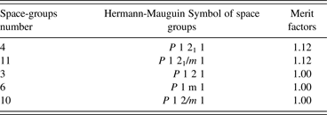
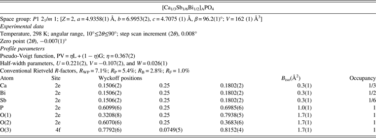
Indexing of the XRPD patterns of CaII 1/3SbV 1/6BiIII 1/2PO4 was performed by means of the computer program Dicvol 04 (Boultif and Louër, Reference Boultif and Louër1991). The first 30 peak positions, with a maximal absolute error of 0.02° (2θ), were used as input data. Monoclinic cells were found with satisfactory figure of merit. The derived unit-cell parameters were subsequently used as starting parameters in the next stage of the refinements. The Check-Group program, which is integrated within the Fullprof-suite program, has been used to obtain the ordered list of possible space groups (Table I). Among five of the possible space groups [i.e.: P21, P21/m, P2, Pm and P2/m], P21/m and the P21 were found with the satisfactory merit factors. In a second step of refinement, the two most probable space groups were tested using The LeBail fitting (profile matching) (Le Bail et al., Reference Le Bail, Duroy and Fourquet1988). A lower reliability factors R B was obtained for both P21/m and the P21 space groups. In fact, in both cases there is only one systematic absence in the diffraction pattern, that is, 0k0 is absent unless k is even. On this evidence, the space group may be either P21/m (No.11) or P21 (No.4). Up to this stage of refinement it did not seem feasible to attempt more than an approximate description of the structure. The final assignment of probable space group is only possible after solving and refining the crystal structure in both P21/m and P21 space groups. In the final stage, structural parameters for all atoms were refined by the Rietveld method using the Fullprof program (Rodriguez-Carvajal, Reference Rodríguez-Carvajal1993). For the lower-symmetry P21 space group, initial starting parameters for the Rietveld refinements of Ca1/3Sb1/6Bi1/2PO4 were based on the approximate structural parameters already reported by Mooney (Reference Mooney-Slater1962) for the high-temperature monoclinic BiPO4 phase (P21 space group). In the case of the P21/m space group, the starting structural parameters for the Rietveld refinements were based on those obtained by Masse and Durif (Reference Masse and Durif1982). During the Rietveld refinement, the occupancy rates of all atoms are not allowed to vary. Since the experimental XRD pattern of Ca1/3Sb1/6Bi1/2PO4 also has diffraction peaks from a small amount (about 6 wt.%) of the β-calcium diphosphate β-Ca2P2O7, the Rietveld refinement was carried out using a two phase model, consisting of Ca1/3Sb1/6Bi1/2PO4 and the β-Ca2P2O7 which crystallizes with the P41 space group (Webb, Reference Webb1966). It should be noted that only high-intensity diffraction peaks of the β-Ca2P2O7 appear as impurity in the XRD pattern. Obtained reliability factors are [R P = 5.7%; R WP = 7.6%; R B = 4.3%; R F = 2.0%] for the P21 space group and [R P = 5.3%; R WP = 7.1%; R B = 2.8%; R F = 1.0%] for the P21/m space group. In both cases the Rietveld refinement led to acceptable reliability factors but a careful comparison of the obtained structural results is in favour of the P21/m space group. Indeed, in the case of the P21/m space group the refinement led to a lowest value of R B factors and in plus the obtained P-O distances values showed that the PO4 tetrahedra are relatively the most regular. Consequently, in the following only the results of the Rietveld refinement in space group P21/m will be retained. Note that the presence of a low percentage of Ca2P2O7 in our sample is in favour of the formation of a negligible amount of amorphous phase. In fact, at this stage of our study we were not able to determine the composition of this amorphous phase and evaluate its percentage vs. the studied phase. Therefore we believe that, in a first approximation, the composition of the studied phase corresponds to that of the stoichiometric proportions which we used at the beginning of its preparation.
Ordered list of possible space groups obtained from the Check-Group program.

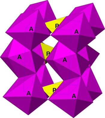
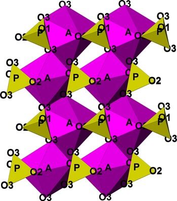
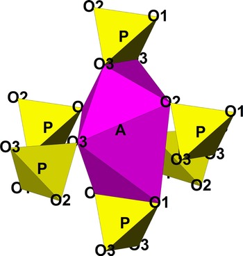
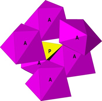
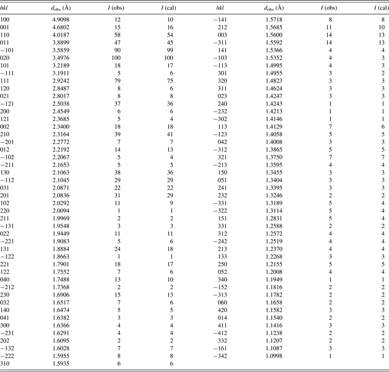
Observed, calculated, and difference XRD patterns are given in Figure 1. Results of refinement without considering the β-Ca2P2O7 as impurity are given for comparison in inset of Figure 1. Structural parameters are given in Table II. X-ray powder diffraction data, obtained from the “observed intensities” of the Rietveld refinement (CuKα 1: 1.5406 Å), is gathered in Table III. Details of the structure of Ca1/3Sb1/6Bi1/2PO4 are represented in Figures 2–5. This structure can be viewed as being composed of alternating edge-sharing AO8 (A = Ca, Sb, Bi) bisdisphenoids and PO4 tetrahedra forming chains parallel to the b axis (Figures 2–4). There are four chains per unit cell. The eight oxygen atoms of each AO8 polyhedron belong to six phosphate PO4 groups (Figure 4). Four of the oxygen atoms come from two bidentate PO4 groups and the other four oxygens belong to the other four monodentate PO4 groups. PO4 tetrahedra are isolated and every one of them is surrounded by six AO8 polyhedra (Figure 5) in such a way that every PO4 tetrahedron is connected, via two of their edges [i.e., O(3)-O(3) and O(1)-O(2)], to two AO8 polyhedra and via their four corners to the other four AO8 polyhedra. Obtained A-O distances values can be divided into two groups. The first group contains four shortest A-O distances with values [A-O(1) = 2.084(4) Å, A-O(2) = 2.331(5) Å, 2 × A-O(3) = 2.2299(4) Å] varying between 2.08 and 2.33 Å whereas the second group is formed by four longest distances with values [A-O(1) = 2.920(6) Å, A-O(2) = 2.916(7) Å, 2 × A-O(3) = 2.671(5) Å] ranging between 2.67 and 2.92 Å. PO4 tetrahedra are relatively regular and the P-O distances values [P-O(1) = 1.542(3) Å, P-O(2) = 1.553(4) Å, 2 × P-O(3) = 1.550(5) Å] match well with those typically observed in orthophosphates. The O-P-O angles vary between 104 and 112°.
Experimental (•••) calculated (—), and difference profile of the XRD pattern of Ca1/3Sb1/6Bi1/2PO4. Refinement without considering the β-Ca2P2O7 as impurity is given in inset.

(Color online) Structure of Ca1/3Sb1/6Bi1/2PO4 along b-axis.

(Color online) View of the Structure of Ca1/3Sb1/6Bi1/2PO4 in the bc plane.

(Color online) View of the AO8(PO4)6 polyhedra (A = Ca, Sb, Bi) in Ca1/3Sb1/6Bi1/2PO4.

(Color online) View of the PO4(AO8)6 polyhedra (A = Ca, Sb, Bi) in Ca1/3Sb1/6Bi1/2PO4.

Results of the Rietveld refinement of Ca1/3Sb1/6Bi1/2PO4.

Powder diffraction dataa of Ca1/3Sb1/6Bi1/2PO4 (CuKα1; λ = 1.5406 Å).

aDiffraction lines with I obs<1 are omitted.
B. Infrared spectroscopy
Infrared spectroscopic study was undertaken in order to obtain further structural information about the nature of bonding in the crystalline solid. The IR spectrum and proposed assignments of obtained sample are shown in Figure 6. According to the factor group analysis, the phosphate PO4 tetrahedron group has Td symmetry and nine internal modes (Γ d = A 1 + E + 2F 2). The IR band positions of the four (ν 1, ν 2, ν 3 and ν 4) modes are close to those expected for orthophosphates materials. The symmetric, triply degenerate PO stretching (ν 3) is observed between 1100 and 1150 cm−1 and the symmetric non degenerate PO stretching modes (ν 1) are observed in the range of 950–1050 cm−1. The triply degenerate, antisymmetric and harmonic OPO bending (ν 4) is observed in the range of 520–680 cm−1 while antisymmetric doubly degenerate OPO bending (ν 2) are located in the 450–510 cm−1 range. The frequencies found at 615 and 630 cm−1 in IR spectrum can be assigned empirically to Sb-O stretching modes involving Sb-O-P linkage. Note that the Sb-O stretching vibrations are probably coupled with the OPO bending ν4 mode. The same Sb–O vibrations are already observed, in the IR spectrum of SbOPO4 at 597 and 652 cm−1 (Sudarsan et al., Reference Sudarsan, Muthe, Vyas and Kulshreshtha2002) and at 580 and 639 cm−1, in the IR and Raman spectra, of Mn0 . 5 MSb(PO4)3 (M = Al, Fe and Cr) phases (Anantharamulu et al., Reference Anantharamulu, Koteswara Rao, Vithal and Prasad2009). The symmetric stretching vibrations ν sPOP, characteristic for the P2O7 polyhedral, is usually (i.e. in a variety of compounds) found to appear strongly in the IR and Raman spectra around 750 cm−1. Consequently, the only one band centered at 725 cm−1 is attributed of the P2O7 groups which come from the minor β-Ca2P2O7 phase. Note that the same vibration is already observed at 728 cm−1 in the IR spectrum of β-Ca2P2O7 (Cornilsen and Condrate, Reference Cornilsen and Condrate1979). Bands observed between 1160 and 1225 can be attributed to a coupling of ν(PO4) vibration and ν(Metal-O) interaction modes.
IR spectrum and proposed assignments bands of Ca1/3Sb1/6Bi1/2PO4.

IV. CONCLUSION
CaII 1/3SbV 1/6BiIII 1/2PO4 compound was prepared and characterized by XRPD and IR spectroscopy. The sample crystallizes in monoclinic high-temperature modification of BiPO4 with the space group P21/m. The Rietveld refinement results show that the structure can be viewed as being composed of alternating edge-sharing AO8 (A = Ca, Sb, Bi) and PO4 tetrahedra forming chains parallel to b-axis. The structural relationship existing between Ca1/3Sb1/6Bi1/2PO4 and monazite structure BiPO4 form is very similar and normally requires only a small rotation of the PO4 tetrahedra. Despite this, in the case of the Ca1/3Sb1/6Bi1/2PO4 material, no spontaneous transformation from the monoclinic high-temperature form to a low-temperature monazite-type was observed. It seems that in this case, the presence of Ca and Sb within the material is more probably suitable for stabilizing the high temperature variety of BiPO4 type-phases. The IR spectroscopic study is in good agreement with the obtained structural data.