A 24-year-old woman presented with several weeks’ history of intermittent chest pain, palpitations, and progressive dyspnoea on exertion. Her past history was significant for asthma, wisdom teeth extraction 3 months before presentation, and an episode of streptococcal throat infection 2 months before presentation, which was treated with antibiotics. She was seen at her local hospital and underwent a transthoracic and transoesophageal echocardiography, which revealed ruptured sinus of Valsalva with a large left-to-right shunt from the aorta into the right atrium. Three sets of blood cultures were sent and these were negative for any signs of infection. She was then referred to our institution to further manage her condition.
Clinical examination revealed normal first and second heart sounds with a diastolic murmur heard loudest along the upper left sternal border, which radiated neither to the carotids nor in the precordium. She had normal temperature of 36°C and there were no peripheral stigmata of infective endocarditis. Her repeated blood cultures and inflammatory markers were again normal. Electrocardiogram demonstrated normal sinus rhythm, and chest radiography did not reveal any abnormalities.
Her outside transthoracic and transoesophageal echocardiography images were evaluated and were found to show a windsock deformity of the right aortic cusp. The right aortic cusp communicated with the right heart chambers, but whether the communication was primarily with the atrium or ventricle could not be fully defined. The flow through the communication was predominately diastolic, but some flow persisted during systole. The aortic valve leaflets were thickened, but the number of aortic cusps could not be identified clearly. The long-axis sinus-to-sinus measurement was normal at 31 millimetres. The left ventricular ejection fraction was normal at 65% with only mild left ventricular enlargement, and the right ventricle had normal size and function.
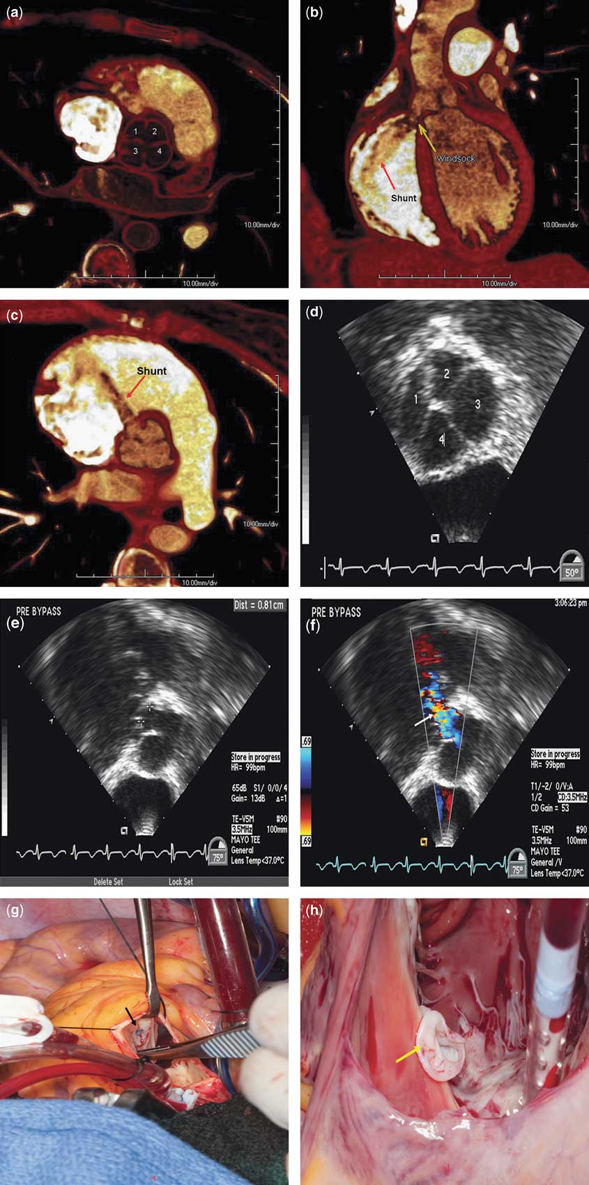
A contrast-enhanced electrocardiogram-gated computed tomography angiogram scan was performed to further characterise the aortic valve and sinus of Valsalva abnormalities, as well as to exclude other abnormalities in the aorta. This revealed a quadricuspid aortic valve, which had two non-coronary cusps (Fig 1a). There was also a ruptured aneurysm of the sinus of Valsalva involving the right anterolateral non-coronary aortic cusp with a 1 centimetre windsock deformity projecting along the tricuspid valve (Fig 1b). There was diastolic shunting through the windsock into the right heart, but no evidence of right ventricular or atrial volume overload (Fig 1b and c).
Electrocardiogram-gated, 128 detector, dual source computed tomography with intravenous contrast material (a), thin-slab volume-rendered image made in a plane through the short axis of the aortic valve shows a quadricuspid aortic valve (labelled). (b) Thin-slab volume-rendered image obtained at end-diastole, reformatted in oblique coronal plane, shows the windsock deformity protruding into the right heart near the level of the tricuspid valve (labelled). The right heart has a greater amount of contrast material than the left heart, allowing visualisation of a jet from the aorta to the right heart (labelled). (c) Short-axis thin-slab volume-rendered image in a plane designed to demonstrate the shunt again shows a jet of blood with less contrast material extending from the aorta and extending into the right heart near the level of the tricuspid valve. Intra-operative transoesophageal echocardiography (d), short-axis view at the aortic level shows quadricuspid aortic valve (labelled). (e) Modified long-axis view at the aortic level shows ruptured sinus of Valsalva (marked with white crosses). (f) With colour Doppler mode, modified long-axis view at the aortic level shows diastolic shunting (arrow) through the ruptured sinus of Valsalva. (g) Intra-operative photograph shows the aortic valve through an aortotomy. The non-coronary cusp is retracted away from the aorta to reveal the ruptured sinus of Valsalva (black arrow). (h) Through a right atriotomy, an intra-operative photograph demonstrates the aortic sinus windsock (yellow arrow) just below the tricuspid valve annulus.
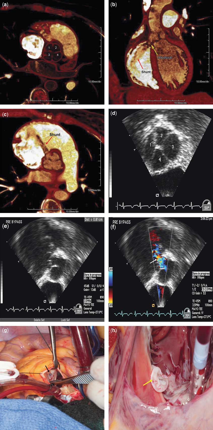
The intra-operative transoesophageal echocardiography confirmed the computed tomography findings (Fig 1d–f, Supplementary Movie S1), following which the patient underwent surgery to correct the ruptured sinus of Valsalva. After median sternotomy, the right atrium was opened and a windsock deformity was observed (Fig 1g and h). The windsock in the right atrium was excised completely and the defects at both the right atrial and aortic ends were closed with pericardial patches. The post-operative transoesophageal echocardiography showed complete repair of the sinus of Valsalva with no further shunt (Supplementary Movies S2 and S3). The patient recovered well and was discharged a few days later with no further complications.
Quadricuspid aortic valve is a congenital cardiac anomaly, and with an incidence between 0.008% and 0.033% at autopsy, and 1% in patients presenting for aortic valve surgery, it remains a rare finding.Reference Timperley, Milner, Marshall and Gilbert 1 Most cases of quadricuspid aortic valve are diagnosed by echocardiography (51%), followed by surgery (22.6%), autopsy (15.6%), and aortography (6.5%).Reference Tutarel 2 On the basis of the size of each individual aortic valve cusp, seven different anatomical variations of quadricuspid aortic valves have been described.Reference Timperley, Milner, Marshall and Gilbert 1 A quadricuspid aortic valve frequently functions abnormally, emphasising the clinical significance of its recognition. Aortic regurgitation is the most common pathology associated with quadricuspid aortic valve, where it is found in approximately 75% of the cases.Reference Tutarel 2 Although generally an isolated anomaly, it is also important to be aware of the various cardiac and non-cardiac anomalies that have been reported in association with the QAV.Reference Timperley, Milner, Marshall and Gilbert 1 Specifically, as seen in our patient, association with aneurysm of sinus of Valsalva (ruptured or not) have been reported.Reference Yang, Rawal, Pillutla and Criley 3 , Reference Pitta, Kondur and Afonso 4 When detected, a quadricuspid aortic valve should be followed closely.Reference Timperley, Milner, Marshall and Gilbert 1 As mentioned earlier, the most common functional abnormality would be regurgitation and this tends to deteriorate in adult life, often requiring surgery for repair or replacement around the sixth decade.Reference Timperley, Milner, Marshall and Gilbert 1 Our case highlighted the important role of multimodality imaging in diagnosing a quadricuspid aortic valve and its associations.
Supplementary materials
For supplementary material referred to in this article, please visit http://dx.doi.org/doi:10.1017/S104795111200042X