Case report
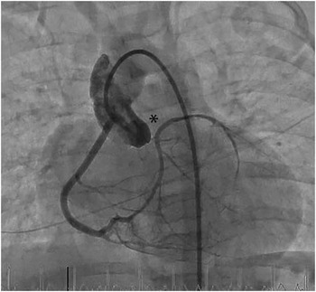
A 6.9 kg, 6-month-old female with Shone’s complex consisting of a diffusely hypoplastic aortic arch with moderate-to-severe coarctation, mildly hypoplastic bicuspid aortic valve with moderate subvalvar narrowing, mildly hypoplastic mitral valve, a peri-membranous ventricular septal defect, and a moderate secundum atrial septal defect underwent surgical repair including aortic arch reconstruction, atrial and ventricular septal defect closure, patent ductus arteriosus ligation, subaortic stenosis resection, and aortic root enlargement at 2 weeks of age. Pre-operative transthoracic and intra-operative transoesophageal echocardiography showed a prominent right coronary artery with a likely small left coronary artery; however, during the operation, only the right coronary ostium was visualised. The epicardial course of the left coronary artery was seen but not the left coronary ostium. There were no peri-operative complications, and the patient was discharged home after an unremarkable post-operative course. On follow-up echocardiograms, the origin of the left coronary artery could not be visualised, raising concern for ostial atresia of the left coronary artery. At 5 months of age, the patient was admitted with respiratory failure and shock secondary to worsening left ventricular outflow tract obstruction and severely depressed left ventricular function. Cardiac catheterisation demonstrated a hypoplastic and bicuspid aortic valve with recurrent severe subaortic stenosis, with peak systolic ejection gradient of 76 mmHg. In addition, coronary angiography showed atresia of the left main coronary artery with flow to a small but patent left coronary artery system via the right coronary artery (Fig 1). On the basis of the position of the left coronary artery bifurcation into the left anterior descending and circumflex coronary arteries, it appeared that the patient may have had anomalous left coronary artery from the pulmonary artery; however, there was no residual connection to the pulmonary artery angiographically. The patient subsequently underwent a Ross/Konno procedure with subaortic stenosis repair without event. Of note, coronary re-vascularisation was not performed. Post-operative echocardiograms including imaging from the follow-up 1 year after surgery revealed normal left ventricular function.
Hand injection of contrast via a 4 French end-hole catheter positioned in the aortic root reveals a bicuspid aortic valve without insufficiency. There is no anterograde flow of contrast into the left coronary artery (*). There is normal anterograde filling of a dilated right coronary artery, which connects to the left main coronary artery. The bifurcation of the left coronary artery into the left main and circumflex coronary arteries appears distant from the aortic root. However, there is no flow of contrast into the left pulmonary artery.

Discussion
Left main coronary artery atresia is a rare coronary artery anomaly with limited data on the incidence, outcome, and optimal management strategy for these patients.Reference Lim and Matherne 1 In one large case series, there were 26 patients with left main coronary atresia and two patients with left main coronary hypoplasia;Reference Musiani, Cernigliaro, Sansa, Maselli and De Gasperis 2 15 of these 28 patients were less than 18 years of age. In this paediatric cohort, eight had surgical repair of their coronary anomaly and all eight were reported as alive and well. Of the remaining seven children who did not undergo coronary artery repair or re-vascularisation, there was an 80% mortality rate in those patients whose outcome was known. This understandably led the authors to recommend re-vascularisation for all patients with left main coronary atresia; however, there were significant confounding variables for the four patients who died without re-vascularisation. Moreover, one patient died post-operatively after an exploratory thoracotomy with an unclear cause of death. There were two patients who died suddenly: one also had right ostial stenosis and the second patient had anterolateral myocardial infarction years before death. The fourth patient died during surgical dilation of the left coronary ostium, which could be considered as a death in the surgical repair group. Clearly, re-vascularisation can be beneficial but is not without risk and may not be completely protective against sudden death. This may be related to other risk factors such as persistent coronary anomalies or underlying dysfunctional myocardial substrate. In addition, a graft to an unobstructed vessel typically develops thrombosis over time, as the open native vessel provides competitive flow.Reference Juleff, Brown, McKain, Glover and Bendick 3 Thus, there is a risk that a graft would become occluded soon after surgical repair.
In our patient, we elected against re-vascularisation and the patient’s ventricular function normalised after the left ventricular outflow tract obstruction was alleviated. In the setting of outflow tract obstruction, there was likely increased myocardial oxygen demand. Theoretically, the collateral circulation to the left coronary artery might have been inadequate to meet this increased demand; however, with relief of the obstruction and return to baseline myocardial demand, the coronary artery flow was adequate. Therefore, although the long-term outcome of this patient has to be assessed, re-vascularisation is not mandatory to restore ventricular function in all patients found to have left main coronary artery atresia.
Acknowledgements
None.
Financial Support
This research received no specific grant from any funding agency, commercial, or not-for-profit sectors.
Conflicts of Interest
None.