Introduction
The sudden appearance of skeletal faunas near the Ediacaran–Cambrian boundary is one of the most conspicuous aspects of the first major diversification of metazoans, a unique event that featured the rise of nearly all extant phyla, including cnidarians, sponges, ctenophores, arthropods, echinoderms, and chordates (Erwin et al., Reference Erwin, Laflamme, Tweedt, Sperling, Pisani and Peterson2011). The diploblastic cnidarians (anthozoans and medusozoans), all or many of which are characterized by nematocysts, mesenteries (septa), and a blind gut with the mouth opening showing radial or biradial symmetry, were a major component of early Cambrian marine communities. Except for a few pelagic forms (Han et al., Reference Han, Hu, Cartwright, Zhao, Ou, Kubota, Wang and Yang2016a), early Cambrian medusozoans of South China were dominated by benthic forms such as carinachitiids (Han et al., Reference Han, Li, Wang, Yang, Guo, Sasaki and Komiya2018), hexangulaconulariids (Conway Morris and Chen, Reference Conway Morris and Chen1992), and the enigmatic olivooids (Dong et al., Reference Dong, Cunningham, Bengtson, Thomas, Liu, Stampanoni and Donoghue2013, Reference Dong, Vargas, Cunningham, Zhang, Liu, Chen, Liu, Bengtson and Donoghue2016; Han et al., Reference Han, Kubota, Li, Yao and Yang2013), which three groups are similar to each other in possessing a tubular periderm having an embryonic apical region and a much larger, postembryonic abapical region bearing well-defined transverse ridges or ribs.
Unlike the more or less radially symmetrical carinachitiids and olivooids (Dong et al., Reference Dong, Cunningham, Bengtson, Thomas, Liu, Stampanoni and Donoghue2013, Reference Dong, Vargas, Cunningham, Zhang, Liu, Chen, Liu, Bengtson and Donoghue2016; Han et al., Reference Han, Kubota, Li, Yao and Yang2013), the family Hexangulaconulariidae (originally Hexaconulariidae He, Reference He1987) (He, Reference He1987; Conway Morris and Chen, Reference Conway Morris and Chen1992) comprises distinctly biradial forms having a compressed, fan-shaped periderm, with each of the two major sides of the postembryonic portion of the periderm exhibiting three or five faces, and thus six or ten faces altogether (Steiner et al., Reference Steiner, Qian, Li, Hagadorn and Zhu2014; Duan et al., Reference Duan, Dong, Porras, Vargas, Cunningham and Donoghue2017). The family currently contains just two genera and species, Arthrochites emeishanensis Chen, Reference Chen1982 (Chen, Reference Chen1982) and Hexaconularia sichuanensis He and Yang, Reference He and Yang1986, which are known from strata of Fortunian age in South China. It has also been proposed that this clade was ancestral to the tetraradial conulariids, that is, to Aciconularia He, Reference He1987 from Cambrian Stage 2 (He, Reference He1987). Here we describe a new hexangulaconulariid, Septuconularia yanjiaheensis n. gen. n. sp. from the Yanjiahe Formation in the Three Gorges area of Hupei Province, China. The periderm of this taxon exhibits 14 faces, the most of any hexangulaconulariid described thus far, and thus provides additional morphological data bearing on the evolution of hexangulaconulariids and conulariids.
Geological setting
The Yanjiahe Formation in the measured Yanjiahe section near the city of Yichang (Fig. 1.1–1.4) is where specimens of S. yanjiaheensis examined in the present study (Figs. 2, 3) were collected. The Yanjiahe Formation measures ~40 m thick and crops out mainly on the southern and western flanks of the Huangling Anticline (Fig. 1.2), where it is particularly well exposed in the vicinity of Yanjiahe village (Fig. 1.3). The Yanjiahe Formation rests unconformably on the dolomitic Baimatuo Member of the Ediacaran Dengying Formation. The contact between the Yanjiahe Formation and the overlying Shuijingtuo Formation is disconformable, representing a relatively minor time gap (Chen, Reference Chen1984; Ding et al., Reference Ding, Li and Chen1992; Guo et al., Reference Guo, Li, Han, Zhang, Zhang, Ou, Liu, Shu, Maruyama and Komiya2008, Reference Guo, Li, Han, Zhang, Zhang, Ou and Shu2009, Reference Guo, Li and Li2014). The Yanjiahe Formation consists of partially dolomitized muddy limestone, calcareous black shale, and minor sandstone and chert. The original sediments were deposited in an intracratonic platformal basin (Zhu et al., Reference Zhu, Zhang, Steiner, Yang, Li and Erdtmann2003). Two thin, mostly chertified phosphorite layers are present in the lower part (Bed 2) and at the top of the Formation (Bed 5), respectively. The presence in these layers of scour structures and intraclasts indicates that they are storm deposits (J. Guo, unpublished observations).
Location and stratigraphy of the Terreneuvian (Cambrian Stage 2) Yanjiahe Formation in the Three Gorges area near Yichang, Hubei Province, China. (1) Sketch map of the People's Republic of China, showing the position of the collecting locality in Hubei Province. (2) Simplified geological map of the Three Gorges area, Hubei Province, showing outcrops of Cambrian strata (gray box in the Yanjiahe area denotes area enlarged for additional details). (3) Detailed geological map of the Yanjiahe area, showing outcrops of the Yanjiahe Formation. (4) Stratigraphic sequence of lower Cambrian strata in the Yanjiahe section, Yangtze Gorges area, indicating the horizons where specimens of Septuconularia yanjiaheensis n. gen. n. sp. were collected.

Septuconularia yanjiaheensis n. gen. n. sp. from the lower Cambrian (Stage 2) Yanjiahe Formation (Bed 5) in the Three Gorges area near Yichang, Hubei Province, China. (1, 2) Side view of the holotype, CUBar90-4; (3–6) CUBar38-2; (7–9) CUBar9-2; (10–14) CURBar5. (15–17) Longitudinal subdivision of the periderm from the base adapertureward into three parts corresponding to the embryonic, juvenile, and adult stages, respectively. (15) CUBar2-1; (16) CUBar9-6; (17) CUBar38-7. aab = apical-abapical boundary; abr = abapical region; am = apertural margin; ap = aperture; apr = apical region; as = adult stage; cf = central face; es = embryonic stage; js = juvenile stage; lf = longitudinal furrow; lfs = lateral faces; lmg = longitudinal median groove; lr = longitudinal ridge; mf = marginal face; tr = transverse rib.

S. yanjiaheensis as for the species by monotypy from the lower Cambrian (Stage 2) Yanjiahe Formation (Bed 5) in the Three Gorges area near Yichang, Hubei Province, China. (1–15) CUBar119-12, CUBar115-31, CUBar9-7, CUBar99-2, CUBar60-4, CUBar38-8, CUBar115-38, CUBar99-9, CUBar99-8, CUBar90-6, CUBar9-1, CUBar38-1, CUBar99-1, CUBar79-1, CUBar10-23, respectively. All photos share the same scale bar to illustrate hypothesized extensional growth of the periderm by addition of transverse ribs.

On the basis of lithological variation, the Yanjiahe Formation has been subdivided into five beds (Fig. 1.4). In the Yanjiahe section, the lowest bed, or Bed 1, is approximately 19.2 m thick and consists of siliciclastic rocks interstratified with sandy dolostone and shale. It contains probable sponge spicules (Chang et al., Reference Chang, Feng, Clausen and Zhang2017, but specific horizon is still in doubt) and the Asteridium–Heliosphaeridium–Comasphaeridium acritarch Assemblage Zone (Yao et al., Reference Yao, Xiao, Yin, Li and Yuan2005; Dong et al., Reference Dong, Xiao, Shen, Zhou, Li and Yao2009; Ahn and Zhu, Reference Ahn and Zhu2017). Bed 2 is 1.9 m thick and consists of siliceous-phosphatic, intraclastic dolostone bearing small shelly fossils of the Anabarites trisulcatus–Protohertzina anabarica Assemblage Zone (Chen, Reference Chen1984; Guo et al., Reference Guo, Li and Li2014). One of the tubular species in this bed, Megathrix longus Yin, Reference Yin1987, has been interpreted as a well-preserved cyanobacterium (Shang et al., Reference Shang, Liu, Yang, Chen and Wang2016). However, Yao et al. (Reference Yao, Xiao, Yin, Li and Yuan2005) and Dong et al. (Reference Dong, Xiao, Shen, Zhou, Li and Yao2009) concluded that M. longus probably was not a cyanobacterium, allying it instead with the Neoproterozoic tubular microfossil Sinocyclocyclicus guizhouensis Xue et al., Reference Xue, Tang and Yu1992, which has been interpreted as a stem-group cnidarian. Bed 3 measures approximately 15.7 m thick and consists of gray-black limestone interbedded with silty shale. The silty shale contains macrofossils such as algae and enigmatic metazoans (Yanjiahella Guo et al., Reference Guo, Li, Han, Ou, Zhou and Zheng2012 and Protoconites minor Chen et al., Reference Chen, Xiao and Yuan1994) (Guo et al., Reference Guo, Li, Han, Zhang, Zhang, Ou, Liu, Shu, Maruyama and Komiya2008, Reference Guo, Li, Han, Zhang, Zhang, Ou and Shu2009, Reference Guo, Li and Shu2010, Reference Guo, Li, Han, Ou, Zhou and Zheng2012), while abundant small shelly fossils (Purella antiqua Zone; Guo et al., Reference Guo, Li and Li2014) and a possible radiolarian (Chang et al., Reference Chang, Feng and Zhang2018) occur in siliceous-phosphatic nodules. Bed 4 is 5.8 m thick and consists of nonfossiliferous carbonaceous limestone. Finally, Bed 5 is about 1.1 m thick and consists of siliceous-phosphatic, intraclastic limestone containing fossils of the Aldanella attleborensis Assemblage Zone (= the A. yanjiaheensis Assemblage Zone [Guo et al., Reference Guo, Li and Li2014], which is equivalent to Cambrian Stage 2 [ca. 528 Ma; Zhu et al., Reference Zhu, Yang, Yuan, Li, Zhang, Zhao, Ahn and Miao2019]). All specimens of S. yanjiaheensis (Figs. 2, 3) examined in the present study were collected from the base of Bed 5.
Materials and methods
Sixty-four specimens of S. yanjiaheensis were obtained from rock samples digested in 10% acetic acid. The specimens were coated with gold and then imaged using an FEI Quanta 400 FEG scanning electron microscope (SEM). Up to 300 microcomputed tomographic (micro-CT) images (VXL format), with a pixel size of 1.1 μm for specimen CUBar90-4, were acquired by CT Lab HX 130 at the Rigaku in Tokyo, Japan, at an accelerating voltage of 50 kV and a current of 80 μA. The micro-CT data were processed using VG Studio 2.2 Max for 3D volume rendering.
Repositories and institutional abbreviations
Fossils investigated in this study are housed in the paleontological collections of Chang'an University (CU) in Xi'an, China. The specimens (provided by J. Han) in Table 2 are housed in the Early Life Institute (ELI), Northwest University, Xi'an, China.
Systematic paleontology
Terminology
Anatomical terminology used herein mostly follows precedents found in Conway Morris and Chen (Reference Conway Morris and Chen1992), Van Iten (Reference Van Iten1992), and Han et al. (Reference Han, Li, Wang, Yang, Guo, Sasaki and Komiya2018).
Phylum Cnidaria Verrill, Reference Verrill1865
Subphylum Medusozoa Petersen, Reference Petersen, Larwood and Rosen1979
Subclass ?Conulata Moore and Harrington, Reference Moore, Harrington and Moore1956
Family Hexangulaconulariidae He, Reference He1987
Included genera
Arthrochites Chen, Reference Chen1982; Hexaconularia He and Yang, Reference He and Yang1986; Septuconularia n. gen.
Emended diagnosis
Rapidly expanding, laterally compressed, biradially symmetrical periderm having a blunt apex and prominent transverse ribs. Each of the two major sides of the periderm bear three, five, or seven faces bounded by longitudinal grooves or ridges.
Genus Septuconularia new genus
Type species
Septuconularia yanjiaheensis n. gen. n. sp., from the Aldanella attleborensis Assemblage Zone of the Yanjiahe Formation (Bed 5; Cambrian Stage 2) in the Yanjiahe section near the city of Yichang, Hubei Province, China.
Diagnosis
As for the species by monotypy.
Occurrence
The A. attleborensis Assemblage Zone in Bed 5 of the Cambrian Yanjiahe Formation (Terreneuvian, Stage 2) in the Yanjiahe section near the city of Yichang, Hubei Province, China.
Etymology
Septu, Latin, referring to the seven faces on each of the two major sides of the periderm; conul-, Latin, cone-shaped periderm.
Remarks
The new genus currently comprises the type species only. Septuconularia n. gen. differs from Hexaconularia and Arthrochites in having 14 faces.
Holotype
CUBar90-4 (Fig. 2.1); the A. attleborensis Assemblage Zone in Bed 5 of the Cambrian Yanjiahe Formation (Terreneuvian, Stage 2) in the Yanjiahe section near Yichang, Hubei Province, China.
Paratypes
A total of 21 relatively well-preserved specimens (CUBar2-1, CUBar9-1, CUBar9-2, CUBar9-6, CUBar9-7, CUBar10-23, CUBar38-1, CUBar38-2, CUBar38-7, CUBar38-8, CUBar60-4, CUBar79-1, CUBar90-6, CUBar99-1, CUBar99-2, CUBar99-8, CUBar99-9, CUBar115-31, CUBar115-38, CUBar119-12, CURBar5) from the same level and locality as the holotype.
Diagnosis
Fan-shaped, biradially symmetrical periderm exhibiting a narrowly lenticular transverse cross section and consisting of a short apical region and a long, tapered, strongly compressed abapical region. Apical region relatively smooth; abapical region with short, irregularly spaced, trochoidal (long.) transverse ribs arrayed on each of the two major sides of the periderm in seven, adapically tapering faces bounded by six, adaperturally diverging longitudinal ridges. Transverse ribs on each face variably offset along a subdued median (midline) groove.
Occurrence
As for the type species.
Description
Fan-shaped tubular periderm with a narrowly lenticular transverse cross section. Available specimens range from 1.7 to 6.3 mm in width and from 1.3 to 5.4 mm in length (Table 1). Periderm strongly tapered, with the apical angle measuring approximately 80°–100° in the plane of greatest width (Table 1). External ornament of the periderm defines a small apical region (apr) and a much larger abapical region (abr) terminating along a slit-like apertural margin that may be incomplete (Figs. 2, 3).
Measurements of S. yanjiaheensis as for the species by monotypy.

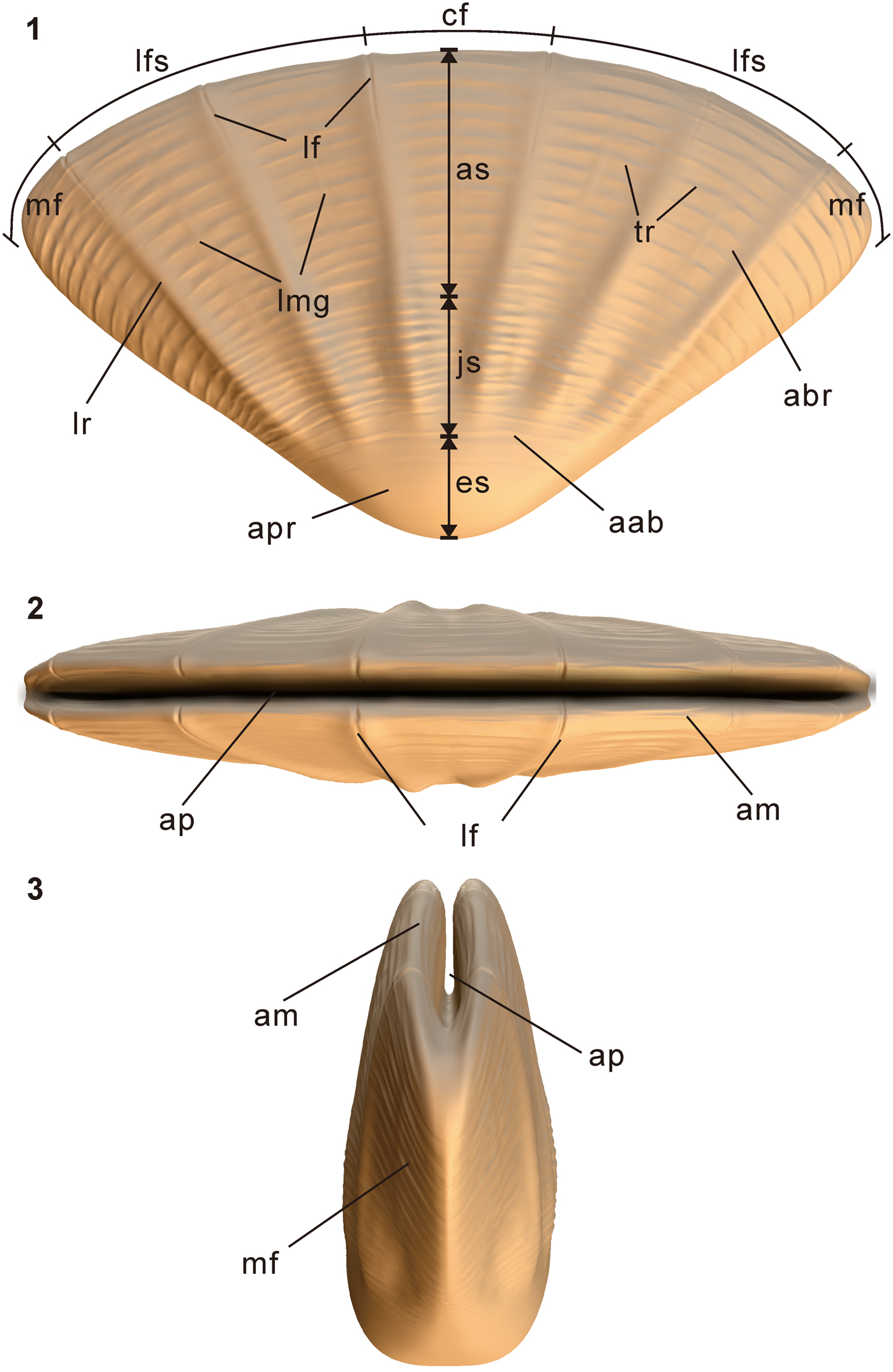
Both sides of the abapical region exhibit seven adapically tapering faces, each bounded by prominent longitudinal ridges (lr; Fig. 2.1, 2.5, 2.10) and crossed by short, irregularly spaced transverse ribs (tr; Fig. 2.3, 2.7, 2.10, 2.11)). Longitudinal ridges measure approximately 40–50 μm in width and extend from the boundary between the apical and abapical regions (aab; Fig. 2.1, 2.2) to the apertural margin (am; Fig. 2.3, 2.5, 2.7). Beginning at a distance of approximately 4–5 ribs from the apertural margin, each longitudinal ridge is divided by a single, narrow, longitudinal furrow (lf) into two, mutually parallel narrow ridges (Fig. 2.4–2.6, 2.12–2.14). Seven faces constituting each side of the periderm collectively consist of two marginal faces (mf), four lateral faces (lfs), and one central face (cf; Fig. 2.1). Faces nearly equal in width, with the apical angle measuring ~11° –17° (Table 1). Along the apertural margin, each face is curved slightly toward the opposite side of the periderm (Fig. 2.4). Faces further subdivided into two equal halves by a single subdued, discontinuous, longitudinal median (midline) groove (lmg; Fig. 2.7, 2.11).
All 14 faces bear numerous, irregularly spaced, trochoidal, adaperturally arching transverse ribs. Height of the transverse ribs variable but generally lower than that of the longitudinal ridges. Most ribs extend across the entire width of the face; others terminate along the facial midline (Figs. 2.7, 3.7, 3.10, 3.13). Some ribs appear as irregular folds that bifurcate toward the longitudinal ridges or longitudinal median grooves (Fig. 2.8, 2.11, 2.13); such ribs curve slightly toward the aperture, numbering approximately 12–13 ribs per 1 mm near the aperture. Transverse ribs in the upper part of the abapical region do not cross the longitudinal ridges. Lower part of the abapical region extends up to 7–8 transverse ribs away from the boundary with the apical region; transverse ribs in the lower part of the abapical region number approximately 20 per 1 mm, in some cases crossing the longitudinal ridges and reaching the midline of the adjacent face or faces, the longitudinal ridges not being as prominent as those in the upper part of the abapical region (Fig. 2.1, 2.2, 2.15–2.17). Viewed from above, apertural margin lenticular in outline, paralleling the transverse ribs and thus gently arched in side view. Apertural margin truncates some of the transverse ribs, suggesting incomplete preservation of the aperture (Fig. 2.4, 2.12).
Apical region preserved in two specimens (Figs. 2.1, 2.2, 3.13), measuring 0.4 mm long and 0.6 mm wide in the holotype, and 0.5 mm long and 1.5 mm wide in CUBar99-1 (Fig. 3.13); apical region cone-shaped, with apical angle measuring 125°. Surface of the apical region mostly smooth but with 4–5 long, continuous, concentric arc-like folds near the boundary with the abapical region (Fig. 2.2). Six longitudinal ridges terminate at the apical-abapical boundary, though with their lines of extension possibly meeting at the apical end (Fig. 2.1, 2.2).
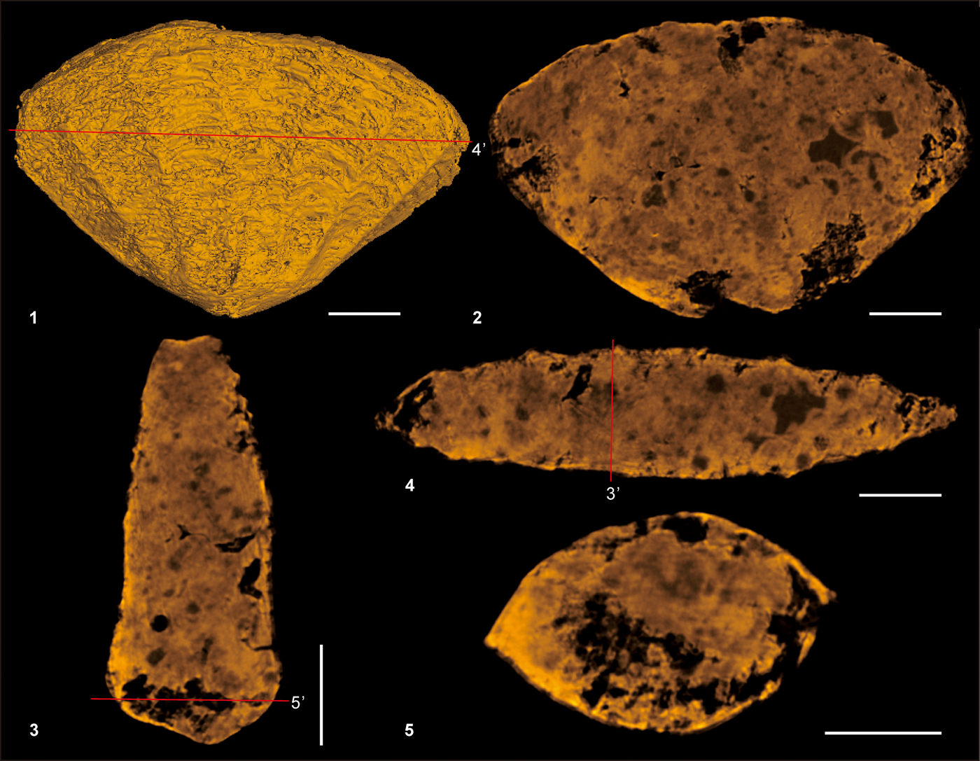
Micro-CT observations reveal that within the plane containing the minimum width of the periderm and the oral-aboral axis, the greatest width of the tube is located at the boundary between the embryonic region and the abapical region, with the width decreasing abapically from that level (Fig. 4; see supplemental data Movie 1). Specimens are partially filled with secondarily phosphatized, irregular debris, with no evidence of relic soft parts. As indicated by the positions of the longitudinal ridges, arrangement of the 14 faces on the two opposite sides of the periderm is not exactly biradial (Fig. 2.4).
Micro-CT tomography of the holotype specimen (CUBar90-4) of Septuconularia yanjiaheensis n. gen. n. sp. (1) 3D volume rendering of the periderm; (2) virtual intersection along the maximum width and the oral–aboral axis of the periderm; (3) virtual intersection close to the minimum width along the oral–aboral axis; (4) virtual horizontal intersection at the level indicated by 4' in (1); (5) virtual horizontal intersection at the level of the boundary between the apical and abapical regions. Scale bars = 100 μm.

Etymology
Yanjiahe, a fossil locality in the Three Gorges area near Yichang, Hupei Province, China.
Preservation
All 64 examined specimens of S. yanjiaheensis show three-dimensional preservation but have undergone secondary phosphatization. Their biradial symmetry probably is a primary anatomical feature, as the specimens occurred together with undeformed cylindrical tubes of other taxa (e.g., Conotheca Missarzhevsky in Rozanov et al., Reference Rozanov1969 and Turcutheca Missarzhevsky in Rozanov et al., Reference Petersen, Larwood and Rosen1969; see Guo et al., Reference Guo, Li and Li2014). The internal cavity of S. yanjiaheensis is generally filled with diagenetic phosphatic debris (Fig. 2.1, 2.4, 2.9, 2.12), and the edges of most specimens are incomplete, indicating that they may have been subjected to transport and abrasion prior to final burial. As in conulariids and other hexangulaconulariids (He, Reference He1987; Han et al., Reference Han, Li, Wang, Yang, Guo, Sasaki and Komiya2018), the apical region is rarely preserved (CUBar90-4 and CUBar38-2), suggesting that this portion of the periderm was relatively thin and fragile. None of the examined specimens preserves the entire aperture, and thus the tube wall in this area probably was very delicate.
Remarks
Together, pronounced biradial symmetry and large apical angles distinguish hexangulaconulariids from all conulariids except Conulariella Bouček, Reference Bouček1928 (Bouček, Reference Bouček1928), which, however, differs from hexangulaconulariids in having only four faces and a well-developed sulcus at each of the four corners. Septuconularia yanjiaheensis is most similar to Hexaconularia sichuanensis in having a strongly compressed conical periderm with more than four faces. However, S. yanjiaheensis differs from both H. sichuanensis and Arthrochites emeishanensis in having or showing: (1) substantially greater size (up to 6 mm in width); (2) 14 faces instead of six; (3) much larger apical angles (generally in excess of 90° [Table 1] as opposed to 75° on average in Hexaconularia [Table 2] and 35° on average in Arthrochites); and (4) irregularly spaced transverse ribs, with the most prominent ribs occurring in the upper part of the abapical region. By contrast, the transverse ribs of Hexaconularia and Arthrochites are much more regular in their spacing, and they may cross the longitudinal furrows (Conway Morris and Chen, Reference Conway Morris and Chen1992; Van Iten et al., Reference Van Iten, Zhu and Li2010). In addition, and as in the lower part of S. yanjiaheensis, the transverse ribs of Arthrochites are continuous and elliptical and are not interrupted along the longitudinal furrows. Additional differences between S. yanjiaheensis and both Hexaconularia and Arthrochites emeishanensis are: (5) the presence of 14 longitudinal ridges in the abapical region of S. yanjiaheensis instead of four prominent longitudinal furrows (though with the 14 longitudinal ridges becoming elevated longitudinal furrows near the aperture); (6) the absence of prominent longitudinal striations between the transverse ribs (present not only in Hexaconularia and Arthrochites but also in conulariids); (7) the presence of closely spaced transverse ribs and intercalated transverse grooves in S. yanjiaheensis versus widely spaced ribs in Hexaconularia and Arthrochites; (8) the convex apertural margin of each face of S. yanjiaheensis (indicated by the course of the apertural transverse ribs) versus the nearly rectilinear margin of Arthrochites; and (9) the central face of S. yanjiaheensis being nearly equal in width to the other faces as opposed to being much wider than the other faces. It should be noted, however, that in some specimens of Arthrochites (Conway Morris and Chen, Reference Conway Morris and Chen1992, fig. 4) and Hexaconularia (Van Iten et al., Reference Van Iten, Zhu and Li2010, text-fig. 1), the central face can be subdivided into three equal sectors, making it similar to the faces of S. yanjiaheensis. To some extent, then, Hexaconularia is morphologically intermediate between Arthrochites and S. yanjiaheensis, though the periderm of Hexaconularia appears to have been much more rigid than that of either Arthrochites or S. yanjiaheensis.
Measurements of Hexaconularia sichuanensis. NXC9-01 quoted from Steiner et al. (Reference Steiner, Li, Qian and Zhu2004, fig. 8.1); He22–99 from Steiner et al. (Reference Steiner, Qian, Li, Hagadorn and Zhu2014, fig. 7.20); GMPKU3132, GMPKU3134, and GMPKU3135 from Duan et al. (Reference Duan, Dong, Porras, Vargas, Cunningham and Donoghue2017, fig. 5(a), (f), (h)); NIGP160430 from Xian et al. (Reference Xian, Zhang, Liu and Zhang2018, fig. 3A); NIGP150159, NIGP150161, and NIGP150162 from Van Iten et al. (Reference Van Iten, Zhu and Li2010, text-fig. 1A, D, E). Other specimens from early Cambrian Kuanchuanpu Formation in Ningqiang and Hexi section (provided by J. Han).

Discussion
Anatomical comparisons with exceptionally well-preserved polypoid medusozoans (carinachitiids and olivooids) from the early Cambrian Kuanchuanpu Formation of South China may provide insights into the ontogeny of S. yanjiaheensis. Thus, the early Cambrian family Carinachitidae He, Reference He1987 (subclass Conulata Moore and Harrington, Reference Moore, Harrington and Moore1956) is characterized by an elongate, rigid periderm consisting of three, four, or five convex faces bordered by broad, deep corner sulci (Conway Morris and Chen, Reference Conway Morris and Chen1992; Liu et al., Reference Liu, Li, Shao, Zheng, Zheng, Wang, Wang and Wang2011; Han et al., Reference Han, Li, Wang, Yang, Guo, Sasaki and Komiya2018). Olivooids consist of a cone-shaped organic periderm showing circular ribs (annulations) and numerous vertical striations (Steiner et al., Reference Steiner, Qian, Li, Hagadorn and Zhu2014). Both the embryonic and apical regions of olivooids are greater than 0.5 mm in diameter, suggesting that these animals produced large eggs containing enough yolk for direct development (Dong et al., Reference Dong, Cunningham, Bengtson, Thomas, Liu, Stampanoni and Donoghue2013, Reference Dong, Vargas, Cunningham, Zhang, Liu, Chen, Liu, Bengtson and Donoghue2016; Han et al., Reference Han, Kubota, Li, Yao and Yang2013, Reference Han, Kubota, Li, Ou and Wang2016b, Reference Han, Li, Kubota, Ou and Toshino2016c; Wang et al., Reference Wang, Han, Vannier, Ou, Yang, Uesugi, Sasaki, Komiya and Sevastopulo2017; Shao et al., Reference Shao, Liu, Duan, Zhang, Zhang, Wang, Zhang and Qin2018). Because the apical region of S. yanjiaheensis is also large, development in this species, too, may have been direct. Furthermore, and in contrast to the more or less spherical apical region of Arthrochites (Conway Morris and Chen, Reference Conway Morris and Chen1992) and olivooids (Liu et al., Reference Liu, Li, Shao, Zhang, Wang and Qiao2014; Steiner et al., Reference Steiner, Qian, Li, Hagadorn and Zhu2014; Han et al., Reference Han, Li, Kubota, Ou and Toshino2016c), the biradial symmetry of the apical region of S. yanjiaheensis suggests that this symmetry was established during the embryonic stage.
The fact that the basal embryonic region of S. yanjiaheensis is strongly differentiated from the rest of the periderm suggests that this species exhibited at least two developmental stages. In addition, the well-differentiated lower and upper parts of the abapical region may represent a relatively transient juvenile stage and a long adult stage, respectively (Fig. 2.1, 2.15–2.17). Similar juvenile-adult differentiation probably occurs in certain specimens of Hexaconularia (see Duan et al., Reference Duan, Dong, Porras, Vargas, Cunningham and Donoghue2017, fig. 5a, b), in which the apical angle changes abruptly from 60° to 85° at a distance of about seven ribs above the apical portion.
It seems probable that growth of the periderm of S. yanjiaheensis involved addition of new material along the apertural margin. Such a mechanism has also been proposed for the growth of conulariids (e.g., Ford et al., Reference Ford, Van Iten and Clark2016) and observed in extant coronate scyphozoans (Werner, Reference Werner1973). In coronates, extensional growth is accompanied by thickening of the periderm through deposition of whole lamellae onto its inner surface, and this process probably accounts for the formation of the numerous microlamellae (Ford et al., Reference Ford, Van Iten and Clark2016) now constituting conulariid fossils. Similar microlamellae were not observed in S. yanjiaheensis and have not been documented in other hexangulaconulariids or in carinachitiids. Thus, while it is likely that extensional growth of the periderm did occur in these organisms, starting with the apical region, there are no certain indicators of rates of growth such as internal growth lines. However, if the spacing of the transverse ribs is indicative of rates of marginal accretion, then during formation of the abapical region, the rate of growth of the periderm appears to have been more or less constant (Fig. 5), as suggested by the more or less regular spacing of the transverse ribs (with 12–13 ribs per millimeter); by contrast, the rate of growth of the apical region appears to have been variable. The apical angles, especially of the median face, remained essentially constant throughout ontogeny (Fig. 6).
Scatter diagrams for selected aspects of the gross morphology of Septuconularia yanjiaheensis n. gen. n. sp. (1) Linear relationship between the maximum width and the longitudinal height of the periderm; (2) linear relationship between the longitudinal height and the number of transverse ribs per 1 mm on a peridermal face.

Comparisons of the apical angles of the central face between Septuconularia yanjiaheensis n. gen. n. sp. and Hexaconularia sichuanensis. (1) Bar chart showing apical angles of adult specimens; (2) bar chart showing apical angles of the central face.

Under the foregoing growth model, the wide end of the periderm was oral, and thus the actual mouth opening may have been slit-like, that is, strongly biradial (Fig. 7; see supplemental data Movie 2). The slit-like aperture may have reduced the chances of ingesting sediment grains, and the narrowly lenticular transverse cross section may have enhanced the stability of the body in strong water currents, the former action of which is suggested by apparent abrasion of the apertural margin.
Three-dimensional reconstructions of Septuconularia yanjiaheensis n. gen. n. sp. with hypothetical aperture, highlighting key anatomical features and terms used herein. (1–3) Lateral, oral, and oblique lateral views, respectively. aab = apical-abapical boundary; abr = abapical region; am = apertural margin; ap = aperture; apr = apical region; as = adult stage; cf = central face; es = embryonic stage; js = juvenile stage; lf = longitudinal furrow; lfs = lateral faces; lmg = longitudinal median groove; lr = longitudinal ridge; mf = marginal face; tr = transverse rib.

Septuconularia yanjiaheensis may be interpreted, then, as a sedentary polypoid medusozoan. Interestingly, its fan-shaped gross morphology with prominent radial ridges is similar to and possibly convergent upon the gross morphology and ornamentation of certain Paleozoic brachiopods (e.g., early Cambrian Diandongia, Lingulellotreta vs Nisusia [Briggs et al., Reference Briggs, Erwin and Collier1994; Zhang et al., Reference Zhang, Robson, Emig and Shu2008]) and bivalve mollusks (e.g., Grammatodon vs Nanonavis [see Cox et al., Reference Cox, Moore and Teichert1969, fig. C6]).
Finally, the results of the present study suggest that carinachitiids (Conway Morris and Chen, Reference Conway Morris and Chen1992, figs. 5.13, 6.1, 6.9–6.13), hexangulaconulariids (Steiner et al., Reference Steiner, Qian, Li, Hagadorn and Zhu2014, fig. 7.11), and conulariids as recently defined (Leme et al., Reference Leme, Simoes, Marques and Van Iten2008) may constitute a monophyletic group within the subphylum Medusozoa. A putative synapomorphy for this group is the possession of an elongate, organic or organophosphatic periderm exhibiting longitudinal furrows, a facial midline, a polygonal transverse cross section, and offset of transverse ribs along the facial midline. The hypothesized sister group of this clade, the olivooids, lacks longitudinal furrows (Fig. 8). The most recent common ancestor of all of these taxa had a thin, possibly organic periderm with flexible ribs, longitudinal striations, and peridermal lappets. The hexangulaconulariids, which arose during the Cambrian Fortunian Age, may have gone extinct before the beginning of Cambrian Age 3 (He, Reference He1987). Comparisons among Arthrochites, Hexaconularia, and S. yanjiaheensis suggest an apparent evolutionary trend involving the stepwise accentuation of biradial symmetry from the Cambrian Fortunian Age to Cambrian Age 2, with S. yanjiaheensis being the most specialized distinguishable species.
Phylogenetic hypothesis for carinachitiids, olivooids, hexangulaconulariids, and conulariids.

Acknowledgments
We thank X. Liu (NWU) for the 3D drawings of hexangulaconulariids. We are grateful to R. Elias, G. Young, and S. Xiao for constructive comments and suggestions. This work was supported by the Strategic Priority Research Program of the Chinese Academy of Sciences (No. XDB26000000), the Natural Science Foundation of China (Nos. 41472015, 41672009, 41621003, 41772010, 41890844, 41890840, 41720104002), and the Ministry of Science and the Overseas Expertise Introduction Centre for Discipline Innovation Project (111) of the Ministry of Education of China (No. D17013).
Accessibility of supplemental data
Data available from the Dryad Digital Repository: https://doi.org/10.5061/dryad.ht8g28b.