Manipulations of the gut microbiota, mainly in mouse models such as germ-free mice, receiving a donor microbiome via faecal transplant, or receiving probiotic supplements, led to stunning discoveries in the past decade, e.g., microbial changes being related to alterations of various important modulatory brain parameters (e.g. neurotrophines, neurotransmitters including dopamine, GABA, serotonin, stress hormones, neuroinflammatory parameters). This was attributed to the existence of the microbiota-gut-brain-axis, whereby the micro-organisms (mostly bacteria) that reside mainly in the gut ‘communicate’ with the brain via diverse mechanisms. These include possible pathways via the autonomic nervous system, the neuroendocrine system, the hypothalamic-pituitary-adrenal (HPA) axis, the immune system, and metabolic pathways. For instance, microbial communities can produce and/or influence neuroactive compounds, including neurotransmitters (GABA, noradrenaline, dopamine, serotonin), and metabolites. Those can travel through circulation and then interact with the host's brain, immune system, endocrine system, or metabolism. Enteric nervous system neuronal cells and vagus nerve afferent fibres can be activated by the microbiota, signalling from gut to brain, and vice versa via, efferences from the brain can signal to the gut (Morais, Schreiber, & Mazmanian, Reference Morais, Schreiber and Mazmanian2021), which may explain, for example, commonly described links between emotional well-being and gut health. Roughly, there are as many cohabitating bacteria as there are human body cells (Sender, Fuchs, & Milo, Reference Sender, Fuchs and Milo2016) making humans ‘hybrid’ organisms. A multitude of theories arose from these discoveries, including hypothesising evolutionary adaptive advantages through the microbiota, shifting, or modulating human phenotypes and behaviours (Henry, Bruijning, Forsberg, & Ayroles, Reference Henry, Bruijning, Forsberg and Ayroles2021). Unsurprisingly thus, the discovery of the microbiota-gut-brain-axis constituted a paradigm shift in neurosciences (Mayer, Knight, Mazmanian, Cryan, & Tillisch, Reference Mayer, Knight, Mazmanian, Cryan and Tillisch2014) and other fields.
The International Scientific Association for Probiotics and Prebiotics consensus statement on the scope and appropriate use of the term probiotics states that those are best defined as ‘live microorganisms that, when administered in adequate amounts, confer a health benefit on the host’ (Hill et al., Reference Hill, Guarner, Reid, Gibson, Merenstein, Pot and Sanders2014). Evidence of the effects of probiotics on rodent brains points particularly towards hippocampal (HC) changes. Specifically, if HC-integrity is challenged by injury, probiotic supplementation can attenuate apoptosis in HC and relatedly the decline in cognitive performance (Mohammadi et al., Reference Mohammadi, Dargahi, Naserpour, Mirzanejad, Alizadeh, Peymani and Nassiri-Asl2019; Rahmati et al., Reference Rahmati, Momenabadi, Vafaei, Bandegi, Mazaheri and Vakili2019). Probiotics have also been shown to be able to alter HC metabolism (O'Hagan et al., Reference O'Hagan, Li, Marchesi, Plummer, Garaiova and Good2017), attenuate the age-typical decline in long-term potentiation (Distrutti et al., Reference Distrutti, O'Reilly, McDonald, Cipriani, Renga, Lynch and Fiorucci2014), and to rescue neurogenesis after antibiotic treatment (Möhle et al., Reference Möhle, Mattei, Heimesaat, Bereswill, Fischer, Alutis and Wolf2016).
However, when it comes to human neuroimaging trials, the translational bottleneck has taken its toll. We identified a total of five neuroimaging studies, all examining functional brain changes (except Bagga et al., Reference Bagga, Aigner, Reichert, Cecchetto, Fischmeister, Holzer and Schöpf2018a, who additionally examined structural connectivity/fibre tract changes). Tillisch et al. (Reference Tillisch, Labus, Kilpatrick, Jiang, Stains, Ebrat and Mayer2013) investigated healthy women aged 18–55 years, with a multi-strain probiotic fermented milk product (n = 12) v. placebo (n = 11) v. no intervention (n = 13). The product was taken twice per day (dose per intake: 1.25 × 10¹⁰ CFU of B. lactis [CNCM I-2494/DN-173 010], 1.2 × 10⁹ CFU each of S. thermophilus [CNCM I-1630] & L. bulgaricus [CNCM I-1632 & I-1519]), for 4 weeks. Pre-post-intervention, an emotional faces attention task functional MRI (fMRI) paradigm was used to study brain responses to emotional stimuli. Results indicated no self-reported changes in anxiety or depression through probiotics, but task-related neuronal network activity decreases were observed in affective, viscero-, and somatosensory cortices. These were related to altered periaqueductal grey-centred (midbrain) resting-state network activity. Overall, findings were interpreted as the probiotics evoking widely distributed changes in functional brain activity potentially related to (improved) regulation of emotion and (pain) sensation. Bagga et al. (Reference Bagga, Aigner, Reichert, Cecchetto, Fischmeister, Holzer and Schöpf2018a) investigated healthy volunteers aged 20–40 years, with a multi-strain commercial probiotic (n = 15) v. placebo (n = 15) v. no intervention (n = 15). The probiotic was a commercially available product (OmniBiotic®, 3 g/day), taken for 4 weeks. Pre-post-intervention, whole-brain resting-state FC changes in broad brain networks were examined on an exploratory basis. Structural connectivity analyses were carried out as well (i.e. changes in fibre tracts). FC increases in areas of the salience network (SN) and decreases in the default mode network (DMN), as well as in the middle and superior frontal gyrus network (MFGN), were observed. There were no changes in structural connections in the brain. The authors interpreted the findings as indicating a possible modulation of behaviour via shifts in attentional control and other higher-order cognitive processes through probiotics. In Bagga et al. (Reference Bagga, Reichert, Koschutnig, Aigner, Holzer, Koskinen and Schöpf2018b), the same study sample was analysed, but this time an emotional decision-making and -recognition memory fMRI task, as well as self-reported mood, were examined. Results indicated significant decreases in subscales of depression (i.e., hopelessness, risk aversion) and increases in positive affect in the probiotics compared to the other two groups. Some of the behavioural emotional decision-making and recognition memory parameters improved in the probiotic group v. placebo or no intervention. Tasked-based neural activity was significantly changed in several areas of the brain: cingulum, precuneus, inferior parietal lobe, thalamus, parahippocampal gyrus, and cerebellum. In another RCT, again, a commercially available multi-strain-probiotic (n = 29) was used (Ecologic®Barrier, 2 g/day) for 4 weeks v. placebo (n = 29), in healthy women, aged 18–40 years (Papalini et al., Reference Papalini, Michels, Kohn, Wegman, van Hemert, Roelofs and Aarts2019). Interestingly, this study revealed possible conditional probiotic effects on brain function (fMRI): those were only visible when a working-memory task was performed in an experimentally induced stressed state (after cold pressor test v. no stress), whereby it was found exclusively in the supplementation group that lower task-related activation in (right) prefrontal cortex was significantly correlated with a better performance under stress after supplementation. This was interpreted by the authors as less cognitive control needed under stress to correctly perform the task in the probiotic group – which was supported by the performance not worsening as much as in the placebo condition (=buffering effect). Deviating from previous imaging studies which all used multi-strain probiotics, another RCT applying magnetencephalography (MEG) in healthy volunteers aged 18–50 years used a single-strain probiotic intervention (n = 20) (1 × 10⁹ CFU/day of B. longum 1714™) v. placebo (n = 20) for 4 weeks (Wang, Braun, Murphy, & Enck, Reference Wang, Braun, Murphy and Enck2019). During a cyberball social stress paradigm, increased theta-&-alpha-band power was observed in frontal and cingulate cortex, as well as supramarginal gyrus, in the probiotic group v. placebo. Oscillatory changes were observed during resting-state MEG pre-post intervention in the probiotic group v. placebo: decreased beta-3-band-power in HC, fusiform gyrus, temporal cortex; increased theta-band-power in frontal and cingulate cortex. Altered resting-state activity in turn was associated with enhanced self-reported vitality and reduced mental fatigue. Broadly, the results may suggest that probiotic supplements may buffer against the effects of acute stress.
Despite the widespread interest in whether and how probiotics affect the brain, to our knowledge no longitudinal RCT has ever investigated neuroplastic brain changes in humans, particularly with a focus on HC. The assumed structural HC change is based on the notion that probiotic interventions can, e.g., have a regulating effect on immunological (pro-/anti-inflammatory) processes, both at a whole-brain level, and on HC specifically, as well as on hippocampal brain-derived neurotrophic factor (BDNF) and cAMP-response element-binding protein (CREB) levels (see review by Tang et al., Reference Tang, Meng, Li, Liu, Li, Chen and Yang2021). BDNF modulates activity-based synaptic plasticity and neurogenesis, and lack thereof is reported to be related to psychiatric disorder, including at a sub-clinical level (Lang, Hellweg, & Gallinat, Reference Lang, Hellweg and Gallinat2004). CREB modulates neuronal differentiation and synaptic plasticity (see Tang et al., Reference Tang, Meng, Li, Liu, Li, Chen and Yang2021). Furthermore, as outlined earlier, first animal studies have evidenced both neuro-protective and neuroplasticity-enhancing effects by probiotics on HC in rodent models (Distrutti et al., Reference Distrutti, O'Reilly, McDonald, Cipriani, Renga, Lynch and Fiorucci2014; Mohammadi et al., Reference Mohammadi, Dargahi, Naserpour, Mirzanejad, Alizadeh, Peymani and Nassiri-Asl2019; Möhle et al., Reference Möhle, Mattei, Heimesaat, Bereswill, Fischer, Alutis and Wolf2016; O'Hagan et al., Reference O'Hagan, Li, Marchesi, Plummer, Garaiova and Good2017; Rahmati et al., Reference Rahmati, Momenabadi, Vafaei, Bandegi, Mazaheri and Vakili2019).
The present trial aspired to apply a rigorous multi-methods approach to fill the exiting research gap on multi-strain probiotics possibly affecting HC-structure and assess potentially altered behavioural variables. HC-driven cognitive functions, such as visual, map-based, object location, and verbal memory, as well as spatial navigation were assessed, to be able to identify links between HC- and behavioural changes. It is also known that HC neurogenesis buffers against the detrimental effects of stress, while hippocampal atrophy is related to mental illness (e.g., see Snyder, Soumier, Brewer, Pickel, & Cameron, Reference Snyder, Soumier, Brewer, Pickel and Cameron2011). Accordingly, mental health status variables (incl. depression, anxiety, stress, and emotion regulation) were assessed as secondary focus of this trial.
Methods
Ethical approval was obtained prior to study onset (German Psychological Society; Approval Number: SK032017). The study was carried out as double-blind, randomised-controlled trial (NIH trial identifier: NCT03478527, registration date: 27 March 2018) with two arms: probiotic v. placebo. Assessors and participants were blind to group allocation. The randomisation was carried out via list-wise (list created with random sequence generator) assignment by a third person, who was unrelated to the study. After finishing data collection and preparation, the randomisation list was handed over to the first author.
Participation criteria and study design
Inclusion criteria were age 18–40 years and right-handedness. Exclusion criteria were self-reported neurological, mental (verified via an adapted version of the Mini International Neuropsychiatry Interview; Sheehan et al., Reference Sheehan, Lecrubier, Sheehan, Amorim, Janavs, Weiller and Dunbar1998), chronic, or severe somatic disorder, veganism and vegetarianism, current or past two months antibiotics intake, self-reported ‘conscious’ probiotic diet/intake, lactose intolerance, and concurrent participation in a drug trial. Participants gave written informed consent prior to study enrolment. Antibiotic intake onset during participation would lead to immediate exclusion.
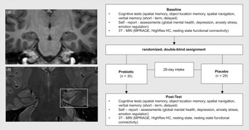
An overview of the study design can be found in Fig. 1. After random group assignment to either the multi-strain probiotic or placebo group (for details see Interventions section below), the participants took the respective preparation for 28 consecutive days. Immediately before and after the intake period, participants were tested by the first author and (medical and psychological) student assistants, who were trained to conduct the assessments. All assessors strictly followed a written test protocol. Assessments included cognitive tasks, self-reports, and MRI. A follow-up, two months after the post-test, was implemented, to follow up on potentially discovered pre-post effects. With a power of 0.90, α = 0.05, between measurements correlation of r = 0.70, and expecting an effect in between small and medium (f = 0.175), (F-test for within-between-interaction, G*Power 3.1.9.7 (http://www.gpower.hhu.de/) a sample of N = 54 was found to be a minimally required sample size to detect changes in the behavioural variables.
Study design.

Participants
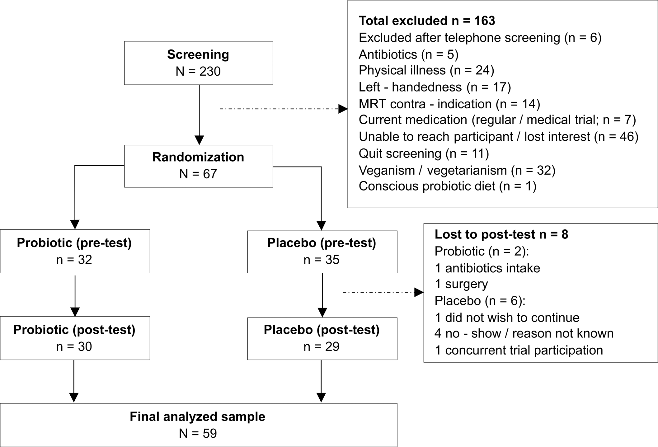
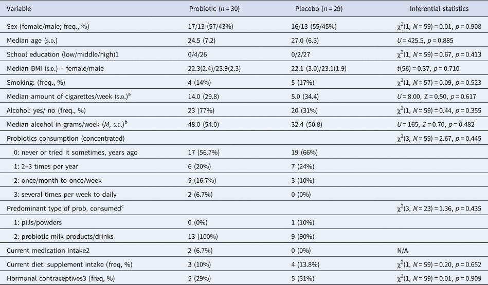
Initially, a total of 230 volunteers contacted the study team for participation. Of those, 163 individuals (≈ 71%) were excluded for different reasons (see Fig. 2 for details). A total of 67 healthy participants (see Table 1 for sample characteristics), were subsequently assessed at the neuroplasticity research laboratory at the University Clinic Hamburg-Eppendorf. Of those, 8 participants dropped out of the study (n = 6 in placebo, n = 2 in verum; corresponding ca. 12% of initially enrolled participants), (see Fig. 2). The total available sample for analysis was N = 59 (nprobiotic = 30; nplacebo = 29). The study started in January 2018 and ended in November 2019 (first in, last out).
CONSORT participant flow chart.

Demographic and other descriptive, health-related data at baseline

a,b,cData reported for respective subsamples of those with reported consumption >0 in probioticor placebo group.
1 German education system; ‘low’ referring to school degree ‘Hauptschule’ (lower secondary school certificate), ‘middle’ referring to school degree ‘Realschule’ (secondary school certificate), high to ‘Abitur’ (A-levels; university-entry certificate).
2 Excluding contraceptives.
3 Within female subgroup within probiotic or placebo group.
Interventions
Participants received daily 4.4 g doses (=1 sachet; corresponding to manufacturer recommendations, suggesting 1–2 sachets per day) of a probiotic (450 × 109 CFU per dose; Vivomixx®), which is composed of eight bacterial strains, including L. paracasei [NCIMB 30439], L. plantarum [NCIMB 30437], L. acidophilus [NCIMB 30442], L. helveticus [NCIMB 30440]), B. lactis [NCIMB 30435] & [NCIMB 30436], B. breve [NCIMB 30441]), and S. thermophilus [NCIMB 30438]. The placebo was a commercially available baby milk-powder (Bebivita® Anfangsmilch), selected due to its similar look and consistency compared to the probiotic. It contained no probiotic bacteria, and, in terms of dosage and composition, only negligible amounts of nutritional components (for details, see Placebo section below and Supplementary Material).
Preparations (probiotic/ placebo powders) were filled into small vials, with 28 vials (4.4 g per vial = 1 dose) per box packed into neutral looking boxes. An instruction label on the box advised participants to keep the preparations cooled in the fridge, and to take one dose per day, dissolved in water, milk, juice, or similar, what they would usually consume at breakfast (cool/room tempered). The intake period was 28 consecutive days. Subjects were instructed not to change their diet, and immediately report antibiotic treatment onset during participation in the trial, with the latter leading to exclusion from the running trial. Participants filled out an eating protocol during one week of their choosing within the 4-week intervention period.
Rationale for probiotic supplement v. placebo selection
Multi-strain probiotic
The multi-strain composition of Vivomixx®, and application over 4 weeks (as in all MRI studies that we were able to identify) in healthy human subjects, to test for effects on the brain, appeared to be a plausible approach, both backed-up by previous findings in animal studies using a similar product, and by to-be-expected beneficial health-effects based on general claims that can be made about some of the strains contained in Vivomixx®.
In a study by Möhle et al. (Reference Möhle, Mattei, Heimesaat, Bereswill, Fischer, Alutis and Wolf2016), the authors were able to show that VSL#3®, applied twice a day, at day two and four after discontinuation of a previous broad-spectrum antibiotic treatment (which lasted for 7 weeks), restituted the intestinal flora and neurogenesis levels (albeit not above baseline) of the animals. It was shown that, through probiotic supplementation, proliferating cells developed in the subgranular zone of the gyrus dentatus of HC. The authors were also able to demonstrate that a potential mediating mechanism of neuroplasticity was a probiotic-induced modulation of neuro-immunological processes, evidenced by changes in levels of Ly6Chi monocytes. Concentration was first reduced in HC tissue after antibiotic treatment, and then raised significantly above baseline levels after probiotic supplementation.
A second study, this time in rats, investigated the ‘archetypal model’ of synaptic plasticity in the HC – long-term potentiation (LTP) (Distrutti et al., Reference Distrutti, O'Reilly, McDonald, Cipriani, Renga, Lynch and Fiorucci2014). Ageing animals were treated with VSL#3®, backed up by two notions: (1) that VSL#3® had been previously shown to provide anti-inflammatory effects, and that (2) age-related decreases in LTP had been found to be related to neuroinflammation. One group of rats received maple syrup, another received VSL#3® at the dose of 12.86 bn living bacteria/kg/day for 6 weeks. VSL#3® was related to attenuations in changes of gene and protein expression associated with inflammatory processes in the rat brain. In addition, the age-related decline of LTP in HC was attenuated through VSL#3®. Importantly, both studies by Möhle et al. (Reference Möhle, Mattei, Heimesaat, Bereswill, Fischer, Alutis and Wolf2016) and Distrutti et al. (Reference Distrutti, O'Reilly, McDonald, Cipriani, Renga, Lynch and Fiorucci2014) evidenced distinct changes of microbial compositions in faecal samples of the probiotic-treated animals, suggesting that the supplement had altered microbial profiles.
Albeit, to our knowledge, the European market equivalent to VSL#3® (Vivomixx®) has never been formally tested in healthy human subjects concerning effects on hippocampal structure, and (relatedly) cognitive performance and mental health status, and albeit (antibiotically treated or ageing) rodent models are certainly not directly transferrable to humans, there are additional arguments why testing this product in healthy human volunteers is relevant. First, ‘psycho-biotic’ effects of several strains which are contained in the Vivomixx® formula, could be reasonably expected. Albeit the EU has no common, formal list of strains or species regarded as probiotics, mostly rejecting health claims made by manufacturers (see: https://www.ipaeurope.org/legal-framework/european-legal-framework/), Canada for instance has published a core-list of species, for which non-strain-specific claims concerning general health benefits may be made. Marked in bold in the following are species from this core-list which are also contained in Vivomixx®: Bifidobacterium (adolescentis, animalis, bifidum, breve and longum) and Lactobacillus (acidophilus, casei, fermentum, gasseri, johnsonii, paracasei, plantarum, rhamnosus and salivarius), (Government of Canada, Health Canada, 2009).
Placebo
Bebivita® Anfangsmilch Pre is recommended by the manufacturer for saturation of infants, ideally in addition to breastfeeding. We chose this product as its appearance, dissolution properties (e.g. in water or juice) and consistency resembled those of Vivomixx® at eye-level, which is an important factor in a placebo-controlled study. The amount of nutrients in this product, contained in 4.4 g/day taken in over 28 days, is negligible for adults, as based on the German Society for Nutrition's guidelines (https://www.dge.de/en/; checked against recommended nutritional reference value tables for adults). Furthermore, the product did not contain probiotics, nor any relevant amounts of prebiotics (please refer to the Supplementary Material, Table 5, p. 9, for an exact list of nutrients contained in 4.4 g of the product).
Cognitive tests
Visuo-spatial short-term working memory was assessed with a computerised version of the Corsi block tapping task (provided by https://www.millisecond.com). A sequence of squares flashing up on a screen was shown, starting with a 2-block-sequence, up until a 16-block-sequence. The participant had two attempts to correctly recall and replicate the sequence by serially clicking on the according squares. The parameter of interest was the total performance score (see Kessels, van Zandvoort, Postma, Kappelle, & de Haan, Reference Kessels, van Zandvoort, Postma, Kappelle and de Haan2000). Spatial memory was assessed using the map sub-tests from the Berlin Intelligence Structure test (BIS-4), (Jäger, Süß, & Beauducel, Reference Jäger, Süß and Beauducel1997). Participants had 30 sec. to memorise a map depicting a route from A to B. Afterwards, they had 40 sec. to draw the memorised route into a new map. The main outcome was the number of correctly recalled route sections. An object location memory task, similar as in Schmiedek, Lövdén, and Lindenberger (Reference Schmiedek, Lövdén and Lindenberger2010), was used. In this task, 12 photorealistic objects appeared sequentially in a 6 × 6 grid on the computer screen. Afterwards, objects had to be ordered in the correct sequence and position on a new grid. The main outcome was the number of correctly placed objects across two main trials. Spatial navigation was assessed using a tunnel task (Gramann et al., Reference Gramann, Onton, Riccobon, Mueller, Bardins and Makeig2010). Participants were instructed to watch a virtual ride through a tunnel with curvy and straight sections. At the end of the tunnel, they had to adjust an arrow to the exact point to the initial tunnel entrance. Allocentric v. egocentric navigational style was first determined in a calibration run comprising 10 trials. The percentage of trials classified by the programme as navigated allo-centrically served as a proxy for change in navigational strategy from pre- to post-test. Thirty main trials followed, whereby the accuracy concerning the habitual navigation strategy was operationalised as mean absolute deviation (in degrees) of the arrow adjustment made by the participant relative to the optimal adjustment (i.e. true entry point of tunnel). The Rivermead Behavioral Memory Test (Wilson, Cockburn, Baddeley, & Hiorns, Reference Wilson, Cockburn, Baddeley and Hiorns1989) was used to assess verbal memory. The total number of information units correctly reproduced from a newspaper-like text read out to the participants, was the main outcome both for the immediate and the delayed recall (after 20 min.).
Self-reports to assess mental health status (emotional well-being)
Questionnaires referred to the past two weeks, and validated German versions of the original tests were used. Depression was assessed with the BDI-II-revised version (Beck, Steer, & Brown, Reference Beck, Steer and Brown2011; Hautzinger, Keller, & Kühner, Reference Hautzinger, Keller and Kühner2006). The Brief Symptom Inventory (BSI) (Derogatis & Melisaratos, Reference Derogatis and Melisaratos1983; Franke, Reference Franke2000) assessed global psychiatric symptom burden. Anxiety and depressive symptoms subscales of the BSI were separately evaluated. The Perceived Stress Scale (PSS) was applied to measure daily life stress (Cohen, Kamarck, & Mermelstein, Reference Cohen, Kamarck and Mermelstein1993; Klein et al., Reference Klein, Brähler, Dreier, Reinecke, Müller, Schmutzer and Beutel2016). Emotion regulation was assessed using the Response Style Questionnaire (RSQ), addressing rumination and distraction (Kühner, Huffziger, & Nolen-Hoeksema, Reference Kühner, Huffziger and Nolen-Hoeksema2016; Nolen-Hoeksema, Reference Nolen-Hoeksema1991).
Additional overlapping self-reports on psychiatric symptoms and emotion regulation were assessed within the scope of this study to exhaust possibilities of identifying existing effects. These measures, including pre-post results, are fully documented in the Supplementary Material for this paper.
MRI data acquisition
Brain scans were performed on a 3T Siemens Magnetom Prisma (Siemens Medical Systems, Erlangen, Germany) using a 64-channel head coil. A regular sagittal MPRAGE (256 slices per slab, FOV = 240 mm, TR = 2500 ms, TE = 2.12 ms, TI = 1100 ms, voxel size = 0.8 mm × 0.8 mm × 0.9 mm) and a high-resolution HC scan consisting of a T1-weighted fast spin echo sequence (TR = 8020 ms, TE = 50 ms, TSE TF = 15, field of view = 175 mm, voxel size = 0.4 mm × 0.4 mm × 2.0 mm, slice thickness = 2 mm) were run. See Fig. 1 for an impression on the difference between a ‘regular’ MPRAGE v. HC-high-resolution image.
Since functional change can occur in the absence of structural changes, we assessed functional connectivity (communication) of HC with other brain regions, using a T2-weighted, BOLD sensitive resting-state EPI sequence, with participants lying relaxed, but awake (eyes open) in the scanner (TR = 2000 ms, TE = 30 ms, image matrix = 64 × 64, FOV = 216 mm, flip angle = 80°, voxel size 3 × 3 × 3 mm3, 36 axial slices).
Hippocampal MRI data pre-processing, analyses, and statistics
Toolboxes were run with Matlab R2017a (MathWorks Inc., Natick, MA). In a first step, the MPRAGE data was pre-processed using CAT12 (Structural Brain Mapping Group, University of Jena – exact version: CAT12.6-rc1[r1429] from 2019-02-08; http://www.neuro.uni-jena.de/cat/index.html), following the recommended (default) settings for longitudinal data, including segmentation into grey and white matter, affine registration and normalisation to MNI space with DARTEL (http://dbm.neur.uni-jena.de/cat12/CAT12-Manual.pdf). Afterwards, the segmented data was smoothed with 8 mm FWHM. A mask of left and right HC, based on the automated anatomical atlas (AAL), (Tzourio-Mazoyer et al., Reference Tzourio-Mazoyer, Landeau, Papathanassiou, Crivello, Etard, Delcroix and Joliot2002), was used as region of interest (ROI) within the REX tool (alpha 0.5 release, https://www.nitrc.org/projects/rex/) to extract rGMV data from the segmented, smoothed grey matter images. SPSS 25 (IBM Corp., 2017) was used to compute a repeated-measures ANOVA, with group as between factor and rGMV of left or right HC as within subject factor (time), and total intracranial volume (determined using CAT12), age and sex as covariates.
On the same MPRAGE scans we determined total left and right hippocampal volume using FreeSurfer 7.0 software (https://surfer.nmr.mgh.harvard.edu/). The segmentation results of FreeSurfer for HC have been shown to be highly correlated with manual tracings confirming methodological reliability (Morey et al., Reference Morey, Petty, Xu, Hayes, Wagner, Lewis and McCarthy2009). On the HC-high-resolution images we used the automated segmentation of hippocampal subfield (ASHS) tool developed by Yushkevich et al. (Reference Yushkevich, Wang, Pluta, Das, Craige, Avants and Mueller2010), with the UPenn PMC Atlas version 2016 (Yushkevich, Wolk, Pluta, & Ding, Reference Yushkevich, Wolk, Pluta and Ding2016), which provides volume estimates of hippocampal subfields traced along the full length of the hippocampus and extrahippocampal cortical structures: cornu ammonis, dentate gyrus, subiculum, entorhinal cortex, parahippocampal gyrus, perirhinal cortex (BA 35, BA 36) and the sulcus. Again, repeated-measures ANOVA was computed to detect any group (probiotic v. placebo) × time (pre-post volumetric data) interactions in SPSS 25, controlled by age, sex, and TIV.
We conducted a seed-based FC analysis, based on the AAL atlas taking the HC as seed to assess changes in communication of HC with other brain regions induced by probiotic supplementation. On the pre-processed images (see Supplementary Material for pre-processing details), the time series of all voxels inside the seed were extracted and averaged. This data was correlated to all other voxels in the brain via Person's correlations, resulting in one seed-based FC map per subject. Fischer's transformation was applied to the individual maps to improve normality. The z-score maps were taken to the second level in SPM12 using a flexible factorial design with subject factor as main effect and the group × time interaction. Mean frame-wise displacement (Power, Barnes, Snyder, Schlaggar, & Petersen, Reference Power, Barnes, Snyder, Schlaggar and Petersen2012) was used as covariate. The resulting maps were thresholded with p < 0.001 uncorrected, with an additional threshold of p ≤ 0.05 FWE corrected on the cluster level. We checked for increases and decreases in FC within group in the probiotic condition, as well as any group by time interaction, assuming stability in the placebo condition.
Whole-brain MRI data analyses and statistics
To assess potential neuroplastic changes besides HC, we conducted an exploratory whole-brain analysis on the grey- and white matter, pre-processed, smoothed images from CAT12 (see previous section). We implemented a flexible factorial design with subject factor as main effect, and group × time as interaction effect in CAT12, following recommended default settings including absolute threshold masking with a threshold of 0.1. Resulting maps were thresholded with p < 0.001 uncorrected, and with an additional threshold of p ≤ 0.05 FWE-corrected on the cluster level in SPM12. We examined both global increases and decreases in rGMV and rWMV as within group changes in the probiotic group as well as any group by time interactions, assuming stability in the placebo condition.
Behavioural data analyses and statistics
Repeated-measures ANOVAs were carried out in SPSS 25 for all variables of interest [group (probiotic v. placebo) × time (pre v. post-test) interactions]. Effect size η 2partial was interpreted as ⩾0.01 small, ⩾0.06 medium, ⩾0.14 large effect. In case of significant behavioural and structural, or functional effects, correlation analyses with brain-related findings would be carried out We additionally computed the Bayes factor for the H0 (BF01) of the interaction effect in JASP (version 0.14.1) to assess evidence in favour of a null effect. BF01 > 1 indicates that, relative to H1, H0 more likely applies to the data, BF01 < 1 indicates the opposite. Values between 0.5 and 1.5 can be regarded as inconclusive, BF01 ⩾ 3 moderate and ⩾10 strong evidence for H0 relative to H1 (van Doorn et al., Reference van Doorn, van den Bergh, Böhm, Dablander, Derks, Draws and Wagenmakers2020). The BF01 for the interaction was computed by dividing BF01 for all main effects plus the interaction by the BF01 for all main effects.
Results
Changes of hippocampal grey matter and functional connectivity
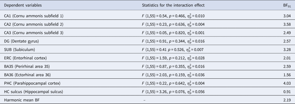
No significant group × time interaction was observed in the REX-based HC estimates (left: F 1,54 = 0.32, p = 0.577, η2p = 0.006; right: F 1,54 = 2.81, p = 0.099, η2p = 0.049). Likewise, no significant interaction was found in the FreeSurfer HC segmentations (left: F 1,54 = 1.51, p = .225, η2p = .027, BF01 = 1.68; right: F 1,54 = 1.27, p = 0.264, η2p = 0.023, BF01 = 1.05). There were also no significant group × time interactions for HC subfields (see Table 2). For descriptive data, see Supplementary material (Table 2).
Hippocampal subfield analyses (combined bilateral subfield volumes) – group × time interaction effects

Note. BF01 refers to the Bayes Factor for H0 relative to H1 concerning the presence of an interaction effect. All results are controlled for sex, age, and TIV. All analyses were conducted by originally assigned groups.
For the FC analysis we used the bilateral, left, and right HC as seed. No significant differences in FC within the probiotic group nor a significant group × time interaction were found in any of the three seeds analyses.
Exploratory analysis of whole-brain changes in grey or white matter
Exploratory whole-brain structural analyses revealed no significant group × time interactions neither for rGMV nor rWMV (with p < 0.001 uncorrected, and an additional threshold of p ⩽ 0.05 FWE-corrected on the cluster level).
Behavioural results
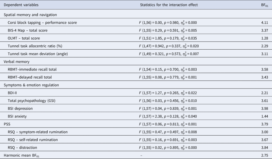
No significant interaction effects were identified (see Table 3). As indicated by the harmonic mean of BF01 approaching 3, evidence for the null hypothesis is of approximately moderate magnitude concerning effects of probiotics on cognition (visuo-spatial, map-based, object location -memory and navigation, verbal memory), psychiatric symptoms (global mental health burden, depression anxiety, and stress) and emotion regulation (rumination, distraction). Descriptive data can be found in online Supplementary Table S3. Further descriptive data and additional analyses on behavioural outcomes can be found in the Supplementary Material (Tables 1 and 4). There were consistent and robust null findings for all additional outcomes assessed in the study.
Mental health and cognition outcomes: group × time interaction effects (pre-to-post)

BIS-4 Map = map subtest from Berlin Intelligence Scale 4, total score (correctly recalled route segments); OLMT = Object Location Memory Task (no. of correctly placed objects both in sequence and position); RBMT = Rivermead Behavioral Memory Test; BDI-II = Beck's Depression Inventory-II revised; GSI = Global Severity Index (Brief Symptom Inventory); BSI depression = Brief Symptom Inventory depression subscale; BSI anxiety = Brief Symptom Inventory anxiety subscale; PSS = Perceived Stress Scale total score; RSQ = Response Style Questionnaire (subscales).
Note. BF01 refers to the Bayes Factor for H0 relative to H1 concerning the presence of an interaction effect (i.e. group × time). All analyses were conducted by originally assigned groups.
Discussion
The present study is the first to assess hippocampal and whole-brain structural, and HC- functional connectivity effects, elicited by probiotic supplementation in young, healthy, adult human volunteers. There were consistent null effects of the applied multi-strain probiotic (Vivomixx®) on hippocampal structure as well as HC functional connectivity, and there were also no grey and white matter volumetric changes at an exploratory whole-brain level. The absence of any effects on HC were also reflected in the null findings in cognitive performance, including visuo-spatial memory, object location memory, spatial navigation performance, or verbal memory, assessed using a range of well-established tests. Using a Bayesian approach, we found consistent evidence in favour of the null hypothesis of small to approximately moderate effect size. This contradicts previous findings from animal studies that suggested a beneficial effect of probiotic supplementation on hippocampal integrity or relatedly cognitive performance, applying a similar multi-strain probiotic as used in the present study (Distrutti et al., Reference Distrutti, O'Reilly, McDonald, Cipriani, Renga, Lynch and Fiorucci2014; Möhle et al., Reference Möhle, Mattei, Heimesaat, Bereswill, Fischer, Alutis and Wolf2016). It needs to be noted though that these studies were conducted in animals that already exhibited age-related or experimentally induced decline/atrophy in HC-function or structure. Our findings are in line with a recent meta-analysis, revealing that healthy subjects (human and animals) may not benefit from probiotic supplementation concerning cognitive performance as opposed to already cognitively impaired subjects (Lv et al., Reference Lv, Ye, Luo, Hu, Wang, Chen and Ding2021). The present study contributes to this pattern of findings, indicating that the applied multi-strain probiotic does not promote (HC) neuroplasticity or any related cognitive improvements in young, healthy adult volunteers.
Concerning the secondary outcome of mental health status (incl. self-reported stress, anxiety, depression, and emotion-regulation), again consistent null-evidence of approximately moderate effect size was found. Meta-analytic evidence of randomised-controlled trials suggests beneficial effects of probiotics in healthy participants, particularly concerning alleviating stress or anxiety. However, there is heterogeneity in applied strains and dosages, studies fall short on methodological rigour, findings are restricted to singular outcomes among several variables, and often only observed in individuals with already pathological symptom levels(e.g., see Chao et al., Reference Chao, Liu, Sutthawongwadee, Li, Lv, Chen and Guo2020). Of note, multi-strain probiotic neuroimaging studies, which were all conducted in healthy young individuals, overall showed inconsistent evidence concerning mental health outcomes. In addition, all significant functional neuroimaging results involved some form of experimentally induced stress, such as by exposure to emotional faces (Bagga et al., Reference Bagga, Reichert, Koschutnig, Aigner, Holzer, Koskinen and Schöpf2018b; Tillisch et al., Reference Tillisch, Labus, Kilpatrick, Jiang, Stains, Ebrat and Mayer2013), cold pressor stress test (Papalini et al. Reference Papalini, Michels, Kohn, Wegman, van Hemert, Roelofs and Aarts2019) or a social exclusion paradigm stressor (Wang et al., Reference Wang, Braun, Murphy and Enck2019).
To sum up: our study and pre-existing neuroimaging studies jointly suggest that multi-strain probiotics may have (transient) effects on brain function, mood, and performance in cognitive tasks that involve emotion, whereby findings may be moderated by stress (e.g. applying a negative-emotion/ stress-inducing paradigm). Effects of multi-strain probiotics on human brain structure in humans, more specifically hippocampus, remain, to our knowledge, unproven until the present day. To further pursue this line of research, several limitations should be considered, resulting in concrete implications and recommendations for future research. This will be outlined in the following paragraph.
Limitations and implications for future research
There are possibly floor effects for cognitive and psychiatric symptom variables due to rigorous screening and the narrow age range of the sample. Neuronal plasticity was expected to be higher in younger individuals and hence to facilitate the detection of any effects, however future trials should perhaps evaluate effects in ageing samples with pre-existing (subclinical) cognitive impairment or elevated mental health symptom/stress levels. Furthermore, albeit our sample size and timeframe as well as dosage of probiotics are comparable to previous fMRI studies that tested mostly multi-strain probiotic supplementation in healthy young-to-middle-aged adults, larger sample sizes and longer treatments might be necessary to detect (more subtle) changes in brain structure.
An additional point of criticism concerns the control of potential moderators, for instance physical exercise. As Marttinen, Ala-Jaakkola, Laitila, and Lehtinen (Reference Marttinen, Ala-Jaakkola, Laitila and Lehtinen2020, p. 1) point out there are ‘reciprocal interactions between physical activity and gut microbiota’, and hence the colonisation of the gut through probiotic intake as well as the metabolic activity of the microbiota might be altered in individuals who exercise regularly. Generally, for young healthy adults effects of exercise on HC volume are often not replicated and meta-analytically non-significant, while significant effects are observed for individuals above 65 years of age (see Wilckens et al., Reference Wilckens, Stillman, Waiwood, Kang, Leckie, Peven and Erickson2021). Overall, this may lead to the conclusion that exercise effects may be more of a preserving/neuroprotective than neurogenetic nature, whereby specifically ageing individuals seem to benefit. All taken together, we suggest that physical exercise could be examined as moderator of probiotic effects in future trials, but this variable is an unlikely candidate as a confounder in the present study on younger adults.
Another crucial point concerns the interpretability of the null findings from the background of whether the administered probiotics changed the microbial profile of participants in the first place. No stool samples were taken, and hence there was no way to confirm that the supplement had effectively altered microbial gut profiles. Accordingly, pre-post-change in microbial profiles in supplemented v. placebo/control groups should be analysed in future studies. Albeit previous animal studies suggest microbial profile alterations induced by a similar product as used in the present study (Distrutti et al., Reference Distrutti, O'Reilly, McDonald, Cipriani, Renga, Lynch and Fiorucci2014; Möhle et al., Reference Möhle, Mattei, Heimesaat, Bereswill, Fischer, Alutis and Wolf2016), those findings are not directly transferrable to human subjects. On a more general level, it remains largely uncertain whether and, if so, to which degree, probiotics can tolerate acids and bile salts while passing the digestive tract, and even the intestines in the first place (Wang et al., Reference Wang, Jiang, Deng, Yi, Wang, Ding and Wong2020). Furthermore, even if they pass these hurdles, probiotics need to overcome colonisation resistance (Han et al., Reference Han, Lu, Xie, Fei, Zheng, Wang and Li2021). Probiotics may however confer indirect effects, such as by producing metabolites that modulate the activity of pre-existing gut microbiota colonies, or by stimulating the intestinal epithelium – however still raising the question of persistence of effects, especially after discontinuation of intake. The form of intake may also influence results, such as protecting probiotics from gastric acid by using microencapsulation technologies (Shori, Reference Shori2017). These questions need rigorous consideration and investigation.
Finally, albeit testing commercially available products can be considered of public interest, single-strain trials, or testing a combination of 2 strains v. strain#1 v. strain#2 v. control, might provide clearer answers to the question which probiotic strains or their combination exactly provide specific benefits to the host. Hereby, adequate strain selection, targeted at specific outcomes (indication), well-delineated hypotheses and testing of mechanism (mediation) and effect modulation (moderation), should be a guiding principle.
Conclusion
Overall, our study found no evidence for effects of a multi-strain probiotic supplement on hippocampal structure or function, overall brain structure (grey and white matter), cognitive performance, or mental health status, in healthy, young adult volunteers. Presumably, effects of multi-strain probiotics in this target group are rather transient and may only show specifically under stress, or in case of pre-existing (at least) subclinical levels of cognitive or mental health problems. Further investigation should consider these issues, and extend the investigation period beyond four weeks, to examine whether HC and/or structural change simply necessitates prolonged supplementation, or time, to occur.
Supplementary material
The supplementary material for this article can be found at https://doi.org/10.1017/S0033291722000988
Financial support
This research received no specific grant from any funding agency, commercial or not-for-profit sectors.
Conflict of interest
None.
Ethical standards
The authors assert that all procedures contributing to this work comply with the ethical standards of the relevant national and institutional committees on human experimentation and with the Helsinki Declaration of 1975, as revised in 2008.