Increases in ventricular systolic function, defined globally as enhanced velocity and force of myocardial contraction, are critical to the cardiac responses to sustained exercise.Reference Rowland1 In a parallel manner, improvements in myocardial relaxation properties augment the rate of diastolic ventricular filling as cardiac output rises. Assessing changes in systolic and diastolic functions in response to an exercise challenge should thus be expected to be an effective means of evaluating myocardial performance in patients with cardiac disease.
In the past, such measurements during exercise have proven challenging, being limited primarily to estimates of changes in ventricular ejection fraction by radionuclide angiography. More recently, Doppler echocardiographic techniques have proven useful in assessing ventricular function in the resting state.Reference Bach2, Reference Dokainish3 Mitral E-wave velocity is a marker of transmitral filling gradient (balance of “upstream” left atrial pressure and “downstream” ventricular relaxation and suction effect), and S- and E′-wave velocities recorded by tissue Doppler imaging serve as markers of myocardial contractility and relaxation, respectively.Reference Nitkin and Witte4 While such measurements have been obtained during maximal exercise tests,Reference Rowland, Garrard, Marwood, Guerra, Roche and Unnithan5–Reference Bougault, Nottin, Doucende and Obert7 the reproducibility of Doppler-derived measures of ventricular function during exercise remains to be established. Recently, Bougault et al demonstrated coefficients of variation of 2.8%, 4.8%, and 5.1% for mitral E, tissue Doppler-E′, and tissue Doppler-S velocities, respectively, at maximal exercise while adult subjects cycled while sitting in a semi-recumbent chair.Reference Bougault, Nottin, Doucende and Obert7
We designed this study to assess the test–retest reproducibility of measurements of transmitral E-wave flow velocity as well as tissue Doppler-S and tissue Doppler-E′ longitudinal myocardial velocities in lean young male subjects pedalling to exhaustion during upright cycling. This assessment included an examination of intra-observer reproducibility for these same variables at rest, submaximal exercise, and maximal exercise.
Methods
In all, 12 adolescent males with a mean age of 14.2 (standard deviation 1.0) years were recruited to perform two identical maximal cycle exercise tests 1–3 weeks apart. All subjects were in good health, non-obese, and taking no medications that would affect cardiovascular function. By questionnaire, the group was generally physically active; 10 had recently participated on community or school sports teams (baseball, football, and lacrosse). The level of sexual maturation was not assessed.
Height and weight were measured by stadiometer and calibrated balance beam scale, respectively. Body mass index was calculated as body weight in kilograms divided by the square of height in metres. Before exercise, a standard echocardiographic assessment (two-dimensional, Doppler) was performed to confirm normal cardiac anatomy and valve function with the subjects in the supine, left lateral position. All echocardiographic measurements in this study were performed using a Philips model iE33 (Andover, Massachusetts, United States of America).
The two exercise tests were performed at approximately the same time of day, using identical equipment, staff, and testing procedures in a normothermic environment (19–20°C, 50–70% relative humidity). Subjects pedalled to exhaustion in the upright sitting position at a cadence of 60 revolutions per minute on a mechanically braked cycle ergometer (Monark model 818, Varberg, Sweden). Initial and incremental workloads were 35 watts, with work stages of 3 minutes.
Peak blood velocity across the mitral valve (E-wave) was measured by pulse wave Doppler echocardiography from an apical four-chamber view at the tips of open mitral valve leaflets using a 2.5-megahertz transducer. Myocardial longitudinal velocities at the lateral aspect of the mitral valve annulus were recorded by pulse wave tissue Doppler imaging using the same transducer in the apical four-chamber view. The 5–15 highest values were measured offline and averaged for each of these measurements. (Mitral A and tissue Doppler-A′ waves were observed to merge with the E- and E′-waves, respectively, at low-level exercise and were not considered in this study).
During data acquisition, the transducer beam was aligned as closely as possible with the cardiac longitudinal axis. These measurements were performed by one author adjusting and fixing the transducer position on the subject’s chest while the other author operated the echocardiographic controls. Subjects were asked to limit body motion, that is avoid exaggerated swinging or leaning forward over the handle bars, but were not otherwise constrained. All echocardiographic measures were obtained during spontaneous breathing.
Mitral flow velocity and tissue Doppler-S and tissue Doppler-E′ measurements were recorded at rest (upright on the cycle), beginning at 1:30 in each exercise stage, and immediately before cessation of exercise at maximal subject effort. Heart rate was determined electrocardiographically at the same times. Fitness was defined by the peak work capacity (highest workload achieved), pro-rated for incomplete stages, and expressed relative to body weight.
Test–retest reproducibility was examined for values at rest, submaximal exercise (70 watts), and maximal exercise. Comparison of data on the two tests was performed by repeated measures analysis of variance, coefficient of variation, and Bland–Altman analysis.Reference Bland and Altman8 Coefficient of variation was calculated as the quotient of the standard deviation of differences between the two tests and the mean velocity value. Limits of agreement by Bland–Altman analysis were defined by 95% confidence limits. Intra-observer test–retest reliability was examined by blinded repeat reading of values at rest, 70 watts (workload no. 2), and maximal exercise for test number 1. Statistical significance was defined as p less than 0.05.
The Institutional Review Board of the Baystate Medical Center approved this study. We obtained informed permission and assent from a parent and subject, respectively.
Results
Mean weight, height, and body mass index for the subjects (test 1) were 62.6 (standard deviation 14.7) kilograms, 173 (standard deviation 8) centimetres, and 21.2 (standard deviation 4.6) kilograms per metre square, respectively. All subjects had normal screening pre-exercise echocardiograms. Peak work capacity (test 1) was 157 (standard deviation 32) watts, or 2.5 (standard deviation 0.6) watts per kilogram. Endurance times on the two tests were 13.57 (standard deviation 2.80) and 13.27 (standard deviation 2.35) minutes, respectively (p greater than 0.05).
By subjective observation, all subjects were judged to have provided a true exhaustive effort. Maximal heart rate values on tests 1 and 2 were 188 (standard deviation 9) and 188 (standard deviation 7) beats per minute, respectively. By endurance time and maximal heart rates then, the subjects were considered to have provided a similar effort on the two tests.
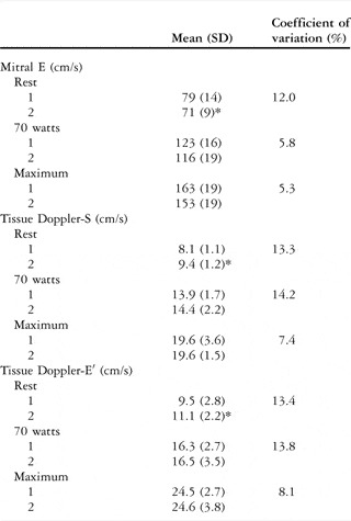
Echocardiographic variables were obtained at maximal exercise for all subjects in both testing sessions. Mean values and coefficients of variation are outlined in Table 1. No significant mean differences were observed between groups for all three variables during submaximal work (70 watts) or maximal exercise (p greater than 0.05). Differences in values at rest between tests 1 and 2 were small but statistically significant. At maximal exercise, coefficients of variation ranged from 5.3% to 8.1%. The average ratios of velocity values at maximal exercise to those at rest on the two tests were 2.10 (standard deviation 0.25) and 2.19 (standard deviation 0.18) for mitral E, 2.56 (standard deviation 0.63) and 2.09 (standard deviation 0.24) for tissue Doppler-S, and 2.78 (standard deviation 0.71) and 2.32 (standard deviation 0.54) for tissue Doppler-E′ (for all comparisons p greater than 0.05).
Mean (SD) values and coefficient of variation on repeat maximal exercise testing for Doppler echocardiographic measures of ventricular systolic and diastolic functions.

*p<0.05
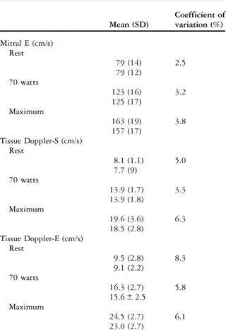
Limits of agreement by Bland–Altman analysis for differences between trials 1 and 2 at maximal exercise were plus 9 and minus 27 centimetres per second for mitral E, plus 3.5 and minus 4.5 centimetres per second for tissue Doppler-E′, and plus 2.9 and minus 2.9 centimetres per second for tissue Doppler-S. Repeated measurements by the same observer, who made the analyses for test 1 values (TWR), are presented in Table 2. We observed no significant differences between any of the mean values in the three measurement conditions. Coefficients of variation at maximal exercise ranged from 3.8% to 6.3%.
Intra-observer reliability for two separate velocity measurements during test 1.

p < 0.05 for all comparisons
Discussion
This study indicates the feasibility and acceptable levels of reproducibility of Doppler echocardiographic techniques in assessing ventricular functional responses to maximal exercise in non-obese adolescent males. No significant differences were observed for mean velocities of mitral E, tissue Doppler-S, and tissue Doppler-E at submaximal work and peak exercise with coefficients of variation at maximal effort of 5.3%, 7.4%, and 8.1%, respectively. Intra-observer reproducibility was high, with coefficients of variation during exercise ranging from 3.2% to 8.3%.
It should be emphasised directly that such findings were observed in a group of athletic, lean young male adolescents. Whether similar supportive findings can be expected in groups of subjects less favourable to ultrasound imaging, that is those who are significantly obese, remains to be established.
Similarly, in the only other study to examine reproducibility of Doppler techniques for measuring ventricular function during maximal exercise by Bougault et al, subjects were restricted to non-obese young adult males described as “moderately to highly echogenic”.Reference Bougault, Nottin, Doucende and Obert7 In that study, coefficients of variation were slightly lower than in this study (2.8–5.1%). The difference may be explained by the fact that these subjects were stabilised in a special semi-supine bench rather than by pedalling in an upright cycling position as in this study.
Doppler measures of ventricular function at maximal exercise in this study mimic those reported in previous investigations using similar methodology. Peak mitral E averaged 152 centimetres per second and 159 centimetres per second in the studies of Rowland et alReference Rowland, Garrard, Marwood, Guerra, Roche and Unnithan5 and Bougault et al,Reference Bougault, Nottin, Doucende and Obert7 respectively, compared to 163 and 153 centimetres per second in the two exercise bouts in this study. Values of tissue Doppler-E′ at maximal exercise were 30 centimetres per second and 33 centimetres per second in those two studies, respectively, somewhat greater than the average of 25 centimetres per second in this report.
In the report by Rowland et al,Reference Rowland, Garrard, Marwood, Guerra, Roche and Unnithan5 mitral E velocity rose by a factor of 2.1 from rest to maximal exercise, identical to that described by Bougault et al.Reference Bougault, Nottin, Doucende and Obert7 This compares to average ratios of 2.1 and 2.2 in the two tests in this study. Bougault et alReference Bougault, Nottin, Doucende and Obert7 reported a mean maximal:rest ratio for tissue Doppler-S of 1.9, while in this study the values were 2.6 and 2.1. Tissue Doppler-E′ ratio averaged 2.8 and 2.3 in the two exercise bouts in this report, compared to 2.3 and 1.7 described by Rowland et alReference Rowland, Garrard, Marwood, Guerra, Roche and Unnithan5 and Bougault et alReference Bougault, Nottin, Doucende and Obert7.
The similar values of maximal:rest ratios in Tissue Doppler-S and tissue Doppler-E′ in this study support the concept that increases in systolic and diastolic function should be expected to parallel each other in response to the cardiac demands of increasing exercise intensity. The number of subjects was, however, too small to permit correlational analysis.
The explanation for the magnitude of variability in test–retest studies of ventricular function with exercise was not addressed in this study. Previous studies of cardiac variables with exercise, including not only myocardial function but stroke volume and cardiac output, typically exhibit variability of 5–10%.Reference Rowland, Melanson, Popowski and Ferrone9–Reference Rowland and Obert11 Effects of body motion and increased ventilation during high-intensity exercise have been assumed to contribute to difficulties in making such measurements echocardiographically. Nevertheless, the consistency of such variation across different measurement modalities raises the possibility that normal biological variation may contribute significantly to the extent of test–retest variability.
In summary, this study revealed acceptable levels of test–retest reproducibility of Doppler measures of ventricular function during maximal upright exercise in healthy adolescent males. These findings support the utility of this technique in assessing myocardial health in young patients with cardiac disease.