INTRODUCTION
Reptiles represent a very diverse group of vertebrates. However, little information is available about their parasites in comparison with mammals and birds. Yet the diversity (numbers of genera and species) of reptilian blood parasites is greater than that of mammals and birds. Lower mobility of terrestrial reptiles, more restricted habitats and greater phyletic age may represent the major factors in the increased taxonomic diversity of their parasites (Telford, Reference Telford2009).
The genus Hepatozoon Miller, 1908 (Apicomplexa: Adeleida), one of six genera of blood parasites belonging to the family Hepatozoidae, is the most common intracellular protozoan parasite found in snakes (Wozniak et al. Reference Wozniak, Telford and McLaughlin1994; Smith, Reference Smith1996). Their heteroxenous life cycle includes an intermediate vertebrate host and a blood-feeding definitive invertebrate host, represented by a wide spectrum of invertebrates including ixodid and argasid ticks, triatomid bugs, leeches, flies, sucking lice, fleas, sandflies and mosquitoes of the genera Culex, Aedes and Anopheles (Smith, Reference Smith1996). Sporogonic development usually occurs in the gut wall or haemocoel of the thorax and abdomen (Sloboda et al. Reference Sloboda, Kamler, Bulantová, Votýpka and Modrý2007) of the haematophagous invertebrate host. In reptiles gamonts parasitize erythrocytes, although in other species of Hepatozoon infecting mammals and birds, they mainly infect leukocytes (Telford, Reference Telford2009). Transmission of the parasite to vertebrates occurs by ingestion of infected invertebrates (primary transmission), ingestion of intermediate prey (secondary transmission) or across the placenta in certain species (Clark, Reference Clark1958; Murata et al. Reference Murata, Inoue, Tateyama, Taura and Nakama1993; Smith, Reference Smith1996; Vilcins et al. Reference Vilcins, Üjvári, Old and Deane2009; Tomé et al. Reference Tomé, Maia and Harris2012).
Despite the high intermediate host diversity, including all tetrapod orders and most classes, for Hepatozoon blood parasites, the morphology of gamonts present in reptile blood cells are often very similar (Telford, Reference Telford1984; Levine, Reference Levine1988; Smith, Reference Smith1996; Harris et al. Reference Harris, Maia and Perera2011). Previously identification of Hepatozoon parasites was based primarily on morphological and morphometric characterization of gamonts as well as sporogonic stages in invertebrates (Sloboda et al. Reference Sloboda, Kamler, Bulantová, Votýpka and Modrý2007; Telford, Reference Telford2009; Maia et al. Reference Maia, Harris and Perera2011). Scientific names of species were sometimes given according to reptile species infected (Ball et al. Reference Ball, Chao and Telford1967; Telford et al. Reference Telford, Wozniak and Butler2001, Reference Telford, Moler and Butler2008, Reference Telford, Moler and Butler2012; Sloboda et al. Reference Sloboda, Kamler, Bulantová, Votýpka and Modrý2007; Telford, Reference Telford2010). For this reason many species have been described, but it is difficult to know how distinct they are, or their phylogenetic relationships.
Another way to characterize Hepatozoon sp. in reptiles is based on molecular methods such as polymerase chain reaction (PCR) using Hepatozoon-specific primers. Universal primers targeting faster-evolving alternative genes remain elusive, and therefore primers which amplify the slowly evolving 18S rRNA gene of Hepatozoon sp. represent the commonest molecular method for parasite detection (Barta et al. Reference Barta, Ogedengbe, Martin and Smith2012).
Molecular detection allows scientists to gain data about phylogenetic relationships between parasites from various reptile species and to examine possible patterns of co-evolution with the vertebrate host; something suggested to occur in other Apicomplexan parasites (Allen and Little, Reference Allen and Little2009). Thus two distinct methods in parasite detection exist. However, since many parasites identified by microscopic observation are often not assessed using molecular markers and vice versa, comparisons are limited.
When sensitivity of molecular methods and microscopic observation of blood smears was analysed, results have varied. Some studies revealed that PCR is more reliable (3- to 10-fold more sensitive) than evaluation of blood smears, possibly due to using slides stained by Giemsa several years after their preparation (Richards et al. Reference Richards, Sehgal, Jones and Smith2002) or screening too small a number of the cells for detection of the parasite (Jarvi et al. Reference Jarvi, Schultz and Atkinson2002; Durrant et al. Reference Durrant, Beadell, Ishtiaq, Graves, Olson, Gering, Peirce, Milensky, Schmidt, Gebhard and Fleischer2006). However, in other assessments microscopy was more sensitive than PCR; both of these methods underestimate prevalence of infection (Valkiūnas et al. Reference Valkiūnas, Iezhova, Križanauskienė, Palinauskas, Sehgal and Bensch2008). In reptiles the absence of blood parasites by microscopic evaluation of blood smears clearly does not always reflect the real status of infection (Üjvári et al. Reference Üjvári, Madsen and Olsson2004; Criado-Fornelio et al. Reference Criado-Fornelio, Rey-Valeiron, Buling, Barba-Carretero, Jefferies and Irwin2007, Reference Criado-Fornelio, Buling, Casado, GIimenez, Ruas, Wendt, da Rosa-Farias, Pinheiro, Rey-Valeiron and Barba-Carretero2009).
The majority of Hepatozoon spp. from reptiles were described only according to examination of blood smears, and most data concern those with hosts from North America. So far, about 25 species including Hepatozoon rarefaciens (Sambon and Seligman, Reference Sambon and Seligman1907), H. fusifex (Ball et al. Reference Ball, Chao and Telford1969), H. caimani mocassini (Laveran, Reference Laveran1902), H. caimani (Carini, Reference Carini1909), H. sipedon (Smith et al. Reference Smith, Desser and Martin1994) and H. caimani punctatus (Telford et al. Reference Telford, Wozniak and Butler2001) were described from this region according to morphology of gamonts and sporozoites. Much less is known about Asian species of Hepatozoon – only H. mesnili (Robin, Reference Robin1936) from Gekko verticillatus and H. octosporei (Shanavas and Ramachandran, Reference Shanavas and Ramachandran1990) from Mabuya carinata were described. African species of parasites are represented by H. ayorgbor (Sloboda et al. Reference Sloboda, Kamler, Bulantová, Votýpka and Modrý2007), H. domerguei (Landau et al. Reference Landau, Chabaud, Michel and Brygoo1970), H. pettiti (Hoare, Reference Hoare1932), H. gracilis (Bashtar et al. Reference Bashtar, Abdel-Ghaffar and Shazly1987), H. langii and H. vascuolatus (As et al. Reference As, Davies and Smit2013).
In addition, parasites identified only as Hepatozoon sp. have been reported in various African reptiles represented by the genus Hemidactylus, Naja, Psammophis, Ptyodactylus, Tarentola and Varanus (Saoud et al. Reference Saoud, Ramdan, Mohammed and Fawzi1995, Reference Saoud, Ramdan, Mohammed and Fawzi1996; Ramdan et al. Reference Ramdan, Saoud, Mohammed and Fawzi1996; Hussein, Reference Hussein2006).
Molecular detection of Hepatozoon sp. from North African reptile species was first determined by Maia et al. (Reference Maia, Harris and Perera2011). Parasites were detected in lizards of the genus Atlantolacerta, Chalcides, Eumeces, Podarcis, Scelarcis and Timon, from Morocco. Snakes from the Maghreb, Mediterranean region and Caucasus (Hemorrhois and Malpolon) also tested positive for the presence of Hepatozoon parasites using the same 18S rRNA marker (Tomé et al. Reference Tomé, Maia and Harris2012).
Surprisingly, given the wide distribution of snakes in Europe, little information is still available about blood parasites from this zoogeographical region, particularly from non-Mediterranean regions.
The aim of the present study was to detect the presence of blood parasites in collected samples, to determine the taxonomy and phylogenetic relationship of parasites in various species of snakes from several regions (Africa, America, Asia and central Europe) using both molecular and microscopic examination methods. In this way we aim to test if different lineages of parasites occur in geographically diverse regions. Further, since snakes may be parasitized by the same genetic lineages as those from their prey (Tomé et al. Reference Tomé, Maia and Harris2012), this can be further assessed by a wider sampling of snake groups.
MATERIALS AND METHODS
Sample collection
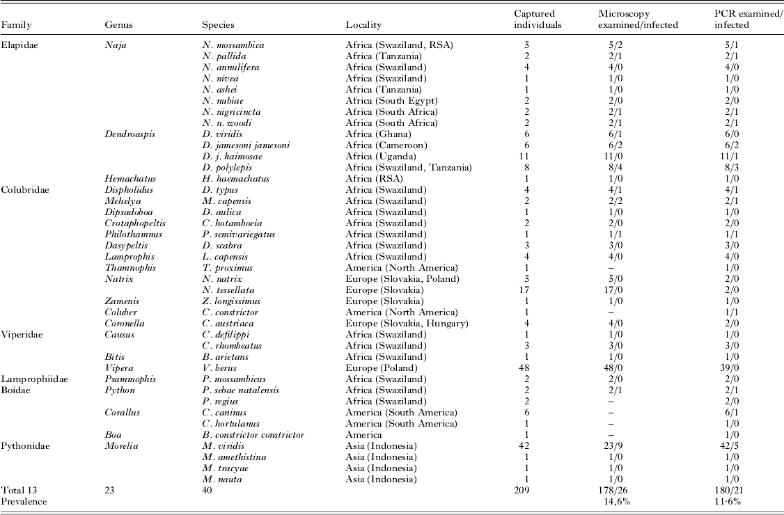
Biological samples from snakes were collected during field expeditions and from snakes captured in nature for commercial purposes. Field expeditions were conducted in southern Africa (Swaziland), central Europe (Poland, Hungary and Slovakia) and North America (USA). Snakes from captivity had been captured in the wild and imported to Slovakia and the Czech Republic as part of the exotic pet trade. These snakes originated from different localities in Africa (Cameroon, Egypt, Ghana, South Africa, Tanzania and Uganda), America (Central and South America), and Asia (Indonesia). Details are given in Table 1.
List of examined snake species

Snakes were captured with the help of snake specialists, by hooking or by hand. Blood from snakes was taken via a ventral puncture of the caudal vein. Blood for molecular analysis was stored in tubes with sodium citrate. Some animals were road kills or perished imported animals, in which case (tail tip, liver or spleen) stored in 70% ethanol was used. Animals captured in the field were immediately released after sampling at the capture place.
Slide examination
Blood smears were made and air-dried immediately in the field. In the laboratory staining was performed using May–Grünwald (10 min) and Giemsa solution (30 min) and examined with a light microscope at ×200 magnification. Approximately 50 microscopic fields on each smear were examined for the presence of blood parasites. When no parasites were detected by this method, the smear was considered negative. Intensity of infection (parasitaemia) was estimated for each positive individual as the percentage of infected red blood cells found in an estimated number of 10 000 cells.
A total of 209 individuals comprising 23 genus and 40 species of snakes were examined for blood parasites (Table 1).
DNA extraction, amplification and sequencing
DNA isolation (blood or tissue) was carried out using a commercial kit (NucleoSpin Blood and Tissue, Macherey-Nagel, Düren, Germany) according to the manufacturer's protocol. Isolated DNA was stored at −20 °C.
PCR reactions were run in a 25 μL reaction mixture from the Taq DNA Polymerase kit (Quiagen, Hilden, Germany) containing 2·5 μL 10×PCR Coral Load PCR Buffer (15 pmol μL−1 MgCl2); 1 μL MgCl2 (25 pmol μL−1); 0·5 μL dNTPs (10 pmol μL−1); 0·5 μL of each primer (10 pmol μL−1) (Integrated DNA Technologies, Leuven, Belgium); 0·125 μL Taq DNA Polymerase (5 U μL−1); 14·875 μL water for molecular biology (Water, Mol Bio grade DN-ase, RN-ase and Protease-free; 5Prime, Hamburg, Germany) and 5 μL of DNA. Verification that the isolated DNA was appropriate for PCR amplification was assessed using primers, which amplify the 12S rRNA (Humair et al. Reference Humair, Douet, Cadenas, Schouls, Van De Pol and Gern2007). Molecular detection of Hepatozoon sp. was made by PCR reactions with HEMO1/HEMO2 primers targeting part of the 18S rRNA gene. The prepared mix was preheated to 95 °C for 5 min. Amplification was performed in 35 cycles at 94 °C for 30 s, 48·8 °C for 30 s and 72 °C for 1 min, followed by a final extension at 72 °C for 5 min (Perkins and Keller, Reference Perkins and Keller2001).
Negative and positive controls were included in each reaction. Amplicons were separated on a 1·5% agarose gel (Sigma-Aldrich, Buchs, Switzerland) in 1× TAE Buffer (40 mm Tris, pH 7·8, 20 mm acetic acid, 2 mm EDTA). The gel was stained by Good View nucleic acid stain (Ecoli, Bratislava, Slovak Republic) and afterwards was visualized using a UV transilluminator. Obtained positive PCR products (approximately 900 bp) were purified by Gen Elute™ PCR Clean-Up Kit (Sigma-Aldrich, Buchs, Switzerland) and sequenced by a commercial sequencing facility (University of Veterinary Medicine, Košice, Slovak Republic), with all fragments sequenced in both directions.
Phylogenetic analyses
Obtained sequences were visualized, edited using MEGA 4 and checked by eye. Checked sequences were BLASTed in GenBank and all of them matched with sequences of Hepatozoon sp. from various hosts. Following Maia et al. (Reference Maia, Harris and Perera2011) and Tomé et al. (Reference Tomé, Maia and Harris2012), 47 related Hepatozoon sequences were downloaded and aligned using Clustal W.
Maximum likelihood (ML) analysis with random sequence addition (100 replicate heuristic searches) was used to estimate evolutionary relationships using the software PAUP v4.0b10 (Swofford, Reference Swofford2002). Support for nodes was estimated using the bootstrap technique (Felsenstein, Reference Felsenstein1985) with 100 replicates. The model of evolution employed was chosen using the AIC criteria carried out in Modeltest 3.06 (Posada and Crandall, Reference Posada and Crandall1998).
Bayesian analysis was implemented using Mr. Bayes v.3.2 (Huelsenbeck and Ronquist, Reference Huelsenbeck and Ronquist2001) with parameters estimated as part of the analysis. The analysis was run for 10 000 000 generations, saving one tree every 1000 generations. The log-likelihood values of the sample point were plotted against the generation time and all the trees prior to reaching stationarity were discarded as burn-in samples. Remaining trees were combined in a 50% majority consensus tree. Haemogregarina balli (Paterson and Desser, Reference Paterson and Desser1976) HQ224959 and Dactylosoma ranarum (Labbé, Reference Labbé1894) HQ224957, HQ 224958 were used as out-groups following Barta et al. (Reference Barta, Ogedengbe, Martin and Smith2012).
RESULTS
Microscopic examination
A total of 178 blood smears obtained from 209 individual snakes, representing 40 species from different localities, were examined: 77 from African snakes, 75 from European snakes and 26 from Asian snakes. The presence of unicellular parasites localized in red blood cells was found in 26 samples (14·6% prevalence) belonging to 12 species of snake (Table 1); all 75 snakes from Europe belonging to five species were infection-free.
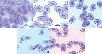
The presence of multiple parasites in a single host cell was also observed. Two individuals within one red blood cell were found in Dendroaspis polylepis and Morelia viridis; moreover, simultaneously both two and three individuals were also found in one specimen of Mehelya capensis (Fig. 1).
The presence of blood parasites in erythrocytes of snakes (A, B, M. capensis; C, D. polylepis; D, E, M. viridis).

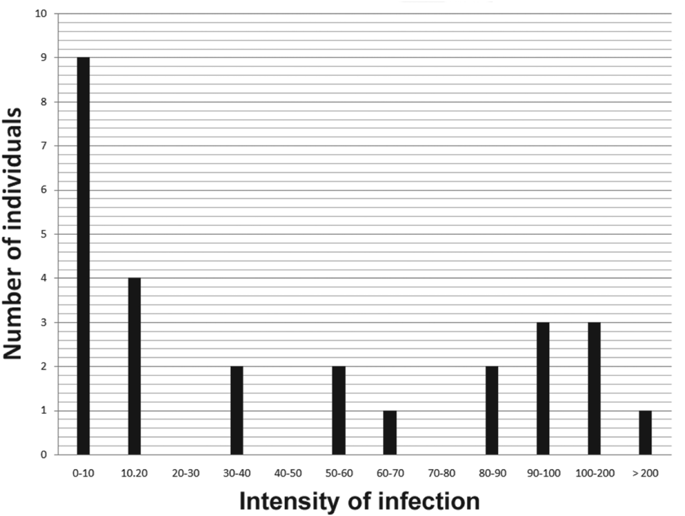
Intensity of infection ranged from one to 1433 infected cells per 10 000 erythrocytes (Fig. 2). Mean intensity was 96 infected cells per 10 000 erythrocytes.
Distribution of number of blood parasites in studied reptiles.

Molecular analysis
Out of 180 samples, the fragment of 18S rRNA was amplified in 21 (prevalence 11·6%) samples by PCR using Hepatozoon-specific primers: 14 African snakes, five Asian snakes and two American snakes. Nine isolates were used for phylogenetic analyses, including Hepatozoon sp. from Coluber constrictor, Corallus caninus, D. polylepis, D. jamesoni kaimosae, D. j. jamesoni, M. capensis, M. viridis, Python sebae natalensis and Philothamnus semivariegatus.
We did not amplify the fragment of 18S rRNA in any of 46 snakes belonging to five species collected in Europe.
In the remaining specimens the sequence quality was too low for them to be included in the phylogenetic analyses.
Comparison of PCR and microscopic examination
Comparison of the two methods used revealed some discrepancies. Parasites found in 10 blood smears by microscopic observation, failed to amplify using these primers. On the other hand, two individual snakes (D. j. kaimosae and M. viridis) were found to be infected through PCR, but no gamonts were observed during the microscopic examination.
Phylogenetic analysis
Both phylogenetic analyses (Bayesian and Maximum likelihood) revealed similar phylogenetic patterns. Results revealed the existence of five haplotypes divided to four main lineages: the first included species of parasites with hosts from Africa and North America (Coluber, Corallus, D. j. jamesoni, Mehelya and Python), the second lineage with an Asian host species (Morelia), and the third and fourth comprised other parasite species with African hosts (D. j. kaimosae, D. polylepis and Philothamnus) (Fig. 3).
Estimate of relationships of Hepatozoon species based on 18S rRNA sequences. Numbers above and below nodes indicated ML bootstrap support and Bayesian posterior probability values, respectively. Sequences amplified in this study are in bold and resulted in existence of five haplotypes divided to four main lineages (indicated by 1–4 behind vertical line).

Analysis revealed that Hepatozoon sp. of snakes from the first lineage clustered in a clade with parasites found in geckos (Ptyodactylus, Quedenfeldtia and Tarentola) from Africa, in rodents (Abrothrix and Clethrionomys) from Chile, Spain and Thailand, but also related to H. ayorgbor isolated from Python regius. A second lineage with a single Hepatozoon sequence from M. viridis from Asia, was not closely related with other currently known haplotypes and represents the sister taxa to the first lineage with African and American species. The third and the fourth lineages included sequences of parasites from African snakes not related with other African haplotypes used in this study and clustered in individual lineages. The third lineage including Hepatozoon sp. from D. j. kaimosae and D. polylepis is grouped together with parasites of the snake Lycognathophis from the Seychelles. Finally, the fourth lineage falls within parasites from geckos (Quedenfeldtia and Tarentola) from Africa and skink (Mabuya) from the Seychelles. Both lineages (3 and 4) represent sister taxa to lineages 1 and 2, which consist of other African, American and Asian species.
None of the obtained sequences were associated with the clade including Hepatozoon sp. isolated from North African lizards, carnivores (H. canis, H. felis and H. americanum), squirrel and pine marten. Analysis revealed no clear geographical pattern of studied Hepatozoon sp., or correlation between phylogenetic relationships observed and those of the vertebrate host.
DISCUSSION
In general, unicellular intracellular blood parasites are distributed worldwide, although there is only scarce information about their occurrence in particular geographical regions. Our results represent partial data, which amend missing information about the incidence of blood parasites in less studied parts of the world. The present study represents the first assessment of the occurrence of blood parasites in the studied snake species collected from Africa, America and Asia.
We examined snakes from five different continents (Africa, Asia, Europe, North America and South America). Although most available information concerns American species of parasites (based on morphology of parasites), their molecular detection has never been assessed in snakes. We detected Hepatozoon sp. in two individuals.
Parasites from host species from Africa have already been detected using both microscopic (Thiroux, Reference Thiroux1910; Landau et al. Reference Landau, Chabaud, Michel and Brygoo1970; Bashtar et al. Reference Bashtar, Abdel-Ghaffar and Shazly1987; As et al. Reference As, Davies and Smit2013) and molecular (Maia et al. Reference Maia, Harris and Perera2011; Tomé et al. Reference Tomé, Maia and Harris2012) techniques. We extended existing data with sequences of Hepatozoon sp. obtained from six additional snake species predominantly from South Africa.
Asian parasites of reptiles represent an area of research which is poorly studied, with little information available about Hepatozoon sp. found in reptiles (Robin, Reference Robin1936; Shanavas and Ramachandran, Reference Shanavas and Ramachandran1990). In this study we identified a lineage of Hepatozoon sp. from the genus Morelia for the first time.
Prevalence of infection in snakes from individual localities achieved different values. Nevertheless, since only five hosts from America were screened, this could be biased by non-representative sampling. Out of 45 Asian snake species, the presence of blood parasites was observed in 10 individuals (prevalence of 22·2%). For African snake species parasites were observed in 18 individuals from 122 examined (prevalence of 14·7%). Prevalence in the present study is higher than that found in North African reptiles by Maia et al. (Reference Maia, Harris and Perera2011) and Tomé et al. (Reference Tomé, Maia and Harris2012) (5 and 8% respectively, based on molecular detection).
Intensity of infection varied from mild (1–20) to intensive infections (100–157). We found extremely high intensity (1433) in one individual from Africa (M. capensis), a snake bred in captivity. It has been reported that high stress conditions such as predation pressure increases the load of haemogregarines in reptiles (Oppliger et al. Reference Oppliger, Clobert, Lecomte, Lorenzon, Boudjemadi and John-Alder1998; Martin and Lopez, Reference Martin and Lopez1999). Our study was conducted on imported snakes and snakes bred in captivity, where stress may be a driving factor for parasite propagation.
Blood smears made from seven individuals were positive, but no parasites were detected using the PCR methodology applied. The same was previously observed only for one individual of the skink Mabuya wrightii by Harris et al. (Reference Harris, Maia and Perera2011), while otherwise all hosts positive for blood smears were successfully amplified. The opposite situation also occurred; two individuals negative by slide evaluation with positive result of PCR; observed also by Criado-Fornelio et al. (Reference Criado-Fornelio, Rey-Valeiron, Buling, Barba-Carretero, Jefferies and Irwin2007, Reference Criado-Fornelio, Buling, Casado, GIimenez, Ruas, Wendt, da Rosa-Farias, Pinheiro, Rey-Valeiron and Barba-Carretero2009). Likewise Üjvári et al. (Reference Üjvári, Madsen and Olsson2004) detected 75% prevalence of Hepatozoon sp. by microscopy evaluation of slides, and 100% prevalence by PCR, but without sequencing all the positives. However, these primers can amplify other Apicomplexan parasites (e.g. Maia et al. Reference Maia, Perera and Harris2012), so without sequencing positives, PCR amplification alone cannot be considered indicative of positive Hepatozoon infection.
Results of phylogenetic analyses show great variation of Hepatozoon sp. within and between hosts. For instance, closely related haplotypes of Hepatozoon sp. were found in snakes derived from two distinct geographical regions, Africa and America. All of them (lineage 1) are clustered together in the same clade with H. ayorgbor and parasites isolated from African geckos, rodents from Spain, Chile and Thailand. Species of Hepatozoon from most vertebrate groups display low host specificity to their vertebrate hosts; transmission to a different host than those for which they are most adapted could result in spatial or temporal isolation of the parasite from its haematophagous vector or vertebrate host (Smith, Reference Smith1996). Closely related haplotypes of Hepatozoon detected in the present study were also observed in North African lizards (Maia et al. Reference Maia, Harris and Perera2011) and Australian squamate families (Üjvári et al. Reference Üjvári, Madsen and Olsson2004). On the other hand, studies suggesting that Hepatozoon are narrowly vertebrate host specific have also been published (Telford et al. Reference Telford, Wozniak and Butler2001, Reference Telford, Moler and Butler2012). The sister lineages (3 and 4) of clade 1 represent another African species of Hepatozoon isolated from D. j. kaimosae, D. polylepis and Philothamnus which are phylogenetically related with parasites from Africa, Australia and the Seychelles.
The results of the phylogenetic relations again seem to support the hypothesis about prey–predator transmission of Hepatozoon sp. (Tomé et al. Reference Tomé, Maia and Harris2012). The diet of snakes from lineage 1 is very miscellaneous, but part of the snake's diet is represented by rodents and parasites from these form part of the same clade. We observed the same host–predation relationship for lineages 3 and 4 where diet of snakes is composed of other snakes and geckos. One exception represents Hepatozoon sp. amplified from genus Morelia, which formed a new lineage. However, this may be simply because of limited sampling – Hepatozoon sp. sequences from rodents and other hosts from these regions are missing in GenBank. Probably more widespread molecular surveying of vertebrates for Hepatozoon parasites could help explain these intercontinental differences between Hepatozoon sp.
We did not observe correlation between parasite phylogeny and that of the vertebrate host. For example, parasites from two subspecies of D. jamesoni are a part of distinct clades. These findings may be related to geographical isolation of these subspecies, since D. j. jamesoni occurs in the western part of Africa; D. j. kaimosae is distributed on the eastern part of Africa along with D. polylepis.
European snakes (Natrix natrix, N. tessellata, Zamenis longissimus, Coronella austriaca and Vipera berus) captured in Slovakia, Hungary and Poland showed no evidence of blood parasites through either microscopic or molecular examination. Tomé et al. (Reference Tomé, Maia and Harris2012) also found no infections in Natrix or Rhinechis. The presence of parasites in a vertebrate host is influenced by many factors including the occurrence of parasites in the region, method of transmission and other biotic and abiotic factors. As proposed by Tomé et al. (Reference Tomé, Maia and Harris2012), Hepatozoon sp. can infect some snakes through ingestion of other vertebrate hosts, such as mice or lizards. However, Hepatozoon sp. related to those found in exotic snakes was already detected in naturally infected Clethrionomys glareolus from Spain (Criado-Fornelio et al. Reference Criado-Fornelio, Ruas, Casado, Farias, Soares, Müller, Brum, Berne, Buling-Saraña and Barba-Carretero2006) and in laboratory-infected mice (Sloboda et al. Reference Sloboda, Kamler, Bulantová, Votýpka and Modrý2008). In Eastern Slovakia, where our samples originated, few studies concerning the incidence of blood parasites in rodents were carried out (Karbowiak et al. Reference Karbowiak, Stanko, Fričová, Wita, Hapunik and Peťko2009, Reference Karbowiak, Fričová, Stanko, Hapunik and Varfalvyová2010) but none of them revealed the presence of Hepatozoon sp., which supports our findings. Several studies carried out through Europe (Baker, Reference Baker1974; Laakkonen et al. Reference Laakkonen, Sukura, Oksanen, Henttonen and Soveri2001; Karbowiak et al. Reference Karbowiak, Rychlik, Nowakowski and Wita2005) revealed relatively high prevalence of Hepatozoon in rodents based on microscopic examination of blood smears. Collecting and examining of snakes at the same localities where Hepatozoon sp. was detected in rodents should be useful and interesting for future research of blood parasites.
Information about haemogregarines of other components of these snakes’ diet – such as fish and amphibians in central Europe – are missing. From our results we might hypothesize the absence of Hepatozoon sp. in central Europe in these groups.
In general, Hepatozoon sp. is transmitted by vectors including mites, ticks, flies, mosquitoes and other insects (Smith, Reference Smith1996). All of the snakes collected in central Europe were screened for the presence of ectoparasites, but no ectoparasites were recorded (pers. observation). Only one publication about ectoparasites of snakes in this region exists, with Lewin and Grabda-Kazubska (Reference Lewin and Grabda-Kazubska1997) observing one tick and one mite on the body of V. berus in Poland. Ixodes ricinus ticks represent the most numerous species in central Europe with a wide spectrum of hosts including rodents, lizards, carnivores, deer, insectivores and birds. Despite its low host specificity it has never been found on the body of central Europe snake species. Moreover Ophionyssus sp., a typical mite for snakes and lizards, has not yet been recorded as parasitizing European snakes. Unlike the situation in Europe, snakes that live in tropical regions serve as hosts for many species of ticks, such as Amblyomma (Vilcins et al. Reference Vilcins, Üjvári, Old and Deane2009). These species are probably more specific to snake hosts in comparison to I. ricinus ticks. An alternative explanation would be that we examined a non-representative sampling of European snake hosts. More snake individuals from central Europe should be tested for the blood parasites to reveal the real status of the infection.
This work represents a pilot study concerning the presence of Hepatozoon sp. in various snake hosts from different regions of the world. Except for snakes from Europe, the presence of Hepatozoon sp. was observed in hosts from Africa, America and Asia. Phylogenetic analysis revealed relationships between Hepatozoon sp. isolated from studied snake hosts with other already studied snake, mammalian and amphibian hosts. The results highlight regions from where more data are needed, particularly Asia, and indicates that Hepatozoon presence in snakes – and possibly their prey – may be patchily distributed in Europe. It further indicates the lack of host specificity of genetic lineages of Hepatozoon parasites in snakes.
ACKNOWLEDGEMENTS
Capturing lizards and sample collection were carried out with official permission from the Middle Danube Valley Inspectorate for Environmental Protection, Nature Conservation and Water Management (Hungary), Ministry of Environment of the Slovak Republic, and issued by the local ethics committee for animal studies in Poznaň (Poland). Thanks to our colleagues, M. Bona, M. Oselský, C. Koen, M. Hromada and P. Tryjanowski, who helped with the field work.
FINANCIAL SUPPORT
This work was financially supported by the project Environmental protection against parasitozoonoses under the influence of global climate and social changes (code ITMS: 26220220116), supported by the Research & Development Operational Programme funded by the ERDF (0·5) and by the Scientific Grant Agency of the Ministry of Education of Slovak Republic and the Slovak Academy of Sciences VEGA 2/0199/11 and 1/0579/12.