




No CrossRef data available.
Article contents
- Abstract
- INTRODUCTION
- GENOMIC AND TECHNICAL RESOURCES FOR PARASITIC NEMATODES
- NEMATODE PHYLOGENY
- CONSERVATION OF NEMATODE GENOME ORGANISATION
- C. ELEGANS AS A MODEL NEMATODE: LESSONS FROM EVOLUTIONARY DEVELOPMENT STUDIES IN FREE-LIVING NEMATODES
- USE OF C. ELEGANS AS A SURROGATE SYSTEM TO STUDY PARASITE GENE REGULATION
- USE OF C. ELEGANS AS A SURROGATE SYSTEM TO STUDY PARASITE GENE FUNCTION
- EXPRESSION OF A PARASITE GENE TO RESCUE A C. ELEGANS MUTANT PHENOTYPE
- EXPRESSION OF A PARASITE GENE TO RESCUE A C. ELEGANS RNAI PHENOTYPE
- CONSTITUTIVE EXPRESSION OF A PARASITE GENE TO PRODUCE A MEASURABLE PHENOTYPE IN TRANSGENIC C. ELEGANS
- INDUCIBLE EXPRESSION OF A PARASITE GENE TO PRODUCE A VISIBLE PHENOTYPE IN TRANSGENIC C. ELEGANS
- CONCLUSION
- References
The use of Caenorhabditis elegans in parasitic nematode research
Published online by Cambridge University Press: 12 May 2005
Article contents
- Abstract
- INTRODUCTION
- GENOMIC AND TECHNICAL RESOURCES FOR PARASITIC NEMATODES
- NEMATODE PHYLOGENY
- CONSERVATION OF NEMATODE GENOME ORGANISATION
- C. ELEGANS AS A MODEL NEMATODE: LESSONS FROM EVOLUTIONARY DEVELOPMENT STUDIES IN FREE-LIVING NEMATODES
- USE OF C. ELEGANS AS A SURROGATE SYSTEM TO STUDY PARASITE GENE REGULATION
- USE OF C. ELEGANS AS A SURROGATE SYSTEM TO STUDY PARASITE GENE FUNCTION
- EXPRESSION OF A PARASITE GENE TO RESCUE A C. ELEGANS MUTANT PHENOTYPE
- EXPRESSION OF A PARASITE GENE TO RESCUE A C. ELEGANS RNAI PHENOTYPE
- CONSTITUTIVE EXPRESSION OF A PARASITE GENE TO PRODUCE A MEASURABLE PHENOTYPE IN TRANSGENIC C. ELEGANS
- INDUCIBLE EXPRESSION OF A PARASITE GENE TO PRODUCE A VISIBLE PHENOTYPE IN TRANSGENIC C. ELEGANS
- CONCLUSION
- References
Abstract
There is increasing interest in the use of the free-living nematode Caenorhabditis elegans as a tool for parasitic nematode research and there are now a number of compelling examples of its successful application. C. elegans has the potential to become a standard tool for molecular helminthology researchers, just as yeast is routinely used by molecular biologists to study vertebrate biology. However, in order to exploit C. elegans in a meaningful manner, we need a detailed understanding of the extent to which different aspects of C. elegans biology have been conserved with particular groups of parasitic nematodes. This review first considers the current state of knowledge regarding the conservation of genome organisation across the nematode phylum and then discusses some recent evolutionary development studies in free-living nematodes. The aim is to provide some important concepts that are relevant to the extrapolation of information from C. elegans to parasitic nematodes and also to the interpretation of experiments that use C. elegans as a surrogate expression system. In general, examples have been specifically chosen because they highlight the importance of careful experimentation and interpretation of data. Consequently, the focus is on the differences that have been found between nematode species rather than the similarities. Finally, there is a detailed discussion of the current status of C. elegans as a heterologous expression system to study parasite gene function and regulation using successful examples from the literature.
Information
- Type
- Research Article
- Information
- Copyright
- © 2004 Cambridge University Press
INTRODUCTION
Caenorhabditis elegans has become one of the most powerful model organisms in developmental biology. Its use in parasitology research was first considered over a decade ago and the last few years have seen an increasing number of parasitology laboratories developing C. elegans as an experimental tool. There have already been a number of excellent reviews discussing the use of C. elegans as a model for parasitic nematodes and this review does not aim to reiterate that material (Blaxter, 1998; Burglin, Lobos & Blaxter, 1998; Bird et al. 1999; Geary & Thompson, 2001; Hashmi, Tawe & Lustigman, 2001; Brooks & Isaac, 2002). Instead, several key issues and applications will be discussed in detail focusing on the use of C. elegans to examine core biological processes expected, at least to some extent, to be conserved in parasitic nematode species. The ultimate aim of such studies is to identify differences between parasitic nematodes and their vertebrate hosts that can be exploited to develop novel methods of parasite control. The emphasis is on animal and, to a lesser extent, human parasites whereas plant parasitic nematodes are not discussed in any detail. Some of the best examples illustrating the use of C. elegans in parasitology research come from work with Haemonchus contortus, a strongylid nematode of sheep, and this is reflected in the material covered.
The utility of C. elegans as a tool to study parasite gene function is now generally accepted and there are many examples which illustrate its use (Veenstra & Roos, 1994; Britton & Murray, 2002; Kampkotter et al. 2003; Couthier et al. 2004). Although there has been some debate about the validity of C. elegans in parasitology research, it is about as credible to suggest it does not have a role as it is to suggest that yeast genetics does not have a role in mammalian biological research. However, as more parasitology researchers seek to exploit the C. elegans system it seems timely to review our current understanding of the conservation of biology between C. elegans and other nematode species. Consequently, the first part of the review examines the extent to which core biology and genome organisation is conserved across the nematode phylum. Examples are predominantly taken from recent research that has used free-living nematodes for evolutionary development studies. These are very relevant to our attempts to extrapolate information from C. elegans to parasites and provide some important fundamental principles. The aim is to illustrate the complexities involved in interpreting data derived from interspecies comparisons and transformation experiments. In order to explore some key issues, a ‘devil's advocate’ approach has been taken by using examples in which significant inter-species differences at the cellular and molecular level underlie superficially similar processes. It is important to emphasise that the aim is not to suggest that all biological processes and molecules will differ significantly between nematode species, quite the contrary, many aspects of development and gene function are highly conserved, even between nematodes and other phyla (Chang & Sternberg, 1999; Putcha & Johnson, 2004). The examples used have been chosen specifically because they illustrate differences and are hopefully thought provoking for those seeking to perform experiments using C. elegans to elucidate parasite gene function. The second part of the review involves a detailed discussion of the current applications of C. elegans as a surrogate expression system to study parasite gene function and regulation. This hopefully demonstrates the value of C. elegans as an experimental tool to study many different aspects of parasite biology.
GENOMIC AND TECHNICAL RESOURCES FOR PARASITIC NEMATODES
The stimulus for using C. elegans in parasitic nematode research stems from the difficulties in working with the organisms themselves. One of the major problems is the lack of in vitro culture systems and the requirement to propagate life cycles in vivo. For some parasites, model laboratory animal host systems are available but for many species this is not the case. As a consequence, technology and genomic resources lag behind those available for most other pathogen groups. There are EST sequencing projects for over 30 different parasitic nematode species and these represent the major genomic resource currently available. These are publicly accessible through curated databases at http://nema.cap.ed.ac.uk and http://www.nematode.net/. These data-sets, along with the availability of associated cloned DNA, are proving to be extremely valuable for many research groups. The value of these EST data-sets, even in the absence of a full genome sequence, is well illustrated by the proteomic analysis of the excretory secretory (E/S) products of H. contortus (Yatsuda et al. 2003). A total of 107 proteins was identified by searching databases of available H. contortus EST sequences with MALD-TOF peptide mass fingerprint and LC-tandem MS data. However, the size of most parasitic EST datasets is relatively small consisting of just a few thousand sequences in many cases. These are clearly biased towards highly transcribed genes and so the majority of genes with regulatory functions are not represented. For example, out of the current 17269 H. contortus EST sequences, in 4145 clusters, there is not a single member of the GATA transcription factor family of which there are 11 different members in C. elegans. Consequently there is a need for more genomic DNA sequences for many of these parasites. At present Brugia malayi is the only parasitic nematode of humans or animals with a genome sequencing project. This is being performed at the TIGR Institute of Genomic Research and aims to produce 8-fold coverage and is currently approximately at 4-fold coverage (available at http//www.tigr.org/tdb/e2k1/bmal/). The only other human or animal parasitic nematode with a genome mapping project is H. contortus. A collaboration between Glasgow University (Dr J. Gilleard), MRC-LMB (Dr P. Dear), The Moredun Institute (Dr F. Jackson) and The Pathogen Sequencing Unit, Sanger Centre (Dr B. Barrell) is constructing an integrated HAPPY, BAC clone map and polymorphic sequence marker map. This should provide a valuable resource for genetic mapping and full genome sequence in the future.
The major reason for a lack of sequencing of parasitic nematode genomes is their relatively large size and consequently the high costs involved. However estimates of nematode genome sizes vary from 300 Mb for Ascaris suum to 50 Mb for H. contortus (Leroy, Duperray & Morand, 2003). Although this is large in comparison to bacterial and many protozoan genomes, it is not so large as to preclude full genome sequencing. Given their medical and economic importance, the sequencing of several nematode genomes should be feasible with current capacities of major genome sequencing centres, particular for those nematodes with smaller genome sizes such as H. contortus. Until full genome sequence is available the application of modern functional genomic approaches will be severely limited in this important group of pathogens.
In recent years there has been some progress on the development of transgenesis for a number of animal and human parasitic nematode species including Litomosoides sigmodontis (Jackstadt et al. 1999), Ascaris suum (Davis et al. 1999), Brugia malayi (Higazi et al. 2002) and Strongyloides stercoralis (Lok & Massey, 2002) and this has been reviewed elsewhere (Brooks & Isaac, 2002; Boyle & Yoshino, 2003). However these approaches, based on bombardment or microinjection, have only achieved transient transfection and have been confined to the demonstration of successful transformation by reporter gene expression. The application of these techniques to the functional analysis of parasite genes has not yet been achieved. The lack of in vitro culture systems for these parasites suggests that the ability to perform stable transformation in a manner that allows sophisticated experimentation is not likely in the immediate future.
Until very recently there was no method of reverse genetics to study parasitic nematode gene function. However, the development of RNA-mediated interference (RNAi) has revolutionised reverse genetic approaches in C. elegans and many other organisms (Fire et al. 1998; Kamath et al. 2003). There are now two published examples of the successful application of RNAi to parasitic nematodes (Hussein, Kichenin & Selkirk, 2002; Aboobaker & Blaxter, 2003b). RNAi was successfully used to suppress the expression of Nippostrongylus brasiliensis–secreted acetylcholinesterases during in vitro culture, although no visible phenotype was detected (Hussein et al. 2002). The expression of three Brugia malayi genes has also been reduced by soaking adult female worms in dsRNA in a microculture system (Aboobaker & Blaxter, 2003b). In this case, RNAi of two genes, β-tubulin (Bm-tub-1) and the RNA polymerase II subunit (Bm-ama-I) resulted in parasite death in culture. RNAi of the third gene, a microfilarial sheath protein (Bm-shp-1), caused a reduction in microfilarial release from adult female worms and abnormalities in microfilarial morphology. Many laboratories are currently trying to apply RNAi approaches to a variety of parasitic nematode species and the long-term prospects for this approach are very good. The simplicity of the technology makes it likely it will be widely adopted and it should be applicable to even the most difficult experimental subjects.
In spite of the progress outlined above, the tools to study parasitic nematode gene function are still relatively crude and the absence of in vitro culture systems means sophisticated experimentation is unlikely in the foreseeable future. Consequently, the powerful genetic, molecular and genomic systems available in C. elegans offer attractive alternative approaches to study parasite gene function.
NEMATODE PHYLOGENY
The phylum Nematoda is extremely large and diverse and the value of C. elegans in parasitic nematode research will vary depending on its phylogenetic distance from particular parasitic nematode species. Hence the evolutionary relationship between C. elegans and different groups of parasites is an important issue. Recent molecular phylogenetic analysis, based on SSU rRNA sequencing, has produced a new view of nematode phylogeny (Blaxter et al. 1998) and this has been reviewed in detail elsewhere (Dorris, De Ley & Blaxter, 1999). Briefly, it is suggested that there are five major clades designated I to V (see Fig. 1). Most of the clades contain a mixture of free-living nematodes as well as animal and plant parasites suggesting that parasitism arose multiple times during evolution. In contrast, Clade III contains only animal parasites suggesting parasitism may be the ancestral state for this group (Blaxter, 2002). Caenorhabditis elegans is in Clade V which contains the strongylid nematodes, many of which are important parasites of man and animals. For example, the human hookworms Necator and Ancylostoma are in Clade V as well as a very large number of important veterinary pathogens. Most notable of these are the Trichostrongyloidea which include the most important parasitic nematode species infecting domestic livestock such as Haemonchus, Ostertagia, Trichostrongylus, Cooperia and Dictyocaulus. It seems reasonable to assume that C. elegans biology will, in general, have more relevance to these parasites than to more distant relatives such as the Clade III parasites including the ascarid and the filarial nematodes. However, this is a complex issue and needs to be addressed on a case by case basis. At present there are very few studies directly comparing aspects of C. elegans biology with analogous processes in particular parasitic nematode species. In contrast, there has been a recent resurgence of research activity involving evolutionary development studies using C. elegans and a number of free-living nematode species. These studies shed light on the extent to which fundamental biological processes are conserved between different members of the nematode phylum. A selection of examples from this work will be used to illustrate current thinking on the inter-species variability of nematode biology at the organismal, cellular and molecular levels before going on to discuss specific applications of C. elegans to study parasitic nematode biology. Fig. 1 shows the phylogenetic relationship of the key free-living and parasitic nematodes discussed in this review and serves to provide a frame of reference for evolutionary relationships.

Fig. 1. Schematic phylogenetic tree showing the evolutionary relationships of the nematode species discussed in this review. It is adapted from Dorris et al. (1999) and is based on small unit rDNA sequence Blaxter et al. (1998). The genera that are underlined are parasitic nematodes and the genera shown in italics are free-living nematodes that have been subject to evolutionary development studies and comparative analysis with C. elegans.
CONSERVATION OF NEMATODE GENOME ORGANISATION
Conservation of synteny between nematode genomes
Synteny refers to the conservation of gene order between organisms. Most genes are free to move within the genome over evolutionary time with the exception of a few special cases such as the Hox gene cluster (Aboobaker & Blaxter, 2003a). The rate of genome rearrangement varies between taxa and in some groups such as the mammals there is a remarkable level of conservation of synteny between quite distantly related species (Wienberg & Stanyon, 1997; Larkin et al. 2003). An understanding of synteny between genomes can lead to a number of useful applications. For example, syntenic relationships can be used to identify orthologues of genes between species where the sequence divergence is too great to allow standard heterologous probing and PCR-based approaches. The first use of this approach in nematodes was the identification of the sex determination gene tra-2 from C. briggsae (Kuwabara & Shah, 1994; Nilsson, Tiensuu & Tuck, 2000). Sex determination genes evolve very rapidly making heterologous probing approaches very difficult (de Bono & Hodgkin, 1996). The tra-2, ppp-1, art-1 and sod-1 genes are arranged in a syntenic cluster between C. elegans and C. briggsae (Kuwabara & Shah, 1994). This has allowed the cloning the C. briggsae tra-1 gene by hybridisation screening of genomic libraries with the more highly conserved downstream genes followed by chromosome walking (Kuwabara & Shah, 1994). This ‘cloning by synteny’ approach has great potential for the isolation of C. elegans homologues from parasitic nematodes, particularly those with few genomic resources, providing there is sufficient synteny. The demonstration of syntenic relationships is also valuable in the confirmation of putative gene orthologues and the inference of gene function where there has been extensive sequence divergence. Hence the extent of both long-range and short-range synteny between particular parasitic nematodes and C. elegans is a crucial issue that has a significant bearing on the way in which genome information from C. elegans can be exploited to study parasite genomes. At present there is a lack of long-range contiguous genomic sequence from parasitic nematodes and so there is very little information on the extent of synteny between C. elegans and parasitic nematode genomes. The free-living nematodes C. briggsae and Pristionchus pacificus and the parasitic nematode B. malayi are currently the only other nematode species for which contiguous genomic sequence encompassing several genes is available.
In terms of nematode phylogeny, C. briggsae is closely related to C. elegans with a divergence time estimated at approximately 80–110 Myr. The C. briggsae genomic sequence is now 98% complete (http://www.sanger.ac.uk/projects/C_briggsae/) allowing a comprehensive analysis of synteny with C. elegans. Comparison of the C. elegans complete genome sequence with 13% of the C. briggsae genome sequence suggested that there have been 4030 rearrangements between their whole genomes since speciation (Coghlan & Wolfe, 2002) This makes the average size of a conserved segment 53 kb in C. briggsae which would be approximately 10 genes, assuming 1 gene per 5 kb. This estimate has been supported by a recently published analysis of the full C. briggsae genome sequence which estimated just over 4000 rearrangements between the two genomes (Stein et al. 2003). Assuming an 80–110 Myr divergence time, the rate of genome rearrangement (0·5–0·7 breakages/Mb per Myr) is remarkably high, being 4–20 fold higher than that seen in closely related Drosophila species (Coghlan & Wolfe, 2002) and some 1400 to 17000 fold the rate calculated for mammalian species (Ranz, Casals & Ruiz, 2001). Hence, although there is extensive co-linearity between the C. elegans and C. briggsae genomes, these results suggest that nematode genomes are more evolutionarily unstable than those of other metazoa. Recent work on P. pacificus and B. malayi support this view. A 126 kb segment of contiguous sequence was obtained from P. pacificus, estimated to have diverged from C. elegans approximately 100–200 Myr ago (Lee et al. 2003). In the sequenced segment 11 of 20 genes had clear C. elegans orthologues and ten of these were located on chromosome III suggesting some degree of synteny. However most of these genes were spread over 12 Mb of the 13 Mb chromosome with only 3 pairs of genes being syntenic (Lee et al. 2003). Hence many intrachromosomal rearrangements had occurred since P. pacificus and C. elegans shared a common ancestor. An 83 kb segment of contiguous genomic sequence was obtained from B. malayi estimated to have shared a common ancestor with C. elegans 300–500 Myr ago (Guiliano et al. 2002). Potential C. elegans orthologues were identified for 11 of the 12 predicted genes with 10 of these being present on chromosome I again suggesting some degree of synteny. Although eight of these were present in a 2·3 Mb region of C. elegans chromosome I there was significant rearrangement in their respective positions. Hence for both B. malayi and P. pacificus there appears to have been a large number of genomic rearrangements since divergence from the C. elegans common ancestor.
There is a need for more comparative studies between C. elegans and representative members of different taxonomic groups before a generalised view of nematode genome synteny can be formed. However the indications are that synteny may be sufficiently conserved in the nematodes most closely related to C. elegans, such as Clade V nematodes, to be of some value in parasite gene discovery. However, for more distantly related nematodes the high rate of genome rearrangement seen in nematodes may limit the potential to identify parasite genes using positional information from the C. elegans genome. A HAPPY map of the Haemonchus contortus genome is currently being constructed (Paul Dear and John Gilleard, unpublished data) which, together with the sequencing of several BAC clone inserts, should shed further light on this issue for the Strongylid parasitic nematodes.
Conservation of gene structure and organisation
C. elegans has polycistronic gene clusters that are similar to bacterial operons (Blumenthal et al. 2002; Blumenthal & Gleason, 2003). These operons produce polycistronic pre-mRNA, transcribed from a promoter that is 5′ to the gene cluster and this is subsequently processed by 3′ end formation and trans-splicing to produce monocistronic mRNAs. The upstream gene of an operon is transpliced with the 22 nucleotide SL1 spliced leader sequence and the majority of downstream genes trans-spliced with SL2. The remainder of the downstream genes are trans-spliced with one of nine other SL2- like leader sequences (Blumenthal et al. 2002). Around 15% of C. elegans genes are grouped into operons, each of which contain between 2 and 8 genes with an intracistronic spacing of 100–400 bp (Blumenthal et al. 2002). The true functional significance of C. elegans operons and the selectional advantage of this form of genome organisation are not yet entirely clear. The extent to which operons are units of co-regulation is still unclear. Although genes in the same operon are more likely to be co-regulated than randomly chosen genes (Lercher, Blumenthal & Hurst, 2003) there are numerous examples of genes that are co-transcribed in the same operon but have different expression patterns, presumably due to differences in post-transcriptional processing (Blumenthal & Gleason, 2003). Although there are now numerous examples of genes with related functions present in the same operon (Huang, Tzou & Sternberg, 1994; Page, 1997; Blumenthal & Gleason, 2003) there are still many more examples of operons containing genes with no obvious functional relationship (Blumenthal & Gleason, 2003). Hence our understanding of the role of nematode operons is far from complete and it is also unclear how widespread the organisation of genes into operons is within the nematode phylum.
SL1 trans-splicing itself is clearly widespread among nematodes and appears to be an ancient character. It has been demonstrated in many nematode species from across the nematode phylum including Haemonchus contortus (Clade V) Strongyloides redivivus (Clade IV), Brugia malayi (Clade III) and Trichinella spiralis (Clade I) (Blaxter & Liu, 1996; Connolly, Trenholme & Smith, 1996).
The sequence of the 22 nucleotide SL1 splice leader is precisely conserved in all these species although the sequence, copy number and genomic organisation of the SL1 encoding genes themselves are variable between species (Blaxter & Liu, 1996). In contrast to the numerous examples of SL transplicing, SL2 trans-splicing has been found in relatively few nematodes to date, namely C. briggsae, Dolichorhabditis (also called Oscheius), Pristionchus pacificus and Haemonchus contortus. Of these, the organisation of genes into operons has only been demonstrated in C. briggsae, Dolichorhabditis/Oscheius and Pristionchus. Examination of the recently completed C. briggsae genome has revealed that 96% of the 900 known C. elegans operons are present in C. briggsae (Blumenthal & Gleason, 2003). Dolichorhabditis/Oscheius (strain CEW1) was the first nematode outside the genus Caenorhabditis in which operons have been found (Evans et al. 1997). Two genes encoding large subunit ribosomal proteins are separated by approximately 100 bp with the upstream gene predominantly SL1 trans-spliced and the downstream gene predominantly SL2 transpliced. Homologues of these genes also appear to be present in the same operon in C. briggsae and C. elegans (Evans et al. 1997). Very recently operons, and SL2 trans-splicing have also been demonstrated in Pristionchus pacificus a more distant relative of C. elegans estimated to have diverged 100–200 Myr ago (Lee & Sommer, 2003). Aldose reductase is a downstream gene in an operon in P. pacificus, as it is in C. elegans, and is trans-spliced with an SL2-like leader sequence. Interestingly however, the other genes of this operon are not orthologous between C. elegans and P. pacificus. Also several genes known to be in operons in C. elegans appear to be present as monocistronic RNAs in P. pacificus (Evans et al. 1997). Hence, although operons and SL2 trans-splicing are features shared by the last common ancestor of the Diplogastridae and the Rhabditidae not all genes present in operons in C. elegans are organised in the same way in P. pacificus.
An SL2-like trans-spliced leader sequence has also been identified in the parasitic strongylid nematode Haemonchus contortus which differs from the C. elegans SL2 sequence by only a 2 bp inversion (Redmond & Knox, 2001). The H. contortus SL2 gene itself has been isolated and has 66% identity with the C. elegans SL2 gene and 74% identity with the Dolichorhabditis/Oeschius SL2 gene. The H. contortus pepsinogen gene is trans-spliced to a mixture of SL1 and the SL2-like leader sequence. This arrangement is often stated to be a defining characteristic of a downstream gene in an operon in C. elegans. Interestingly however, there is no evidence of a gene within the 2 kb of genomic sequence upstream of the 5′ end of this H. contortus pepsinogen gene. Furthermore, a promoter has been identified in this 2 kb region of H. contortus genomic DNA which, when cloned upstream of a reporter gene and transfected into C. elegans, can drive appropriate tissue specific expression (Britton et al. 1999). Hence it appears that H. contortus pepsinogen is not a downstream gene in an operon in spite of being SL2 transpliced. Low-level SL2 trans-splicing has also been reported in the first gene in an operon in both C. elegans (Hough, Lingam & Bass, 1999) and Dolichorhabditis/Oeschius (Evans et al. 1997) suggesting that the identification of SL2 trans-splicing in itself does not prove the presence of an operon. The most distant nematode from C. elegans to date for which there is evidence of operons is Brugia malayi (Clade 3) (Giuliano & Blaxter, personal communication). Interestingly, the downstream genes in this case were trans-spliced with SL1 whereas SL2 trans-spliced leader sequences could not be demonstrated.
Hence there is evidence, albeit limited at present, that operons may be present quite widely within the nematode phylum. The small number of examples to date may simply reflect the limited genomic sequence available from nematodes other than C. elegans and C. briggsae. However the extent to which the organisation of particular operons are conserved between nematode species is at present unclear.
C. ELEGANS AS A MODEL NEMATODE: LESSONS FROM EVOLUTIONARY DEVELOPMENT STUDIES IN FREE-LIVING NEMATODES
The relevance of C. elegans biology to that of parasitic nematodes is a subject of continuous debate. On the one hand, the remarkable extent to which core developmental and cellular mechanisms are conserved across large evolutionary distances has been one of the key revelations in biology over the last decade. This argues that a significant amount of biological information from C. elegans research should be highly relevant to that of parasitic nematodes. On the other hand C. elegans is a free-living nematode existing in a completely different environment from that of parasitic nematodes. It is obvious that there will be many biological processes that are specific to particular groups of parasites. This is clearly reflected by EST sequencing projects that have revealed tremendous genomic diversity between parasites across the nematode phylum. For example, 50% of Trichuris muris ESTs have no match in C. elegans (Blaxter et al. 1998) and 34% of 152 genes from the filarial nematode Litomosoides sigmodontis are not found either in C. elegans or in other filarial EST datasets (Allen et al. 2000). Clearly the relevance of C. elegans to parasitic nematode research depends on the particular aspect of biology that is being considered. Many key aspects of biology clearly differ between C. elegans and parasitic nematodes such as mode of feeding, exposure to host immune system, migration within the host and use of an intermediate host, to name but a few. Consequently there are severe limitations to the use of C. elegans as a model for those aspects of biology that are specifically associated with parasitism. It seems equally obvious that many aspects of biology will be shared between C. elegans and parasitic nematodes but are divergent from mammalian host species. It is in these areas that C. elegans offers greatest promise as a tool to study parasite biology and the implications for the identification of novel control targets are intuitively obvious.
In recent years our view of nematode diversity has changed considerably and it has become apparent that the nematode phylum is both evolutionarily ancient and extremely diverse. This is a critical issue for those seeking to use C. elegans as a model for other nematode species. The relationship between C. elegans and C. briggsae provides a good illustration of this point. These two nematode species are virtually indistinguishable in morphology and, being soil-dwelling, would appear to occupy a very similar environmental niche. The best estimate of divergence times of these two species, based on a comparison of 338 sets of orthologous genes, is between 80–110 Myr (Stein et al. 2003). This compares to an estimated human/mouse divergence time of 65–75 Myr (Waterston et al. 2002). These evolutionary relationships suggest that biological data cannot be automatically extrapolated between these two nematode species that appear so similar on superficial examination. Recent evolutionary development studies, using a number of free-living nematode species, have also uncovered previously unapparent diversity in core developmental processes between what are considered to be closely related nematodes species. Free-living nematodes such as members of the genera pristionchus and Oscheius are being developed as ‘satellite organisms’ of C. elegans to study mechanisms of evolutionary change. Like C. briggsae, these nematodes are anatomically very similar and often inhabit very similar environments to C. elegans and so might be expected to be more similar to C. elegans than parasitic nematodes of equivalent phylogenetic distance. These evolutionary studies give an indication of the amount of variation from C. elegans that might be expected in a particular taxonomic group without the additional complications of a parasitic life-style. They also shed light on which tissues and processes are most likely to be conserved between nematode species over a given evolutionary distance. For example molecular mechanisms of sex determination tend to be poorly conserved (de Bono & Hodgkin, 1996; Haag, Wang & Kimble, 2002) whereas mechanisms of endodermal differentiation are more highly conserved (Goldstein, 2001). The examples discussed below have been chosen because they highlight the variation in developmental processes that can occur between nematode species and illustrate the importance of careful experimentation before information is extrapolated from C. elegans to parasitic nematodes.
Evolution of cell fate specification and cell lineage patterns
A striking example of developmental variation between nematode species is seen in mechanisms of early cell fate specification. One of the key conclusions from the classic work of Sulston and co-workers was that the cell lineage of C. elegans was almost entirely invariant (Sulston et al. 1983). Blastomeres of the early C. elegans embryo display regular reproducible differences in size, position and pattern of division to produce predetermined sets of differentiated descendants. Although cell signalling is increasingly found to play important roles during development of C. elegans, the fates of the early blastomeres are largely set by maternally-derived intrinsic cell factors (McGhee, 1995). For many years it was suggested that the uniform lineage pattern of C. elegans was a general feature of ‘nematode’ development and that it may be a consequence of the relatively small number of cells in the organism. However, more recent work on other nematode species has shown this is not the case. Intracellular labelling of the marine nematode Enoplus brevis (Clade II) with lineage-tracing fluorescent dyes has shown that its cell lineage is not fixed (Voronov & Panchin, 1998). Up to the 8 cell stage, blastomeres are visibly indistinguishable and, with the exception of the endoderm precursor, have no fixed lineage pattern (Voronov & Panchin, 1998). In spite of this, the final cell composition is invariant with developmental decisions presumably being heavily dependent on early cell signalling events.
Enoplus brevis is phylogenetically distant from C. elegans but a closer relative, Acrobeloides nanus (Cephalobidae, Clade IV), also has a strikingly different mode of cell fate specification in the early embryo (Wiegner & Schierenberg, 1998, 1999). In C. elegans specification of the gut founder cell E depends on an inductive signal from an adjacent blastomere at the 4 cell stage of embryogenesis (Goldstein, 1992). In contrast, no such inductive signal occurs in A. nanus. Indeed the production of an E lineage seems to be the default fate of all blastomeres in the three cell A. nanus embryo but the fate is restricted to a single blastomere by inhibitory lateral signalling (Wiegner & Schierenberg, 1998, 1999). Hence, a completely different mode of early cell fate specification results in very similar final body plans in these two nematode species.
Recent work has also shown that the cell lineage patterns during mid and late embryogenesis can differ markedly between nematodes that are relatively phylogenetically close to C. elegans. C. elegans has a polyclonal mode of lineage development where a particular block of the cell lineage (i.e. a group of lineally related cells) gives rise to numerous different cell types (Sulston et al. 1983). In contrast, Halicephalobus sp. (Panagrolaimidae, in nematode Clade IV) has a predominantly monoclonal mode of lineage development where blocks of the cell lineage (i.e. groups of lineally related cells) give rise to cells of one particular tissue type. These different modes of lineage pattern have different benefits and drawbacks and imply that very different development mechanisms operate. The benefit of the monoclonal pattern of development of Halicephalobus is that it requires a more simple system of cell fate specification than does the polyclonal pattern of C. elegans (Houthoofd et al. 2003). The drawback of the monoclonal lineage pattern is that differentiated cells of a particular type are born in regional groups in the embryo and then need to migrate to their final positions. In contrast, the polyclonal lineage pattern of C. elegans appears to be arranged so that cells originate close to their final position. Hence the C. elegans lineage pattern requires a complex mechanism of cell fate specification but a simple system of cell migration control whereas the Halicephalobus lineage pattern requires a more simple mechanism of cell fate specification but a complex signalling system to direct the cross-traffic of large numbers of terminally-differentiated migrating cells. One might predict that this difference would be reflected in the numbers of genes involved in cell fate specification compared to cell signalling in the respective genomes.
Analysis of the lineage of Pellioditis marina (a clade V nematode), an even closer relative of C. elegans, reveals it has a much more similar lineage pattern to that of C. elegans with 95·5% lineage homology (Houthoofd et al. 2003). However, even in this very close relative, there is only 76·4% cell fate homology. In other words almost a quarter of cells with homologous lineage histories have different terminal fate assignments (Houthoofd et al. 2003). Interestingly, the extent of conservation of the lineage origin varies between tissues; the intestine and body wall muscles being highly conserved while the pharynx, epidermis and nervous system are more varied. Whether these differences in the developmental conservation of particular tissue types will be consistent across the nematode phylum will have to await studies on additional nematode species. However, it appears that the endoderm is the most conserved of all the tissue types in developmental terms. In all nematodes examined to date, the endoderm is derived from a clonal lineage from a single blastomere. Also the process of gastrulation is extremely similar in C. elegans and other nematodes even those as phylogenetically distant as C. elegans and E. brevis (Skiba & Schierenberg, 1992). Gastrulation normally starts at the 24–30 blastomere stage when the two endoderm precursor cells (daughters of the original endoderm blastomere) migrate from the surface of the embryo to the inside (Voronov & Panchin, 1998). We have examined endoderm development in the trichostrongylid parasitic nematode Haemonchus contortus (Strongylida, Clade V) and found that gastrulation follows this pattern precisely (Gilleard & Couthier, unpublished data). Also the timing of the H. contortus endodermal cell divisions with respect to the stage of embryogenesis is extremely similar to that of C. elegans, as is the final embryonic endodermal cell number of 20.
In summary, it appears that early embryogenesis, early cell fate specification and the pattern and mode of the cell lineage are very variable between nematode species. The development of the endodermal lineage appears to be more highly conserved. Clearly there is a need for more comparative studies between C. elegans and parasitic nematodes to enable researchers to make informed judgements regarding the relevance of C. elegans to different developmental processes in different parasitic nematode tissues.
Evolution of molecular function
Evolutionary differences in the development of organs, tissue and cells between different nematode species will presumably be reflected in differences in molecular function and in the ways in which different molecules and pathways are employed. Comparative studies on vulval development between C. elegans and the free-living nematode Pristionchus pacificus (Clade V) provide an extremely illuminating example of how molecular function can evolve between two closely related nematode species. Although vulval development may not seem of obvious interest to parasitologists, the work provides an important paradigm for the evolution of gene function that is extremely relevant to those aiming to extrapolate experimental data between different nematode species.
The C. elegans vulva is an extremely useful system for investigating pattern formation and dissecting signal transduction pathways (Greenvald, 1997). It is derived from six specialised cells of the ventral hypodermis (P3.p–P8.p), the so-called ‘vulval equivalence group’ (Fig. 2). These six cells are initially equipotent but respond to several interacting signalling systems, including an inductive Epidermal Growth factor (EGF)-mediated signal from the gonadal anchor cell, to adopt primary (1 °), secondary (2 °) or tertiary (3 °) fates. The descendants of these six cells undergo a complex morphogenesis to form the vulva. The remaining ventral hypodermal cells (P1.p, P2.p and P9.p–P11.p) fuse with the rest of the hypodermal syncitium during the L1 stage. The hox gene lin-39 is a transcriptional regulator that plays a key role at two separate points in C. elegans vulval development (Clark, Chisholm & Horvitz, 1993; Clandinin, Katz & Sternberg, 1997; Maloof & Kenyon, 1998). First, lin-39 expression is required early in larval development to prevent the cells of the vulval equivalence group fusing to the hypodermal syncitium (Clark et al. 1993). This has been shown by the observation that vulval precursor cells fuse to the rest of the hypodermal synctium in lin-39 loss-of-function mutants to result in a vulvaless phenotype. The second function of lin-39 is to act downstream of the EGF-RAS-MAPK signalling pathway in the vulval precursor 1 ° fate cell during vulval induction in the L4 and is absolutely required for the adoption of the 1 ° fate (Clandinin, et al. 1997; Maloof & Kenyon, 1998). At a superficial level, genetic studies on P. pacificus suggest that lin-39 function has been highly conserved during evolution. Loss-of-function lin-39 mutants in P. pacificus have a vulvaless phenotype similar to that seen in C. elegans (Eizinger & Sommer, 1997; Eizinger, Jungblut & Sommer, 1999). However more detailed analysis has shown that the role of lin-39 has changed during evolution. The vulval precursor cells of P. pacificus lin-39 mutants, instead of undergoing cell fusion to the hypodermal syncitium as in C. elegans, undergo programmed cell death (Eizinger & Sommer, 1997; Eizinger et al. 1999). Therefore the function of lin-39 has changed from the inhibition of cell fusion in C. elegans to the inhibition of cell death in P. pacificus. Furthermore, unlike in C. elegans where lin-39 is required twice during vulval development, in P. pacificus, lin-39 is only required once (Sommer et al. 1998). This has been demonstrated by showing that vulval development is normal in P. pacificus lin-39, ced-3 double mutants, suggesting that the prevention of programmed cell death is the only role of lin-39 in P. pacificus vulval development (Sommer et al. 1998). Hence the P. pacificus lin-39 is not required for the vulval precursor cells to respond to the gonadal signal during vulval induction as is the case in C. elegans. This example illustrates how, at the molecular level, the function of a central regulatory gene is quite different in two quite closely related nematodes even though it is involved in the same process with essentially the same outcome.

Fig. 2. Schematic representation of the cells of the ventral larval hypodermis that form the vulva in C. elegans. The vulval equivalence group (P3.p–P8.p) are the precursor cells that form the vulval whereas the remaining cells (marked V) fuse to the hypodermal syncitium to contribute to the hypodermis of the adult worm.
There are several other genes involved in the regulation of vulval development where clear functional differences between closely related nematode species have been identified. Mab-5 is a homeotic gene that is involved in specification of the P(7,8).p vulval precursor cells in both C. elegans and P. pacificus (Jungblut & Sommer, 1998). Genetic evidence suggests that this gene is used twice during vulval development in P. pacificus and only once in C. elegans, precisely the opposite situation to that seen for the lin-39 gene (Jungblut & Sommer, 1998). RNAi studies have shown that the Notch-like receptor glp-1 has an essential role in vulval development in C. briggsae but not in C. elegans (Rudel & Kimble, 2001).
The examples outlined above clearly demonstrate how processes that on superficial examination appear to be highly conserved between nematode species, can differ significantly at the molecular level. They also show that orthologous genes can be utilised in different regulatory pathways and can differ in their precise molecular mechanism between closely related nematode species. However, it is important to emphasise that the examples discussed above have been specifically chosen because they illustrate differences between C. elegans and other nematodes. There are equally other examples where the details of molecular mechanisms are highly conserved. For example, in addition to their roles in vulval development, lin-39, mab-5 and glp-1 also have very highly conserved roles in the development of the ventral nerve cord, male tail bursal rays and germ line respectively (Eizinger & Sommer, 1997; Eizinger et al. 1999; Rudel & Kimble, 2001). The important point is that even for closely related nematodes, it is difficult to predict the likelihood of a particular molecule or process being functionally conserved. Hence there is a need for careful experimentation and comparative analysis when attempting to extrapolate information from C. elegans to parasitic nematode species.
Evolution of gene regulation
Expression patterns of orthologous genes are often conserved, even between distantly related organisms (Davidson, 2001). However, it is apparent that individual cis regulatory elements evolve relatively rapidly and accumulate multiple changes even over a few million years (Ludwig et al. 2000; Stone & Wray, 2001). The mechanisms by which expression patterns are maintained in the face of evolving regulatory elements are poorly understood. This issue has an important bearing on the use of C. elegans as an in vivo system to assay parasite promoter and regulatory element function. There appears to be little functional conservation of cis regulatory elements between C. elegans and distantly related organisms such as Drosophila (Ruvinsky & Ruvkun, 2003). Most Drosophlia enhancer elements that have been tested are not properly recognised in C. elegans. On the other hand, many cis regulatory elements appear to be functionally conserved between C. elegans and its close relative C. briggsae (Krause et al. 1994; Maduro & Pilgrim, 1996; Gilleard, Barry & Johnstone, 1997; Duggan, Ma & Chalfie, 1998). Although parasitic nematode species are more distantly related to C. elegans that is C. briggsae, at least some cis regulatory elements are conserved (Qin et al. 1998; Britton et al. 1999; Shabalina & Kondrashov, 1999; Gomez-Escobar et al. 2002). The use of C. elegans to study parasite promoter function is discussed in detail in a later section of this review. However, as with the evolution of gene function, comparative studies between C. elegans and free-living nematodes, particularly C. briggsae, provide important insights into how gene regulation evolves between nematode species. This has important implications for the interpretation of experiments that use C. elegans as a transgenic system to study parasite promoter function. The comparison of cis regulatory elements between C. briggsae and C. elegans can be considered ‘a best case scenario’ in terms of what we can anticipate for conservation of gene regulation between C. elegans and parasitic nematodes. The evolutionary distance between C. elegans and C. briggsae is thought to be such that their genomes are saturated for selectively neutral mutations and so sequence conservation is considered to indicate functional significance (Shabalina & Kondrashov, 1999; Kent & Zahler, 2000). This is supported by functional studies that demonstrate a significant degree of sequence conservation between many C. elegans and C. briggsae regulatory elements (Krause et al. 1994; Gilleard, Barry & Johnstone, 1997; Harfe & Fire, 1998). However, although regulatory elements can often be identified as conserved blocks of sequence, these blocks are not precisely conserved and in extreme cases regulatory regions can function appropriately in interspecies transformation experiments in spite of very minimal sequence conservation (Marshall & McGhee, 2001; Ruvinsky & Ruvkun, 2003). Hence in the early stages of species divergence, the functional equivalence of enhancers is maintained in spite of the accumulation of sequence differences. This view is consistent with the observation that there is generally little sequence homology between parasite promoters capable of functioning in transgenic C. elegans and the 5′ flanking sequence of the C. elegans orthologue (Gilleard, J. S. and Britton, C., unpublished observations).
Although the regulation of a large number of genes appears to be conserved between C. elegans and C. briggsae, there can sometimes be important differences, even between these closely related nematodes (Marshall & McGhee, 2001; Wang & Chamberlin, 2002; Ruvinsky & Ruvkun, 2003). An interesting example is the lin-48 gene which in C. elegans is expressed in hindgut cells, several neural support cells and the excretory duct cell. In C. briggsae it is expressed in all the same cells except for the excretory duct cell. A regulatory element (Ire2) was found to be essential for excretory duct cell expression in C. elegans. This was not in itself sufficient for excretory duct cell expression but appeared to act in conjunction with several other elements a short distance downstream. Surprisingly, in spite of the lack of excretory duct cell expression in C. briggsae, the Ire2 element was conserved upstream of the C. briggsae lin-48 gene. Furthermore the C. briggsae Ire2 element could direct excretory cell expression in transgenic C. elegans when placed upstream of the other C. elegans elements required for expression. Hence this element had not only retained function but also had the ability to interact with C. elegans regulatory elements that were not conserved in C. briggsae. The ability of the C. briggsae Ire2 element to direct excretory duct cell expression in C. elegans when apparently not utilised for this purpose in C. briggsae is intriguing. A detailed comparison of the regulation of the C. elegans and C. briggsae ges-1 genes has revealed some similar results (Marshall & McGhee, 2001). The endogenous ges-1 gene is expressed in the gut of both species but is expressed in the phraynx and rectum only in C. briggsae. In spite of this, a cryptic enhancer downstream of the C. elegans ges-1 gene, in the same position as the one found in C. briggsae, is capable of activating pharynx and rectum expression in transgenic worms. The function of this cryptic enhancer is revealed in transgenic C. elegans upon deletion of a pair of GATA motifs. Hence, as in the case of lin-48, a functional enhancer has been conserved when apparently not utilised for directing the expression of the endogenous gene in one of the two species. The reason for this is unclear since one might expect that, since these two genomes are saturated for silent mutations, an enhancer that is not utilised would have accumulated sufficient mutations to lose function. One possibility is that both the lin-48 Ire2 element and the ges-1 cryptic pharyngeal enhancer may have roles in other aspects of the expression pattern that are shared between the species and hence still be evolutionarily constrained. The lin-48 Ire2 enhancer has indeed been shown to be necessary for expression of lin-48 in tissues other than the excretory cell although this does not appear to be the case for the ges-1 cryptic enhancer.
The lesson from these impressive studies is that the not all aspects of gene regulation are precisely conserved even between closely related nematode species. Functional regulatory elements can be retained which are effectively historical remnants of gene regulation machinery and may not be directly relevant to the final expression pattern of the endogenous gene. This is, at least in part, a consequence of the modular nature of cis regulatory elements and the fact that the same element may have multiple roles in different aspects of a gene's expression pattern. These findings have important implications for the use of C. elegans as a surrogate transformation system to assay parasite gene promoters and to determine putative parasite gene expression patterns.
USE OF C. ELEGANS AS A SURROGATE SYSTEM TO STUDY PARASITE GENE REGULATION
The ease with which C. elegans can be transformed with DNA, along with the availability of a range of sophisticated expression vectors, has the made the analysis of expression patterns of C. elegans genes a routine procedure. The expression patterns of genes are examined by placing Green Fluorescent Protein (GFP) and lacZ reporter genes under the control of putative regulatory regions in the appropriate expression vectors. A range of vectors, primarily produced in Dr Andrew Fire's laboratory (Carnegie Institute of Washington), are available for this and information on these can be accessed at http://www.ciwemb.edu. Injection of the DNA construct into the adult syncitial gonad leads to the formation of tandem multicopy extrachromosomal arrays that are inherited in a non-Mendelian fashion in a similar way to free duplications. Once transgenic lines are established, the expression of the reporter gene can be easily examined. This approach has become the standard way to examine the expression pattern of C. elegans genes and is actually quicker and easier than generating specific antibodies to localise the endogenous polypeptide. The relative ease of the approach has allowed it to be used for the detailed dissection of regulatory regions of many C. elegans genes (Okkema et al. 1993; Egan et al. 1995; Gilleard et al. 1997). The potential use of C. elegans to determine parasite gene expression patterns and to analyse parasite gene promoters functionally is obvious. A number of parasite promoters has now been assayed using this method (Table 1) and it seems timely to review the ‘state of play’ of this approach. The key issue is whether the expression pattern of a parasite gene is faithfully reproduced by a reporter gene in transgenic C. elegans. This can be broken down into two separate questions: firstly, what regions of a parasite genomic locus are required to encompass all components of the cis regulatory apparatus? Secondly, to what extent are cis regulatory regions of particular parasite genes sufficiently conserved to function appropriately in C. elegans?

It is extremely important to include all regulatory elements of a gene in a reporter gene construct because incomplete promoter regions can lead to either incomplete or ectopic reporter gene expression depending on whether positive or negative regulatory elements are missing (Aamodt, Chung & McGhee, 1991; Egan et al. 1995). Due to the lack of data on parasitic nematode gene regulation we have to rely on the more detailed knowledge of C. elegans gene regulation to make judgements about construct design. For the majority of monocistronic C. elegans genes, the key regulatory elements are located 1–2 kb upstream of the initiator ATG and in most cases this is sufficient to drive appropriate expression of reporter genes. The majority of parasitic nematodes examined to date appear to have genome sizes in the range of 0·5 to 2×10⁸ bp in comparison to the 1×10⁸ bp C. elegans genome (Leroy et al. 2003). If the total gene number for these species is approximately the same as C. elegans, the similarity of gene density would suggest the arrangement of gene regulatory elements might be equally compact in parasitic nematodes. This hypothesis is supported by the ability of relatively short regions of 5′ flanking sequence from a number of parasite genes to drive apparently appropriate reporter gene expression in transgenic C. elegans (Qin et al. 1998; Britton et al. 1999). It is important to emphasise that there are examples of C. elegans genes where important regulatory elements are found more than 6 kb upstream, within introns and even 3′ to the gene (Grandien & Sommer, 2001; Marshall & McGhee, 2001). Also it is important to be aware of the possibility that the gene being examined is downstream in an operon and so the regulatory regions might be far upstream. Consequently, C. elegans researchers generally try to use the maximum amount of 5′ sequence that can be cloned into an expression vector and perform translational fusions in the first, or preferably the second exon, to include at least the first intron. The upper limit of fragment size that can be cloned into most of the C. elegans expression vectors is 6–7 kb. It is advisable that a similar approach is adopted for parasite genes providing sufficient genomic sequence can be obtained. An alternative PCR fusion-based approach for the construction of reporter gene constructs is becoming increasingly popular with C. elegans researchers (Hobert, 2002). The advantage of this approach, which avoids any cloning procedures, is that it is very quick and simple and that the size of the promoter fragment used is not limited by plasmid cloning procedures. The fragment size is only limited by the genomic sequence available and the limitations of long-range PCR. The method involves the generation of two PCR fragments (the promoter region of interest and the appropriate region of a reporter gene expression vector) that overlap by virtue of amprimer tag sequence. These are then fused by a second PCR reaction with nested primers. Amplicons are not purified at any stage and are injected directly into the hermaphrodite gonad. We have used this technique to examine the expression directed by a 3·3 kb promoter fragment of the H. contortus elt-2 gene in transgenic C. elegans and it appears to work just as effectively as cloning-based approaches (Couthier, A., Smith, J. and Gilleard, J. S., personal communication). Another point of consideration concerns the choice of reporter gene expression vector, either for cloning or as a template for the PCR-fusion-based method. Some vectors include sequence encoding a nuclear localisation signal fused to the reporter molecule and some do not. The nuclear localisation of the reporter gene is often beneficial since it generally gives a more defined expression pattern and enables cell numbers to be easily counted. However a non-nuclear localised construct, which results in cytoplasmic localisation of the reporter molecule, can provide valuable additional information. For example, visualisation of cell shape often aids cell identification and axons can be visualised in neuronal patterns. In cases of complex expression patterns it can be very useful to use both types of expression construct.
Several parasitic nematode promoters have now been examined in C. elegans (Table 1). The H. contortus isotype 1 β-tubulin gene (tub-1) was the first example of a parasite gene being expressed in C. elegans under the control of its own cis regulatory elements (Kwa et al. 1995). This was used to provide a functional assay to test putative benzimidazole resistance-conferring alleles from the parasitic nematode H. contortus. Transformation of the C. elegans ben-1 benzimidazole resistant mutant strain with susceptible alleles of the H. contortus tub-1 gene increased the sensitivity of the strain to benzimidazoles. Although experiments were not performed to examine the expression pattern of the parasite gene directly, the ability to modulate the drug sensitivity of C. elegans strongly implies the transgene was appropriately expressed in the correct target tissues. Less than 2 kb of tub-1 flanking sequence was required in the gene construct in order to achieve this. Promoters from two other H. contortus genes as well as a T. circumcincta promoter were assayed using transcriptional fusion reporter constructs in C. elegans (Britton et al. 1999). In all three cases, the spatial tissue distribution of reporter gene expression was as expected from previous knowledge of the parasite gene function. The promoters of two genes that encoded enzymes predicted to be involved in digestion (AC-1 and pep-1) resulted in gut-specific reporter gene expression and a cuticle collagen gene promoter resulted in hypodermal-specific reporter expression (colost-1). We have examined the expression pattern driven by a 3·3 kb 5′ fragment of the H. contortus elt-2 gene, a GATA type transcription factor (Couthier, A., Smith, J. and Gilleard, J. S., unpublished data). In this case we know, from immunolocalisation studies, that the endogenous parasite gene is expressed in the gut throughout development and also in cells either side of the pharynx in late embryos and larval stages (Couthier et al. 2004). The Hcelt-2 reporter gene was expressed in the endoderm and four neuronal cells adjacent to the pharynx in transgenic C. elegans. Hence, in all the above examples, the spatial regulation of genes is well conserved between C. elegans and the strongylid nematodes. In contrast, the regulatory elements of these genes did not result in reporter gene expression that accurately reproduced the temporal expression of the endogenous parasite genes. The AC-1 and pep-1 genes are only expressed in the L4 and adult parasite stages whereas both promoters directed reporter expression in all stages of transgenic C. elegans, including embryos. The T. circumcincta colost-1 gene is expressed in all parasite stages but the promoter only directed reporter gene expression in the L4 stage in transgenic C. elegans. Similarly, the endogenous Hcelt-2 gene is expressed in the gut lineage from early embryogenesis throughout post-embryonic development to the adult stage but the promoter only produced gut expression in the post-embryonic stages. Indeed, only one out of the ten parasite promoters examined to date has produced stage-specific reporter gene expression in transgenic C. elegans that matches that of the endogenous parasite gene. Hence, it appears that the evolutionary divergence of temporal regulatory elements may be greater than spatial regulatory elements and that the C. elegans regulatory machinery is unable to correctly ‘interpret’ temporal regulatory information in many parasite promoters, even from relatively closely related parasites.
Several filarial nematode promoters (Clade III) have also been examined in transgenic C. elegans (Table 1). Although the data-set is small, it appears that spatial regulation may not be as tightly conserved with C. elegans as is the case for the strongylid nematodes. The alt-1 and alt-2 (abundant larval transcript) genes are two genes present in all filarial species that are abundantly expressed in the infective L3 stage and account for >1% of L3 message (Gregory et al. 2000). The expression of alt-1 is entirely restricted to the L3 and alt-2 expression largely so. The polypeptides are located in the glandular oesophagus in both B. malayi and Onchocerca volvulus (Joseph, Huima & Lustigman, 1998; Gregory et al. 2000). However the alt-1 and alt-2 promoters both direct reporter gene expression in the larval and adult gut in transgenic C. elegans (Gomez-Escobar et al. 2002). Hence these promoters do not reproduce the correct spatial or temporal expression pattern in C. elegans. The promoter of the O. volvulus GST1a glutathione-S-transferase gene appears to be an intermediate example in that it reproduces the parasite expression pattern in C. elegans but also expression in additional tissues. Immunogold EM studies have only shown GST1 expression in the parasite hypodermis but both hypodermal and pharyngeal expression is produced by reporter gene fusions in C. elegans (Wildenburg, Liebau & Henkle-Duhrsen, 1998). The regulation of two other filarial genes appears to be more accurately conserved in C. elegans. The B. malayi phy-1 promoter directs hypodermal C. elegans GFP reporter gene expression in all life-cycle stages which matches the expression profile in the parasite (Winter, Myllyharju & Page, 2003). Also the ability of 0·54 kb of 5′ flanking sequence upstream of the B. pahangi hsp-83 gene to direct CAT reporter gene in C. elegans has been examined (Thompson et al. 2001). CAT activity was detected in transgenic worms following a 35 °C heat shock but not in worms grown at 21 °C or following a milder 28 °C heat shock. Although the tissue specificity of the transgene expression was not examined, the study demonstrates that this B. pahangi heat shock promoter responds to C. elegans trans acting factors controlling the heat shock response in the appropriate manner.
In summary, although there are insufficient studies for reliable generalisations, the current data suggest that there is a high degree of conservation of cis regulatory elements that control tissue specific expression between C. elegans and strongylid nematodes (Clade V). In contrast, temporal regulation appears to be less conserved. The situation is less clear for more distantly related nematodes such as the Filariae (Clade III) with some promoters appearing to be conserved and others not. However it is possible that these results may reflect the nature of the genes examined rather reflecting evolutionary significance. For example phy-1 and hsp-83 polypeptides are more highly conserved with their C. elegans homologues than are alt-1 and alt-2 which may, at least in part, explain the differences in the conservation of their regulation. Also, a maximum of 3·3 kB of 5′ flanking sequence has been used for the parasite promoters examined to date and important positive or negative regulatory elements could be missing in some cases. More parasite gene promoters need to be tested in C. elegans in order to answer these questions. Detailed analysis of parasite promoters to identify individual regulatory elements using the C. elegans surrogate system has not yet been attempted. Consequently, the extent to which individual cis regulatory sequences are conserved between C. elegans and particular parasite species is not clear. Certainly, there are no extensive regions of sequence homology when the 5′ flanking sequence of H. contortus genes are aligned with the equivalent region of their C. elegans homologues (Gilleard, J. S. and Britton, C., unpublished observations). This is consistent with the view that individual cis regulatory elements evolve relatively rapidly even when gene regulation is maintained (Ludwig et al. 2000; Stone & Wray, 2001). The only functional result to suggest that individual elements may be sufficiently maintained to be recognised at the sequence level is the demonstration that a GATA motif is essential for the function of the promoter of both the C. elegans cpr-1 gene and its H. contortus homologue in transgenic C. elegans (Britton et al. 1999).
USE OF C. ELEGANS AS A SURROGATE SYSTEM TO STUDY PARASITE GENE FUNCTION
The use of C. elegans as a surrogate expression system to examine parasite gene function is one of the most promising applications of C. elegans in parasitic nematode research. The real power of this approach is that once a parasite gene has been functionally expressed in C. elegans, the full range of molecular, genetic and genomic tools available in C. elegans can be used to study its function. This allows detailed in vivo analysis of parasite genes in a way not currently possible in parasite systems themselves. However, as with all heterologous systems, the validity of such experiments needs to be carefully considered. It is pertinent to illustrate a number of important principles using examples from comparative studies between C. elegans and other free-living nematodes before experiments involving parasite genes are discussed.
The most common in vivo test of gene function in C. elegans is to introduce the gene of interest into the appropriate mutant C. elegans strain to determine whether it can restore wild type phenotype. This approach is routinely used by C. elegans researchers to help identify genes responsible for particular mutant phenotypes and provides a very powerful assay of gene function. Although phenotypic rescue is used to provide evidence, it does not in itself prove that mutations in a particular gene are responsible for a particular mutant phenotype. It is important to be aware that the phenotype produced by a mutation in one gene can, in some cases, be rescued by transformation with a wild type copy of a different gene. For example, if gene A is epistatic to gene B (e.g. acts downstream in the same pathway), overexpression of A may rescue the phenotype produced by a loss-of-function mutation in gene B. A good example of this is the ability of the egl-18 (previously elt-5) GATA transcription factor to partially rescue the lin-39 mutant phenotype (Koh et al. 2002). Alternatively, it is possible for two genes to share sufficient functional properties that one is capable of rescuing the phenotype caused by a mutation in the other (Gao & Kimble, 1995; Li & Greenwald, 1997). The ability of the C. elegans apx-1 gene to rescue the C. elegans lag-2 mutant phenotype, when expressed under the control of the lag-2 promoter, provides a good illustration of this (Gao & Kimble, 1995). The genes apx-1 and lag-2 are transmembrane receptors that are related in sequence and function. However, whilst lag-2 is involved in transducing multiple cell–cell signals in different tissues and at different times of development (Lambie & Kimble, 1991), apx-1 is only involved in a single cell–cell interaction during early embryogenesis (Mello, Draper & Priess, 1994). In spite of this, apx-1 is able to replace lag-2 function if expressed in the appropriate cells. This and other examples (Li & Greenwald, 1997) demonstrate the fact that the ability of a gene to rescue a mutant phenotype does not necessarily prove gene identity. Hence, the ability of a parasite gene to rescue the phenotype of a particular C. elegans mutant, does not necessarily infer an orthologous relationship.
It is also important to be aware that the ability of a parasitic nematode gene to rescue a C. elegans mutant phenotype does not necessarily mean that the gene functions in an identical manner and in an identical pathway in both species. The ability of the P. pacificus lin-39 gene to rescue the C. elegans lin-39 mutant phenotype provides a good illustration of this (Grandien & Sommer, 2001). As described in an earlier section, in spite of very effective interspecies transgenic rescue there are significant differences in the way in which lin-39 functions during the regulation of vulval development in the two species. In C. elegans, lin-39 inhibits cell fusion of vulval precursor cells and transduces an EGF-MAP kinase based signal whereas its only apparent function in P. pacificus is to prevent programmed cell death of the vulval precursor cells. Hence, although this gene has evolved quite different molecular roles there is still sufficient shared functionality for interspecies rescue to work. Conversely, the inability of a parasitic nematode gene to rescue a C. elegans mutant phenotype does not necessarily suggest it is involved in a different process. It may be that interacting molecules and downstream targets have coevolved such that the parasite gene cannot function in the C. elegans molecular environment in spite of having very similar functions in a similar pathway in the parasite. The genes involved in sex determination provide an excellent example of this; fog-3, tra-1 and tra-2 all play similar roles in sex determination in different species of the genus Caenorhabditis. However, C. remanei fog-3 and C. briggsae tra-1 and tra-2 cannot rescue the appropriate C. elegans mutants even when expressed under the control of C. elegans regulatory sequences (Stothard & Pilgrim, 2003). Clear evidence for the coevolution of these molecules exists since a direct interaction of TRA-1 and TRA-2 occurs in both C. elegans and C. briggsae but the molecules cannot interact between species (Haag, Wang & Kimble, 2002). Hence interspecies mutant rescue experiments need to be interpreted with care and in conjunction with other experimental data, such as comparison of expression patterns, to allow meaningful conclusions to be drawn.
Although there are currently only four published examples of functional expression of parasite genes in transgenic C. elegans, they clearly illustrate the potential of the approach. It is worth considering each in turn since they have used different methodology and represent different applications.
EXPRESSION OF A PARASITE GENE TO RESCUE A C. ELEGANS MUTANT PHENOTYPE
The H. contortus isotype 1 β-tubulin gene (tub-1) was the first parasite gene to be functionally expressed in C. elegans (Kwa et al. 1995). The functional assay was the rescue of the C. elegans ben-1 mutant phenotype and this work represents a classic example of how an interspecies mutant rescue experiment can allow the function of a parasite gene to be studied in some detail. Previous work had shown that mutations in the C. elegans ben-1 β-tubulin gene conferred resistance to benzimidazole anthelmintics (Driscoll et al. 1989). Along with equivalent work on fungal β-tubulins, this led to the hypothesis that similar mutations might be responsible for benzimidazole resistance in parasitic nematodes. However this hypothesis could not be functionally tested in parasites due to a lack of transgenic technology. Consequently, Roos and co-workers transformed the C. elegans ben-1 mutant with isotype 1 β-tubulin (tub-1) alleles from both benzimidazole susceptible and benzimidazole resistant Haemonchus contortus isolates. The H. contortus tub-1 allele from the susceptible isolate conferred increased thiabendazole susceptibility to the resistant isolate C. elegans ben-1 mutant. This result not only demonstrated functional expression of the parasite β-tubulin gene but showed it was capable of conferring benzimidazole resistance in vivo. In contrast, the tub-1 allele from a benzimidazole resistant H. contortus isolate had no effect on the ben-1 mutant strain's susceptibility to thiabendazole. However, when this H. contortus resistant allele was first modified by site-directed mutagenesis to change a Tyr to Phe at amino acid position 200, it too could confer increased thiabendazole susceptibility to the C. elegans ben-1 mutant strain. Hence this work also proved the functional significance of a particular point mutation of a parasite gene. Another interesting observation from this work involved the differential effects of benomyl on H. contortus and C. elegans. This benzimidazole is quite toxic to C. elegans but has little effect on parasites such as H. contortus. This could be due to a number of different reasons including differences in drug uptake, metabolism or target binding. Interestingly, although the benzimidazole susceptible H. contortus tub-1 allele increased the sensitivity of C. elegans ben-1 mutants to thiabendazole it had no effect on their sensitivity to benomyl. This strongly implies that differences in benomyl sensitivity between C. elegans and the parasites relates to differences in the binding of the drug to the respective β-tubulin polypeptides. This work provides a paradigm for the use of C. elegans to investigate the molecular basis of differential susceptibility of different parasite species to particular drugs.
Another aspect of this work worth noting is that the C. elegans ben-1 mutant strain was rescued by transformation of an H. contortus tub-1 genomic clone. Correct cis-, and possibly trans-, splicing as well as 5′ and 3′ end formation of the parasite pre-RNA to produce mature mRNA would be necessary to achieve rescue. This implies that not only are the regulatory elements of the H. contortus tub-1 gene sufficiently conserved for the parasite promoter to direct appropriate expression in C. elegans but also other aspects of transcript processing are conserved. This is supported by the expression of a genomic copy of the H. contortus pepsinogen gene under the control of the C. elegans cpr-5 promoter in transgenic C. elegans (Redmond et al. 2001). Although the parasite pepsinogen polypeptide expressed from the transgene was not tested for function, RT-PCR and immunocytochemistry demonstrated that expression from the parasite genomic DNA resulted in correctly processed mRNA. Hence it appears that for strongylid nematodes, such as H. contortus, RNA processing is sufficiently conserved with C. elegans to allow functional expression from a genomic copy of the parasite gene.
EXPRESSION OF A PARASITE GENE TO RESCUE A C. ELEGANS RNAI PHENOTYPE
The tub-1/ben-1 example outlined above illustrates the power of mutant phenotype rescue as an assay of parasite gene function. However, in many cases, this approach can be problematic. For example, there may not be an appropriate mutant strain available for the gene of interest or, if the mutant phenotype is lethal, it will require balancing with an appropriate genomic rearrangement (Edgley et al. 1995). This can require time consuming and relatively complex genetic manipulations to produce an appropriate C. elegans mutant strain in which to perform rescue experiments. A conceptually simple but elegant alternative is to rescue a C. elegans RNAi phenotype with the parasite gene (Britton & Murray, 2002). The effectiveness of this approach relies on the fact that a very high degree of sequence homology is required for a dsRNA molecule to interfere with an endogenous transcript. Hence, in general, the degree of sequence identity between homologous genes in C. elegans and parasites is insufficient to allow dsRNA corresponding to a gene in one species to interfere with the endogenous transcript in the other. This means that a parasite gene can be expressed efficiently from a C. elegans transgene even in the presence of dsRNA corresponding to and interfering with the C. elegans orthologue. Britton & Murray (2002) used this approach to rescue the embryonic lethal phenotype by RNA-mediated interference of the C. elegans cathepsin-L protease cpl-1. A 275 bp dsRNA fragment corresponding to the C. elegans cpl-1 gene, resulted in 99·7% embryonic lethality of F1 progeny when microinjected into the hermaphrodite gonad of wild type C. elegans. However, the same dsRNA fragment only resulted in 2·1% F1 embryonic lethality when microinjected into the gonad of transgenic worms expressing the H. contortus cpl-1 homologue under the control of the C. elegans cpl-1 promoter. Hence the H. contortus cpl-1 gene can functionally replace the C. elegans cpl-1 gene knocked out by RNAi. The variety of ways in which RNAi can be applied to C. elegans, by feeding or soaking for example, makes this a very simple and versatile approach. Also the availability of the full genome sequence and bacterial libraries expressing dsRNA for every C. elegans gene makes it immediately applicable to any parasite gene for which a C. elegans homologue can be clearly identified. It seems likely that this approach will be widely adopted in functional studies of parasite genes.
CONSTITUTIVE EXPRESSION OF A PARASITE GENE TO PRODUCE A MEASURABLE PHENOTYPE IN TRANSGENIC C. ELEGANS
The previous two examples both involve expressing a parasite gene in C. elegans to complement loss-of-function of a C. elegans homologue. This allows conservation of function to be tested and a functional assay for further studies to be developed. A conceptually different approach is simply to overexpress a parasite gene in wild type C. elegans and observe the effect. The advantage of this approach is that it does not require the identification of a clear C. elegans homologue and so it can, in principle, be applied to parasite genes that are functionally divergent from C. elegans genes. The major requirement is to have a hypothesis regarding the possible function of the parasite gene to allow the consequences of overexpression in transgenic C. elegans to be investigated with appropriate assays. A recent example of this approach is the expression of the Onchocerca volvulus (Ov-GST-3) glutathione-S-transferase gene in C. elegans (Kampkotter et al. 2003). Glutathione-S-transferases are involved in major cellular detoxification systems including protection against reactive oxygen species (ROS) (Salinas & Wong, 1999). Part of the parasite killing mechanism of the immune system involves the generation of ROS from inflammatory cells (Rosen et al. 1995) and it has been postulated that parasites use antioxidant enzymes to protect themselves against the host immune response (Callahan, Crouch & James, 1988). The O. volvulus GST (Ov-GST-3) has been implicated in this process because its expression in the parasite is upregulated in response to external oxidative stress to a much greater degree than to internal oxidative stress (Liebau et al. 2000). This has led to the hypothesis that it might have a role in defence against host-derived ROS as opposed to simply protecting against ROS derived as a result of the parasites own metabolic processes. However, although the expression data suggest an interesting correlation, it is not possible to test whether Ov-GST-3 has the ability to protect against in vivo oxidative stress in the parasite. Hence Kampkotter et al. (2003) generated transgenic C. elegans lines expressing Ov-GST-3 under the control of the C. elegans let-858 promoter which directs relatively high levels of expression in most C. elegans tissues (Kelly et al. 1997). Transgenic worms expressing Ov-GST-3 were more resistant to both internal (applied by exposure to juglone) and external (applied by exposure to hypoxanthine/xanthine oxidase) oxidative stress. This effect was abrogated following interference of transgene expression by RNAi, demonstrating the protective effect was due to expression of the parasite GST in C. elegans tissues. Interestingly the protection against the external oxidative stress was higher than that against the internal oxidative stress consistent with Ov-GST function being to protect the worm from ROS generated outside the worm. This work not only confirms the functional ability of Ov-GST-3 to protect against oxidative stress but provides a powerful laboratory tool to investigate its function in vivo. This approach has a lot of potential to develop in vivo assays for the function of many different types of parasite gene. For example, specific parasite chemoreceptors or olfactory receptors could be overexpressed in the appropriate C. elegans neurones and the effect of potential ligands assayed.
INDUCIBLE EXPRESSION OF A PARASITE GENE TO PRODUCE A VISIBLE PHENOTYPE IN TRANSGENIC C. ELEGANS
Many genes produce lethal phenotypes when overexpressed in C. elegans making it impossible to establish transgenic strains using constitutively active promoters. One approach to circumvent this problem is to use an inducible promoter to control transgene expression. The commonly used C. elegans inducible promoters are those of the small heat shock genes hsp 16-48 and hsp 16-41 (Stringham et al. 1992). These promoters are inactive at 20–25 °C, the normal growth temperature of C. elegans, but at 32–34 °C they direct high levels of expression in many tissues. This allows transgenic lines to be established in worms grown at normal temperatures and then transgenic expression to be subsequently induced by exposing the worms to a brief heat shock. Although there are subtle differences in the expression patterns directed by these promoters, they direct high levels of expression in a wide variety of tissues in response to heat shock. The promoter of hsp 16-41 appears to drive expression in response to heat shock in every major cell type except the germline (Stringham et al. 1992). Expression vectors are available which allow the coding sequences of any gene of interest to be expressed under the control of these promoters (Mello & Fire, 1995). A major advantage of this system is that the transgene expression can be induced at any time during embryonic or postembryonic development and the functional consequences examined. This approach has been predominantly used by C. elegans researchers to study the function of regulatory molecules such as transcription factors (Fukushige, Hawkins & McGhee, 1988; Zhu et al. 1998; Gilleard & McGee, 2001; Quintin et al. 2001) and it has recently been used to study the function of a Haemonchus contortus GATA type transcription factor Hcelt-2 (Couthier et al. 2004). This is a homologue of the C. elegans elt-2 gene which encodes a transcription factor that has a central role in the regulation of endodermal differentiation and gut specific gene expression in C. elegans (Fukushige, Hawkins & McGhee, 1998). The aim of the work was to determine the extent of the functional conservation between the parasite and C. elegans elt-2 homologues and to develop an in vivo assay to enable a detailed functional study of this parasite transcription factor. Transgenic C. elegans strains were constructed in which the Hcelt-2 coding sequence was placed under the control of the hsp 16-41 promoter allowing inducible expression of the parasite transcription factor in C. elegans. Pulses of heat shock as short as 40 minutes were shown to induce levels of HcELT-2 expression that were easily detectable by Western blotting. When Hcelt-2 expression was induced in early (30–40 cell stage) C. elegans embryos, cell divisions continued in the absence of morphogenesis resulting in embryos that consisted of an amorphous ball of 200–300 cells. These cells expressed a range of endodermal markers but not a number of non-endodermal markers showing that they had adopted an ‘endodermal-like’ phenotype. In other words, the forced expression of the Hcelt-2 gene in early C. elegans embryos resulted in them developing into a ball of endodermal-like cells. Therefore, the H. contortus elt-2 gene is capable of changing the fate of C. elegans blastomeres so that they produce endodermal cells at the expense of other cell types. The effects of expressing the H. contortus and C. elegans elt-2 genes ectopically were very similar and indeed the two genes appeared to be equally effective at activating endodermal differentiation in C. elegans embryos (Couthier et al. 2004). This suggests a high degree of conservation of both elt-2 function and of the gene regulatory mechanisms involved in endodermal differentiation between C. elegans and H. contortus. Interestingly, in spite the ability of the H. contortus ELT-2 polypeptide to function appropriately in C. elegans embryos, there is only 26·8% overall sequence identity between the C. elegans and H. contortus ELT-2 polypeptide sequences. This sequence identity was largely confined to the DNA binding domain (82% identity) with very little sequence conservation outside this region.
The ectopic expression assay was then used to undertake a structure-function analysis of both the C. elegans and H. contortus ELT-2 polypeptides (Smith, J. and Gilleard, J. S., unpublished data). The principle of these experiments was to assay the effect of particular deletions, domain swaps and single residue mutations on the ability of the C. elegans and H. contortus polypeptides to activate endodermal differentiation in the ectopic expression assay. The objective was to determine the extent to which the molecular function of ELT-2 was conserved between the two species and to identify key functional domains and residues in the parasite polypeptide. In the experiments conducted so far, where the same deletions and mutageneses have been performed on the C. elegans and H. contortus polypeptides, the same effect on function has been obtained, suggesting a high degree of conserved function at the molecular level. A number of interesting general conclusions have also emerged from these experiments. Curiously, although the DNA binding domain is the only region of extensive polypeptide sequence identity between the two species, the deletion analysis has shown that it is not sufficient to activate endodermal differentiation. This is confirmed by domain swap experiments, where the ELT-2 DNA binding domain has been replaced with that of other non-endodermal GATA factors. These experiments suggest that the tissue specific functionality of ELT-2 is determined by regions outside the DNA binding domain. Since there is only limited sequence identity between the C. elegans and H. contortus ELT-2 polypeptides outside the DNA-binding domain, we mutated residues in the small conserved regions in an attempt to identify the key functional domains. Of most interest was a novel zinc finger region that has 50% amino acid identity between the C. elegans and H. contortus ELT-2 polypeptides (Couthier et al. 2004) and which the ectopic expression experiments suggest is essential for ELT-2 to activate endodermal differention. Since none of the vertebrate GATA factors contain such a domain, further investigation of its role may uncover nematode-specific aspects of endodermal GATA factor function that might identify novel drug targets. In contrast, disruption of several other small domains conserved between the C. elegans and H. contortus polypeptides had little effect on the ability of the polypeptide to activate endodermal differentiation. This leaves two unanswered questions. Firstly why are these small domains conserved if they are not critical for ELT-2 to activate endodermal differentiation? One possibility is that they are involved in other aspects of gene function that are not uncovered by the ectopic expression assay and phenotypic rescue experiments might help test this. Secondly, why are the N and C terminal regions of the polypeptide necessary for the ELT-2 polypeptide to activate endodermal differentiation in the absence of conserved residues of functional significance? The answer to this may involve conservation of tertiary structure between the C. elegans and H. contortus ELT-2 polypeptides that is not revealed by simple comparison of primary amino acid sequences. It would be interesting to compare the 3D structures of the two polypeptides to investigate this hypothesis. These experiments illustrate how the expression of a parasite gene in C. elegans allows detailed analysis of its function which is simply not possible in the parasite system itself. They also illustrate the importance of functional analysis to complement interspecies sequence comparisons to identify functional domains and residues in polypeptides.
CONCLUSION
C. elegans is being used increasingly as a tool for parasitic nematode research and the examples discussed in the second part of this review unequivocally demonstrate its value and validity. Recent evolutionary development studies have been highly informative regarding how gene regulation and function evolves between different nematode species. However, our understanding of these issues is still far from complete. Furthermore, most comparative studies and interspecies transformation experiments have been performed using free-living nematode species and there is a need more comparative work with parasitic nematodes. The lesson from the examples discussed above is that detailed comparative analysis and careful experimental interpretation are critical when using C. elegans in parasitology research.
References
REFERENCES
AAMODT, E. J.,
CHUNG, M. A. &
McGHEE, J. D. (
1991).
Spatial control of gut-specific gene expression during Caenorhabditis elegans development.
Science
252,
579–582.CrossRefGoogle Scholar
ABOOBAKER, A. &
BLAXTER, M. (
2003 a).
Hox gene evolution in nematodes: novelty conserved.
Current Opinion in Genetics and Development
13,
593–598.Google Scholar
ABOOBAKER, A. A. &
BLAXTER, M. L. (
2003 b).
Use of RNA interference to investigate gene function in the human filarial nematode parasite Brugia malayi.
Molecular and Biochemical Parasitology
129,
41–51.Google Scholar
ALLEN, J. E.,
DAUB, J.,
GUILIANO, D.,
McDONNELL, A.,
LIZOTTE-WANIEWSKI, M.,
TAYLOR, D. W. &
BLAXTER, M. (
2000).
Analysis of genes expressed at the infective larval stage validates utility of Litomosoides sigmodontis as a murine model for filarial vaccine development.
Infection and Immunity
68,
5454–5458.CrossRefGoogle Scholar
BIRD, D. M.,
OPPERMAN, C. H.,
JONES, S. J. &
BAILLIE, D. L. (
1999).
the caenorhabditis elegans genome: A Guide in The Post Genomics Age.
Annual Review of Phytopathology
37,
247–265.CrossRefGoogle Scholar
BLAXTER, M. (
1998).
Caenorhabditis elegans is a nematode.
Science
282,
2041–2046.CrossRefGoogle Scholar
BLAXTER, M. (
2002).
Molecular analysis of nematode evolution. In
Parasitic Nematodes. Molecular Biology, Biochemistry and Immunology
(ed. Kennedy, M. & Harnett, W.), pp.
1–25.
CABI publishing,
Oxford.
BLAXTER, M. L.,
DE LEY, P.,
GAREY, J. R.,
LIU, L. X.,
SCHELDEMAN, P.,
VIERSTRAETE, A.,
VANFLETEREN, J. R.,
MACKEY, L. Y.,
DORRIS, M.,
FRISSE, L. M.,
VIDA, J. T. &
THOMAS, W. K. (
1998).
A molecular evolutionary framework for the phylum Nematoda.
Nature
392,
71–75.CrossRefGoogle Scholar
BLAXTER, M. &
LIU, L. (
1996).
Nematode spliced leaders – ubiquity, evolution and utility.
International Journal for Parasitology
26,
1025–1033.Google Scholar
BLUMENTHAL, T.,
EVANS, D.,
LINK, C. D.,
GUFFANTI, A.,
LAWSON, D.,
THIERRY-MIEG, J.,
THIERRY-MIEG, D.,
CHIU, W. L.,
DUKE, K.,
KIRALY, M. &
KIM, S. K. (
2002).
A global analysis of Caenorhabditis elegans operons.
Nature
417,
851–854.CrossRefGoogle Scholar
BLUMENTHAL, T. &
GLEASON, K. S. (
2003).
Caenorhabditis elegans operons: form and function.
Nature Review Genetics
4,
112–120.CrossRefGoogle Scholar
BOYLE, J. P. &
YOSHINO, T. P. (
2003).
Gene manipulation in parasitic helminths.
International Journal for Parasitology
33,
1259–1268.CrossRefGoogle Scholar
BRITTON, C. &
MURRAY, L. (
2002).
A cathepsin L protease essential for Caenorhabditis elegans embryogenesis is functionally conserved in parasitic nematodes.
Molecular and Biochemical Parasitology
122,
21–33.CrossRefGoogle Scholar
BRITTON, C.,
REDMOND, D. L.,
KNOX, D. P.,
McKERROW, J. H. &
BARRY, J. D. (
1999).
Identification of promoter elements of parasite nematode genes in transgenic Caenorhabditis elegans.
Molecular and Biochemical Parasitology
103,
171–181.CrossRefGoogle Scholar
BROOKS, D. R. &
ISAAC, R. E. (
2002).
Functional genomics of parasitic worms: the dawn of a new era.
Parasitology International
51,
319–325.CrossRefGoogle Scholar
BURGLIN, T. R.,
LOBOS, E. &
BLAXTER, M. L. (
1998).
Caenorhabditis elegans as a model for parasitic nematodes.
International Journal for Parasitology
28,
395–411.CrossRefGoogle Scholar
CALLAHAN, H.,
CROUCH, R. &
JAMES, E. (
1988).
Helminth anti-oxidant enzymes: a protective mechanism against host oxidants?
Parasitology Today
4,
218–225.Google Scholar
CHANG, C. &
STERNBERG, P. W. (
1999).
C. elegans vulval development as a model system to study the cancer biology of EGFR signaling.
Cancer Metastasis Review
18,
203–213.Google Scholar
CLADININ, T. R.,
KATZ, W. S. &
STERNBERG, P. W. (
1997).
Caenorhabditis elegans hom-c genes regulate the response of vulval precursor cells to inductive signal.
Developmental Biology
182,
150–161.CrossRefGoogle Scholar
CLARK, S. G.,
CHISHOLM, A. D. &
HORVITZ, H. R. (
1993).
Control of cell fates in the central body region of C. elegans by the homeobox gene lin-39.
Cell
74,
43–55.Google Scholar
COGHLAN, A. &
WOLFE, K. H. (
2002).
Fourfold faster rate of genome rearrangement in nematodes than in Drosophila.
Genome Research
12,
857–867.CrossRefGoogle Scholar
CONNOLLY, B.,
TRENHOLME, K. &
SMITH, D. F. (
1996).
Molecular cloning of a myod-like gene from the parasitic nematode, Trichinella spiralis.
Molecular and Biochemical Parasitology
81,
137–149.CrossRefGoogle Scholar
COUTHIER, A.,
SMITH, J.,
McGARR, P.,
CRAIG, B. &
GILLEARD, J. S. (
2004).
Ectopic expression of a Haemonchus contortus GATA transcription factor in Caenorhabditis elegans reveals conserved function in spite of extensive sequence divergence.
Molecular and Biochemical Parasitology
133,
241–253.CrossRefGoogle Scholar
DAVIDSON, E. (
2001).
Genomic Regulatory Systems: Development and Evolution.
Academic Press,
San Diego, CA.
DAVIS, R. E.,
PARRA, A.,
LOVERDE, P. T.,
RIBEIRO, E.,
GLORIOSO, G. &
HODGSON, S. (
1999).
Transient expression of DNA and RNA in parasitic helminths by using particle bombardment.
Proceedings of the National Academy of Sciences, USA
96,
8687–8692.CrossRefGoogle Scholar
DE BONO, M. &
HODGKIN, J. (
1996).
Evolution of sex determination in Caenorhabditis: unusually high divergence of tra-1 and its functional consequences.
Genetics
144,
587–595.Google Scholar
DORRIS, M.,
DE LEY, P. &
BLAXTER, M. L. (
1999).
Molecular analysis of nematode diversity and the evolution of parasitism.
Parasitology Today
15,
188–193.CrossRefGoogle Scholar
DRISCOLL, M.,
DEAN, E.,
REILLY, E.,
BERGHOLZ, E. &
CHALFIE, M. (
1989).
Genetic and molecular analysis of a Caenorhabditis elegans beta-tubulin that conveys benzimidazole sensitivity.
Journal of Cell Biology
109,
2993–3003.CrossRefGoogle Scholar
DUGGAN, A.,
MA, C. &
CHALFIE, M. (
1998).
Regulation of touch receptor differentiation by the Caenorhabditis elegans mec-3 and unc-86 genes.
Development
125,
4107–4119.Google Scholar
EDGLEY, M.,
BAILLIE, D.,
RIDDLE, D. &
AM, R. (
1995).
Genetic Balancers. In
Caenorhabditis elegans Modern Biological Analysis of an Organism
(ed. H. F. Epstein & D. C. Shakes), vol.
48, pp. 148–182.
Academic Press,
San Diego.
EGAN, C. R.,
CHUNG, M. A.,
ALLEN, F. L.,
HESCHL, M. F.,
VAN BUSKIRK, C. L. &
Mcghee, J. D. (
1995).
A gut-to-pharynx/tail switch in embryonic expression of the Caenorhabditis elegans ges-1 gene centers on two GATA sequences.
Developmental Biology
170, 397–419.CrossRefGoogle Scholar
EIZINGER, A.,
JUNGBLUT, B. &
SOMMER, R. J. (
1999).
Evolutionary change in the functional specificity of genes.
Trends in Genetics
15,
197–202.CrossRefGoogle Scholar
EIZINGER, A. &
SOMMER, R. J. (
1997).
The homeotic gene lin-39 and the evolution of nematode epidermal cell fates.
Science
278,
452–455.CrossRefGoogle Scholar
EVANS, D.,
ZORIO, D.,
MacMORRIS, M.,
WINTER, C. E.,
LEA, K. &
BLUMENTHAL, T. (
1997).
Operons and SL2 trans-splicing exist in nematodes outside the genus Caenorhabditis.
Proceedings of the National Academy of Sciences, USA
94,
9751–9756.CrossRefGoogle Scholar
FIRE, A.,
XU, S.,
MONTGOMERY, M. K.,
KOSTAS, S. A.,
DRIVER, S. E. &
MELLO, C. C. (
1998).
Potent and specific genetic interference by double-stranded RNA in Caenorhabditis elegans.
Nature
391,
806–811.CrossRefGoogle Scholar
FUKUSHIGE, T.,
HAWKINS, M. G. &
McGHEE, J. D. (
1998).
The GATA-factor elt-2 is essential for formation of the Caenorhabditis elegans intestine.
Developmental Biology
198,
286–302.CrossRefGoogle Scholar
GAO, D. &
KIMBLE, J. (
1995).
APX-1 can substitute for its homolog LAG-2 to direct cell interactions throughout Caenorhabditis elegans development.
Proceedings of the National Academy of Sciences, USA
92,
9839–9842.CrossRefGoogle Scholar
GEARY, T. G. &
THOMPSON, D. P. (
2001).
Caenorhabditis elegans: how good a model for veterinary parasites?
Veterinary Parasitology
101,
371–386.Google Scholar
GILLEARD, J. S.,
BARRY, J. D. &
JOHNSTONE, I. L. (
1997).
Cis regulatory requirements for hypodermal cell-specific expression of the Caenorhabditis elegans cuticle collagen gene dpy-7.
Molecular Cell Biology
17,
2301–2311.CrossRefGoogle Scholar
GILLEARD, J. S. &
McGHEE, J. D. (
2001).
Activation of hypodermal differentiation in the Caenorhabditis elegans embryo by GATA transcription factors ELT-1 and ELT-3.
Molecular Cell Biology
21,
2533–2544.CrossRefGoogle Scholar
GOLDSTEIN, B. (
1992).
Induction of gut in Caenorhabditis elegans embryos.
Nature
357,
255–257.CrossRefGoogle Scholar
GOLDSTEIN, B. (
2001).
On the evolution of early development in the Nematoda.
Philosophical Transactions of the Royal Society London B Biological Sciences
356,
1521–1531.CrossRefGoogle Scholar
GOMEZ-ESCOBAR, N.,
GREGORY, W. F.,
BRITTON, C.,
MURRAY, L.,
CORTON, C.,
HALL, N.,
DAUB, J.,
BLAXTER, M. L. &
MAIZELS, R. M. (
2002).
Abundant larval transcript-1 and -2 genes from Brugia malayi: diversity of genomic environments but conservation of 5′ promoter sequences functional in Caenorhabditis elegans.
Molecular and Biochemical Parasitology
125,
59–71.CrossRefGoogle Scholar
GRANDIEN, K. &
SOMMER, R. J. (
2001).
Functional comparison of the nematode Hox gene lin-39 in C. elegans and P. pacificus reveals evolutionary conservation of protein function despite divergence of primary sequences.
Genes and Development
15,
2161–2172.Google Scholar
GREENVALD, I. (
1997).
Development of the Vulva. In
C. elegans II
(ed. Riddle, D., Blumenthal, T., Meyer, B. J. & Preiss, J. R.), pp.
519–542.
Cold Spring Harbour Laboratory Press.
GREGORY, W. F.,
ATMADJA, A. K.,
ALLEN, J. E. &
MAIZELS, R. M. (
2000).
The abundant larval transcript-1 and -2 genes of Brugia malayi encode stage-specific candidate vaccine antigens for filariasis.
Infection and Immunity
68,
4174–4179.CrossRefGoogle Scholar
GUILIANO, D. B.,
HALL, N.,
JONES, S. J.,
CLARK, L. N.,
CORTON, C. H.,
BARRELL, B. G. &
BLAXTER, M. L. (
2002).
Conservation of long-range synteny and microsynteny between the genomes of two distantly related nematodes.
Genome Biology
3, RESEARCH0057,
1–14.Google Scholar
HAAG, E. S.,
WANG, S. &
KIMBLE, J. (
2002).
Rapid coevolution of the nematode sex-determining genes fem-3 and tra-2.
Current Biology
12,
2035–2041.CrossRefGoogle Scholar
HARFE, B. D. &
FIRE, A. (
1998).
Muscle and nerve-specific regulation of a novel NK-2 class homeodomain factor in Caenorhabditis elegans.
Development
125,
421–429.Google Scholar
HASHMI, S.,
TAWE, W. &
LUSTIGMAN, S. (
2001).
Caenorhabditis elegans and the study of gene function in parasites.
Trends in Parasitology
17,
387–393.CrossRefGoogle Scholar
HIGAZI, T. B.,
MERRIWEATHER, A.,
SHU, L.,
DAVIS, R. &
UNNASCH, T. R. (
2002).
Brugia malayi: transient transfection by microinjection and particle bombardment.
Experimental Parasitology
100,
95–102.CrossRefGoogle Scholar
HOBERT, O. (
2002).
PCR fusion-based approach to create reporter gene constructs for expression analysis in transgenic C. elegans.
Biotechniques
32,
728–730.Google Scholar
HOUGH, R. F.,
LINGAM, A. T. &
BASS, B. L. (
1999).
Caenorhabditis elegans mRNAs that encode a protein similar to ADARs derive from an operon containing six genes.
Nucleic Acids Research
27,
3424–3432.CrossRefGoogle Scholar
HOUTHOOFD, W.,
JACOBSEN, K.,
MERTENS, C.,
VANGESTEL, S.,
COOMANS, A. &
BORGONIE, G. (
2003).
Embryonic cell lineage of the marine nematode Pellioditis marina.
Developmental Biology
258,
57–69.CrossRefGoogle Scholar
HUANG, L. S.,
TZOU, P. &
STERNBERG, P. W. (
1994).
The lin-15 locus encodes two negative regulators of Caenorhabditis elegans vulval development.
Molecular Biology of the Cell
5,
395–411.CrossRefGoogle Scholar
HUSSEIN, A. S.,
KICHENIN, K. &
SELKIRK, M. E. (
2002).
Suppression of secreted acetylcholinesterase expression in Nippostrongylus brasiliensis by RNA interference.
Molecular and Biochemical Parasitology
122,
91–94.CrossRefGoogle Scholar
JACKSTADT, P.,
WILM, T. P.,
ZAHNER, H. &
HOBOM, G. (
1999).
Transformation of nematodes via ballistic DNA transfer.
Molecular and Biochemical Parasitology
103,
261–266.CrossRefGoogle Scholar
JOSEPH, G. T.,
HUIMA, T. &
LUSTIGMAN, S. (
1998).
Characterization of an Onchocerca volvulus L3-specific larval antigen, Ov-ALT-1.
Molecular and Biochemical Parasitology
96,
177–183.CrossRefGoogle Scholar
JUNGBLUT, B. &
SOMMER, R. J. (
1998).
The Pristionchus pacificus mab-5 gene is involved in the regulation of ventral epidermal cell fates.
Current Biology
8,
775–778.CrossRefGoogle Scholar
KAMATH, R. S.,
FRASER, A. G.,
DONG, Y.,
POULIN, G.,
DURBIN, R.,
GOTTA, M.,
KANAPIN, A.,
LE BOT, N.,
MORENO, S.,
SOHRMANN, M.,
WELCHMAN, D. P.,
ZIPPERLEN, P. &
AHRINGER, J. (
2003).
Systematic functional analysis of the Caenorhabditis elegans genome using RNAi.
Nature
421,
231–237.CrossRefGoogle Scholar
KAMPKOTTER, A.,
VOLKMANN, T. E.,
DE CASTRO, S. H.,
LEIERS, B.,
KLOTZ, L. O.,
JOHNSON, T. E.,
LINK, C. D. &
HENKLE-DUHRSEN, K. (
2003).
Functional analysis of the glutathione S-transferase 3 from Onchocerca volvulus (Ov-GST-3): a parasite GST confers increased resistance to oxidative stress in Caenorhabditis elegans.
Journal of Molecular Biology
325,
25–37.CrossRefGoogle Scholar
KELLY, W. G.,
XU, S.,
MONTGOMERY, M. K. &
FIRE, A. (
1997).
Distinct requirements for somatic and germline expression of a generally expressed Caenrorhabditis elegans gene.
Genetics
146,
227–238.Google Scholar
KENT, W. J. &
ZAHLER, A. M. (
2000).
Conservation, regulation, synteny, and introns in a large-scale C. briggsae–C. elegans genomic alignment.
Genome Research
10,
1115–1125.Google Scholar
KOH, K.,
PEYROT, S. M.,
WOOD, C. G.,
WAGMAISTER, J. A.,
MADURO, M. F.,
EISENMANN, D. M. &
ROTHMAN, J. H. (
2002).
Cell fates and fusion in the C. elegans vulval primordium are regulated by the EGL-18 and ELT-6 GATA factors – apparent direct targets of the LIN-39 Hox protein.
Development
129,
5171–5180.Google Scholar
KRAUSE, M.,
HARRISON, S. W.,
XU, S. Q.,
CHEN, L. &
FIRE, A. (
1994).
Elements regulating cell- and stage-specific expression of the C. elegans MyoD family homolog hlh-1.
Developmental Biology
166,
133–148.Google Scholar
KUWABARA, P. E. &
SHAH, S. (
1994).
Cloning by synteny: identifying C. briggsae homologues of C. elegans genes.
Nucleic Acids Research
22,
4414–4418.Google Scholar
KWA, M. S.,
VEENSTRA, J. G. &
ROOS, M. H. (
1994).
Benzimidazole resistance in Haemonchus contortus is correlated with a conserved mutation at amino acid 200 in beta-tubulin isotype 1.
Molecular and Biochemical Parasitology
63,
299–303.CrossRefGoogle Scholar
KWA, M. S.,
VEENSTRA, J. G.,
VAN DIJK, M. &
ROOS, M. H. (
1995).
Beta-tubulin genes from the parasitic nematode Haemonchus contortus modulate drug resistance in Caenorhabditis elegans.
Journal of Molecular Biology
246,
500–510.CrossRefGoogle Scholar
LAMBIE, E. J. &
KIMBLE, J. (
1991).
Two homologous regulatory genes, lin-12 and glp-1, have overlapping functions.
Development
112,
231–240.Google Scholar
LARKIN, D. M.,
EVERTS-VAN DER WIND, A.,
REBEIZ, M.,
SCHWEITZER, P. A.,
BACHMAN, S.,
GREEN, C.,
WRIGHT, C. L.,
CAMPOS, E. J.,
BENSON, L. D.,
EDWARDS, J.,
LIU, L.,
OSOEGAWA, K.,
WOMACK, J. E.,
DE JONG, P. J. &
LEWIN, H. A. (
2003).
A cattle-human comparative map built with cattle BAC-ends and human genome sequence.
Genome Research
13,
1966–1972.Google Scholar
LEE, K. Z.,
EIZINGER, A.,
NANDAKUMAR, R.,
SCHUSTER, S. C. &
SOMMER, R. J. (
2003).
Limited microsynteny between the genomes of Pristionchus pacificus and Caenorhabditis elegans.
Nucleic Acids Research
31,
2553–2560.CrossRefGoogle Scholar
LEE, K. Z. &
SOMMER, R. J. (
2003).
Operon structure and trans splicing in the nematode Pristionchus pacificus.
Molecular Biology and Evolution,
20,
2097–2103.CrossRefGoogle Scholar
LERCHER, M. J.,
BLUMENTHAL, T. &
HURST, L. D. (
2003).
Coexpression of neighboring genes in Caenorhabditis elegans is mostly due to operons and duplicate genes.
Genome Research
13,
238–243.CrossRefGoogle Scholar
LEROY, S.,
DUPERRAY, C. &
MORAND, S. (
2003).
Flow cytometry for parasite nematode genome size measurement.
Molecular and Biochemical Parasitology
128,
91–93.CrossRefGoogle Scholar
LI, X. &
GREENWALD, I. (
1997).
HOP-1, a Caenorhabditis elegans presenilin, appears to be functionally redundant with SEL-12 presenilin and to facilitate LIN-12 and GLP-1 signaling.
Proceedings of the National Academy of Sciences, USA
94,
12204–12209.CrossRefGoogle Scholar
LIEBAU, E.,
ESCHBACH, M. L.,
TAWE, W.,
SOMMER, A.,
FISCHER, P.,
WALTER, R. D. &
HENKLE-DUHRSEN, K. (
2000).
Identification of a stress-responsive Onchocerca volvulus glutathinone S-transferase (Ov-GST-3) by RT-PCR differential display.
Molecular and Biochemical Parasitology
109,
101–110.CrossRefGoogle Scholar
LOK, J. B. &
MASSEY, H. C. Jr. (
2002).
Transgene expression in Strongyloides stercoralis following gonadal microinjection of DNA constructs.
Molecular and Biochemical Parasitology
119,
279–284.CrossRefGoogle Scholar
LUDWIG, M. Z.,
BERGMAN, C.,
PATEL, N. H. &
KREITMAN, M. (
2000).
Evidence for stabilizing selection in a eukaryotic enhancer element.
Nature
403,
564–567.CrossRefGoogle Scholar
MADURO, M. &
PILGRIM, D. (
1996).
Conservation of function and expression of unc-119 from two Caenorhabditis species despite divergence of non-coding DNA.
Gene
183,
77–85.CrossRefGoogle Scholar
MALOOF, J. N. &
KENYON, C. (
1998).
The Hox gene lin-39 is required during C. elegans vulval induction to select the outcome of Ras signaling.
Development
125,
181–190.Google Scholar
MARSHALL, S. D. &
McGHEE, J. D. (
2001).
Coordination of ges-1 expression between the Caenorhabditis pharynx and intestine.
Developmental Biology
239,
350–363.CrossRefGoogle Scholar
McGHEE, J. D. (
1995).
Cell fate decisions in the early embryo of the nematode Caenorhabditis elegans.
Developmental Genetics
17,
155–166.CrossRefGoogle Scholar
MELLO, C. C.,
DRAPER, B. W. &
PRIESS, J. R. (
1994).
The maternal genes apx-1 and glp-1 and establishment of dorsal-ventral polarity in the early C. elegans embryo.
Cell
77,
95–106.CrossRefGoogle Scholar
MELLO, C. C. &
FIRE, A. (
1995).
DNA transformation. In
Caenorhabditis elegans: Modern Biological Analysis of an Organism
(ed. H. F. Epstein & D. C. Shakes), vol.
48, pp. 452–480.
Academic Press,
San Diego.
NILSSON, L.,
TIENSUU, T. &
TUCK, S. (
2000).
Caenorhabditis elegans lin-25: a study of its role in multiple cell fate specification events involving Ras and the identification and characterization of evolutionarily conserved domains.
Genetics
156,
1083–1096.Google Scholar
OKKEMA, P. G.,
HARRISON, S. W.,
PLUNGER, V.,
ARYANA, A. &
FIRE, A. (
1993).
Sequence requirements for myosin gene expression and regulation in Caenorhabditis elegans.
Genetics
135,
385–404.Google Scholar
PAGE, A. P. (
1997).
Cyclophilin and protein disulfide isomerase genes are co-transcribed in a functionally related manner in Caenorhabditis elegans.
DNA and Cell Biology
16,
1335–1343.CrossRefGoogle Scholar
PUTCHA, G. V. &
JOHNSON, E. M. (
2004).
‘Men are but worms:’ (small star, filled) neuronal cell death in C. elegans and vertebrates.
Cell Death and Differentiation
11,
38–48.CrossRefGoogle Scholar
QIN, L.,
SMANT, G.,
STOKKERMANS, J.,
BAKKER, J.,
SCHOTS, A. &
HELDER, J. (
1998).
Cloning of a trans-spliced glyceraldehyde-3-phosphate-dehydrogenase gene from the potato cyst nematode Globodera rostochiensis and expression of its putative promoter region in Caenorhabditis elegans.
Molecular and Biochemical Parasitology
96,
59–67.CrossRefGoogle Scholar
QUINTIN, S.,
MICHAUX, G.,
McMAHON, L.,
GANSMULLER, A. &
LABOUESSE, M. (
2001).
The Caenorhabditis elegans gene lin-26 can trigger epithelial differentiation without conferring tissue specificity.
Developmental Biology
235,
410–421.CrossRefGoogle Scholar
RANZ, J. M.,
CASALS, F. &
RUIZ, A. (
2001).
How malleable is the eukaryotic genome? Extreme rate of chromosomal rearrangement in the genus Drosophila.
Genome Research
11,
230–239.CrossRefGoogle Scholar
REDMOND, D. L.,
CLUCAS, C.,
JOHNSTONE, I. L. &
KNOX, D. P. (
2001).
Expression of Haemonchus contortus pepsinogen in Caenorhabditis elegans.
Molecular and Biochemical Parasitology
112,
125–131.CrossRefGoogle Scholar
REDMOND, D. L. &
KNOX, D. P. (
2001).
Haemonchus contortus SL2 trans-spliced RNA leader sequence.
Molecular and Biochemical Parasitology
117,
107–110.CrossRefGoogle Scholar
ROSEN, G. M.,
POU, S.,
RAMOS, C. L.,
COHEN, M. S. &
BRITIGAN, B. E. (
1995).
Free radicals and phagocytic cells.
Faseb Journal
9,
200–209.CrossRefGoogle Scholar
RUDEL, D. &
KIMBLE, J. (
2001).
Conservation of glp-1 regulation and function in nematodes.
Genetics
157,
639–654.Google Scholar
RUVINSKY, I. &
RUVKUN, G. (
2003).
Functional tests of enhancer conservation between distantly related species.
Development
130,
5133–5142.CrossRefGoogle Scholar
SALINAS, A. E. &
WONG, M. G. (
1999).
Glutathione S-transferases – a review.
Current Medicinal Chemistry
6,
279–309.Google Scholar
SHABALINA, S. A. &
KONDRASHOV, A. S. (
1999).
Pattern of selective constraint in C. elegans and C. briggsae genomes.
Genetics Research
74,
23–30.Google Scholar
SKIBA, F. &
SCHIERENBERG, E. (
1992).
Cell lineages, developmental timing, and spatial pattern formation in embryos of free-living soil nematodes.
Developmental Biology
151,
597–610.CrossRefGoogle Scholar
SOMMER, R. J.,
EIZINGER, A.,
LEE, K. Z.,
JUNGBLUT, B.,
BUBECK, A. &
SCHLAK, I. (
1998).
The Pristionchus hox gene Ppa-lin-39 inhibits programmed cell death to specify the vulva equivalence group and is not required during vulval induction.
Development
125,
3865–3873.Google Scholar
STEIN, L. D.,
BAO, Z.,
BLASIAR, D.,
BLUMENTHAL, T.,
BRENT, M. R.,
CHEN, N.,
CHINWALLA, A.,
CLARKE, L.,
CLEE, C.,
COGHLAN, A.,
COULSON, A.,
D'EUSTACHIO, P.,
FITCH, D. H.,
FULTON, L. A.,
FULTON, R. E.,
GRIFFITHS-JONES, S.,
HARRIS, T. W.,
HILLIER, L. W.,
KAMATH, R.,
KUWABARA, P. E.,
MARDIS, E. R.,
MARRA, M. A.,
MINER, T. L.,
MINX, P.,
MULLIKIN, J. C.,
PLUMB, R. W.,
ROGERS, J.,
SCHEIN, J. E.,
SOHRMANN, M.,
SPIETH, J.,
STAJICH, J. E.,
WEI, C.,
WILLEY, D.,
WILSON, R. K.,
DURBIN, R. &
WATERSTON, R. H. (
2003).
The genome sequence of Caenorhabditis briggsae: A platform for comparative genomics.
PLoS Biology
1,
E45.CrossRefGoogle Scholar
STONE, J. R. &
WRAY, G. A. (
2001).
Rapid evolution of cis-regulatory sequences via local point mutations.
Molecular Biology and Evolution
18,
1764–1770.CrossRefGoogle Scholar
STOTHARD, P. &
PILGRIM, D. (
2003).
Sex-determination gene and pathway evolution in nematodes.
Bioessays
25,
221–231.CrossRefGoogle Scholar
STRINGHAM, E. G.,
DIXON, D. K.,
JONES, D. &
CANDIDO, E. P. (
1992).
Temporal and spatial expression patterns of the small heat shock (hsp 16) genes in transgenic Caenorhabditis elegans.
Molecular Biology of the Cell
3,
221–233.CrossRefGoogle Scholar
SULSTON, J. E.,
SCHIERENBERG, E.,
WHITE, J. G. &
THOMSON, J. N. (
1983).
The embryonic cell lineage of the nematode Caenorhabditis elegans.
Developmental Biology
100,
64–119.CrossRefGoogle Scholar
THOMPSON, F. J.,
COCKROFT, A. C.,
WHEATLEY, I.,
BRITTON, C. &
DEVANEY, E. (
2001).
Heat shock and developmental expression of hsp83 in the filarial nematode Brugia pahangi.
European Journal of Biochemistry
268,
5808–5815.CrossRefGoogle Scholar
VORONOV, D. A. &
PANCHIN, Y. V. (
1998).
Cell lineage in marine nematode Enoplus brevis.
Development
125,
143–150.Google Scholar
WANG, X. &
CHAMBERLIN, H. M. (
2002).
Multiple regulatory changes contribute to the evolution of the Caenorhabditis lin-48 ovo gene.
Genes and Development
16,
2345–2349.CrossRefGoogle Scholar
WATERSTON, R. H.,
LINDBLAD-TOH, K.,
BIRNEY, E.,
ROGERS, J.,
ABRIL, J. F.,
AGARWAL, P.,
AGARWALA, R.,
AINSCOUGH, R.,
ALEXANDERSSON, M.,
AN, P.,
ANTONARAKIS, S. E.,
ATTWOOD, J.,
BAERTSCH, R.,
BAILEY, J.,
BARLOW, K.,
BECK, S.,
BERRY, E.,
BIRREN, B.,
BLOOM, T.,
BORK, P.,
BOTCHERBY, M.,
BRAY, N.,
BRENT, M. R.,
BROWN, D. G.,
BROWN, S. D.,
BULT, C.,
BURTON, J.,
BUTLER, J.,
CAMPBELL, R. D.,
CARNINCI, P.,
CAWLEY, S.,
CHIAROMONTE, F.,
CHINWALLA, A. T.,
CHURCH, D. M.,
CLAMP, M.,
CLEE, C.,
COLLINS, F. S.,
COOK, L. L.,
COPLEY, R. R.,
COULSON, A.,
COURONNE, O.,
CUFF, J.,
CURWEN, V.,
CUTTS, T.,
DALY, M.,
DAVID, R.,
DAVIES, J.,
DELEHAUNTY, K. D.,
DERI, J.,
DERMITZAKIS, E. T.,
DEWEY, C.,
DICKENS, N. J.,
DIEKHANS, M.,
DODGE, S.,
DUBCHAK, I.,
DUNN, D. M.,
EDDY, S. R.,
ELNITSKI, L.,
EMES, R. D.,
ESWARA, P.,
EYRAS, E.,
FELSENFELD, A.,
FEWELL, G. A.,
FLICEK, P.,
FOLEY, K.,
FRANKEL, W. N.,
FULTON, L. A.,
FULTON, R. S.,
FUREY, T. S.,
GAGE, D.,
GIBBS, R. A.,
GLUSMAN, G.,
GNERRE, S.,
GOLDMAN, N.,
GOODSTADT, L.,
GRAFHAM, D.,
GRAVES, T. A.,
GREEN, E. D.,
GREGORY, S.,
GUIGO, R.,
GUYER, M.,
HARDISON, R. C.,
HAUSSLER, D.,
HAYASHIZAKI, Y.,
HILLIER, L. W.,
HINRICHS, A.,
HLAVINA, W.,
HOLZER, T.,
HSU, F.,
HUA, A.,
HUBBARD, T.,
HUNT, A.,
JACKSON, I.,
JAFFE, D. B.,
JOHNSON, L. S.,
JONES, M.,
JONES, T. A.,
JOY, A.,
KAMAL, M.,
KARLSSON, E. K.,
KAROLCHIK, D.,
KASPRZYK, A.,
KAWAI, J.,
KEIBLER, E.,
KELLS, C.,
KENT, W. J.,
KIRBY, A.,
KOLBE, D. L.,
KORF, I.,
KUCHERLAPATI, R. S.,
KULBOKAS, E. J.,
KULP, D.,
LANDERS, T.,
LEGER, J. P.,
LEONARD, S.,
LETUNIC, I.,
LEVINE, R.,
LI, J.,
LI, M.,
LLOYD, C.,
LUCAS, S.,
MA, B.,
MAGLOTT, D. R.,
MARDIS, E. R.,
MATTHEWS, L.,
MAUCELI, E.,
MAYER, J. H.,
McCARTHY, M.,
McCOMBIE, W. R.,
McLAREN, S.,
McLAY, K.,
McPHERSON, J. D.,
MELDRIM, J.,
MEREDITH, B.,
MESIROV, J. P.,
MILLER, W.,
MINER, T. L.,
MONGIN, E.,
MONTGOMERY, K. T.,
MORGAN, M.,
MOTT, R.,
MULLIKIN, J. C.,
MUZNY, D. M.,
NASH, W. E.,
NELSON, J. O.,
NHAN, M. N.,
NICOL, R.,
NING, Z.,
NUSBAUM, C.,
O'CONNOR, M. J.,
OKAZAKI, Y.,
OLIVER, K.,
OVERTON-LARTY, E.,
PACHTER, L.,
PARRA, G.,
PEPIN, K. H.,
PETERSON, J.,
PEVZNER, P.,
PLUMB, R.,
POHL, C. S.,
POLIAKOV, A.,
PONCE, T. C.,
PONTING, C. P.,
POTTER, S.,
QUAIL, M.,
REYMOND, A.,
ROE, B. A.,
ROSKIN, K. M.,
RUBIN, E. M.,
RUST, A. G.,
SANTOS, R.,
SAPOJNIKOV, V.,
SCHULTZ, B.,
SCHULTZ, J.,
SCHWARTZ, M. S.,
SCHWARTZ, S.,
SCOTT, C.,
SEAMAN, S.,
SEARLE, S.,
SHARPE, T.,
SHERIDAN, A.,
SHOWNKEEN, R.,
SIMS, S.,
SINGER, J. B.,
SLATER, G.,
SMIT, A.,
SMITH, D. R.,
SPENCER, B.,
STABENAU, A.,
STANGE-THOMANN, N.,
SUGNET, C.,
SUYAMA, M.,
TESLER, G.,
THOMPSON, J.,
TORRENTS, D.,
TREVASKIS, E.,
TROMP, J.,
UCLA, C.,
URETA-VIDAL, A.,
VINSON, J. P.,
VON NIEDERHAUSERN, A. C.,
WADE, C. M.,
WALL, M.,
WEBER, R. J.,
WEISS, R. B.,
WENDL, M. C.,
WEST, A. P.,
WETTERSTRAND, K.,
WHEELER, R.,
WHELAN, S.,
WIERZBOWSKI, J.,
WILLEY, D.,
WILLIAMS, S.,
WILSON, R. K.,
WINTER, E.,
WORLEY, K. C.,
WYMAN, D.,
YANG, S.,
YANG, S. P.,
ZDOBNOV, E. M.,
ZODY, M. C. &
LANDER, E. S. (
2002).
Initial sequencing and comparative analysis of the mouse genome.
Nature
420,
520–562.Google Scholar
WIEGNER, O. &
SCHIERENBERG, E. (
1998).
Specification of gut cell fate differs significantly between the nematodes Acrobeloides nanus and Caenorhabditis elegans.
Developmental Biology
204,
3–14.CrossRefGoogle Scholar
WIEGNER, O. &
SCHIERENBERG, E. (
1999).
Regulative development in a nematode embryo: a hierarchy of cell fate transformations.
Developmental Biology
215,
1–12.CrossRefGoogle Scholar
WIENBERG, J. &
STANYON, R. (
1997).
Comparative painting of mammalian chromosomes.
Current Opinion in Genetics and Development
7,
784–791.CrossRefGoogle Scholar
WILDENBURG, G.,
LIEBAU, E. &
HENKLE-DUHRSEN, K. (
1998).
Onchocerca volvulus: ultrastructural localization of two glutathione S-transferases.
Experimental Parasitology
88,
34–42.CrossRefGoogle Scholar
WINTER, A. D.,
MYLLYHARJU, J. &
PAGE, A. P. (
2003).
A hypodermally expressed prolyl 4-hydroxylase from the filarial nematode Brugia malayi is soluble and active in the absence of protein disulfide isomerase.
Journal of Biological Chemistry
278,
2554–2562.CrossRefGoogle Scholar
YATSUDA, A. P.,
KRIJGSVELD, J.,
CORNELISSEN, A. W.,
HECK, A. J. &
DE VRIES, E. (
2003).
Comprehensive analysis of the secreted proteins of the parasite Haemonchus contortus reveals extensive sequence variation and differential immune recognition.
Journal of Biological Chemistry
278,
16941–16951.CrossRefGoogle Scholar
ZHU, J.,
FUKUSHIGE, T.,
McGHEE, J. D. &
ROTHMAN, J. H. (
1998).
Reprogramming of early embryonic blastomeres into endodermal progenitors by a Caenorhabditis elegans GATA factor.
Genes and Development
12,
3809–3814.CrossRefGoogle Scholar
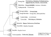
Fig. 1. Schematic phylogenetic tree showing the evolutionary relationships of the nematode species discussed in this review. It is adapted from Dorris et al. (1999) and is based on small unit rDNA sequence Blaxter et al. (1998). The genera that are underlined are parasitic nematodes and the genera shown in italics are free-living nematodes that have been subject to evolutionary development studies and comparative analysis with C. elegans.
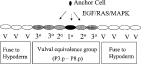
Fig. 2. Schematic representation of the cells of the ventral larval hypodermis that form the vulva in C. elegans. The vulval equivalence group (P3.p–P8.p) are the precursor cells that form the vulval whereas the remaining cells (marked V) fuse to the hypodermal syncitium to contribute to the hypodermis of the adult worm.
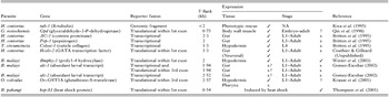
Table 1. Published examples of the use of C. elegans as a heterologous transformation system to study the ability of putative regulatory elements of parasite genes to direct reporter gene expression. [check ] – indicates that reporter gene expression matches that expected from knowledge of the parasite gene function and/or expression pattern. x – indicates that reporter gene expression differs from that expected from knowledge of the parasite gene function and/or expression pattern
 You have
Access
- 51
- Cited by