Variability in practice can be considered to foster clinical innovation, and allow for individualized therapeutic plans and independence of practitioners. The Institute of Medicine, however, has issued a report suggesting that variability in patterns of practice are “illogical”, and should be avoided whenever possible.1 Perhaps nowhere in the field of congenital cardiac disease is variability in practice more apparent than in the management of hypoplastic left heart syndrome. Scientific meetings, as well as the peer reviewed literature, are replete with examples of controversies and uncertainties in management, including type of intervention, techniques for intraoperative support, medical therapies, nutritional support, and protocols for follow-up. At “Heart Week in Florida: 2007”, differences in strategies for management were debated by surgeons, cardiologists, anaesthesiologists and intensivists, with fervent opinions expressed, supported by extensive clinical experience, but reflecting minimal in terms of an evidence base for most of the controversial issues. The purpose of this report, therefore, is to describe this variability in practice at a large number of centres that manage neonates with hypoplastic left heart syndrome, with an emphasis on practice patterns before, during and after the first stage of the Norwood sequence of operations, as well as to suggest changes in future strategies for research.
Methods
We constructed an internet-based survey using commercially available software (www.surveymonkey.com). Following a small pilot feasibility trial, the survey was sent in March of 2007 to colleagues at 55 international centres thought to admit and manage neonates with hypoplastic left heart syndrome. Contacts within the centres were chosen based upon personal knowledge by one of the authors (GW), using email addresses available through cts.net and pcics.org. Participants were encouraged to obtain input from their colleagues in cardiac surgery, intensive care, cardiology and anaesthesia when completing the survey. Questions were designed to assess the differences in patterns of practice regarding counselling, preoperative medical strategies, surgical and cardiopulmonary bypass techniques, and postoperative management.
Answers required categorical responses or a subjective Likert scale with 4 or 5 points. Practices were investigated for a hypothetical, “straight-forward” neonate with hypoplastic left heart syndrome (below). In addition to the questions regarding medical and surgical management, the physicians at each centre were asked to report the number of surgeons currently performing the first stage of the Norwood sequence for reconstructive surgery in their programme, the estimated number of first stage procedures, primary cardiac transplantations or “hybrid” procedures performed for hypoplastic left heart syndrome and its variants per year. Information was also requested regarding the location of delivery of care in their institution.
We proposed a hypothetical case, as follows:
• A neonate is born at 39 weeks gestation weighing 3.2 kilograms with no other congenital anomalies, the second child to an intact family. APGAR scores were 8 at one minute and 9 at 5 minutes. A prenatal diagnosis had been made, and an amniocentesis revealed a normal karyotype. The postnatal echocardiogram confirmed atresia of the aortic and mitral valves, and an extremely diminutive left ventricle. The baby is non-dysmorphic, clinically well, is spontaneously breathing room air with a natural airway and a saturation of oxygen of 85%. There are no infectious or non-cardiac issues.
Results are shown as summary statistics. Given the subjective nature of many of the potential responses, as well as the lack of external validation, formal statistical comparisons were not made, nor were analyses performed correlating strategies for management with any measure of outcome, such as mortality or length of stay in the hospital. For graphical representation of the responses, questions utilizing a Likert scale have been shaded (“frequently”, “occasionally”, “rarely”), or kept solid (“essentially always” or “essentially never”) to represent the subjective nature of the responses.
Results
Responses were received from 53 of the 55 centres identified for potential participation. The results from one centre were censored, as it was subsequently determined that all neonates with a diagnosis of hypoplastic left heart syndrome were transferred to another centre for surgical management. The final data, therefore, is based on returns from 52 centres, as noted in the Appendix A. One additional data set was received from a participant that did not identify the institution. This data was censored as, due to limitations of software, and the subsequent inability to determine if the data was duplicative from another surveyed centre, or from one of the two centres that did not contribute data.
Centre volume and care model
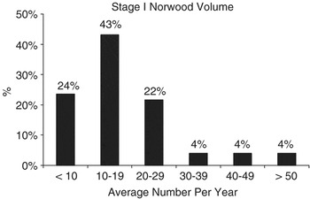
The median number of surgeons performing the first stage of reconstructive surgery at each centre was 2, with a range from 1 to 6. The average number of first stage procedures performed annually is shown in Figure 1. One centre did not respond to this question. In addition to the first stage of reconstructive surgery, primary cardiac transplantation was performed, on average, 1 to 3 times each year in 12 centres, and from 4 to 6 times per year in 2 centres. Hybrid palliation was performed 1 to 3 times per year in 18 centres, 4 to 10 times per year in 7 centres, and more than 10 times per year in one centre.
The average annual number of first stage procedures performed at the 51 centres; one centre left this item blank.

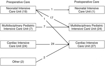
A variety of models for care were documented by the respondents. Approximately half of the centres provided postoperative care in units specifically designated for paediatric cardiac intensive care, and half in multidisciplinary units providing paediatric intensive care. The most common model was for preoperative and postoperative care to be given in a unit for paediatric cardiac intensive care, achieved in 24 centres, followed by preoperative care in a neonatal intensive care unit, and postoperative care in a multidisciplinary unit, as carried out in 17 centres. The location of the patients within the hospital before and after surgery is shown in Figure 2.
The location of neonates with hypoplastic left heart syndrome at the 51 centres; one centre left this item blank.

Preoperative strategies
Counselling
Almost nine-tenths of centres (86.5%) responded that they “recommend” a first stage procedure to families. In the survey the word “recommend” was capitalized to provide emphasis, and to suggest an active proposal from the team. No centre recommended primary cardiac transplantation, a “hybrid” approach, or non-intervention. In 7 centres (14.5%), it emerged that there was discussion of some or all of the above options, but ultimately the families decided upon the appropriate strategy, rather than having one particular option recommended by the practitioners.
Preoperative management
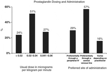
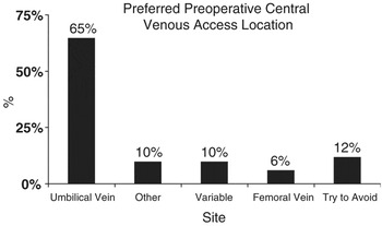
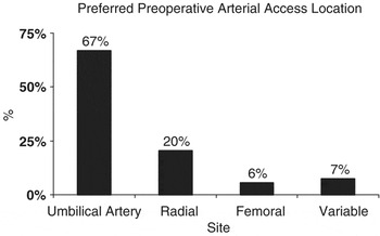
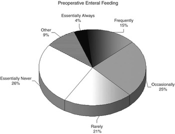
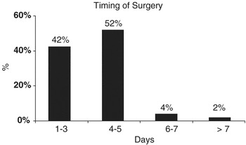
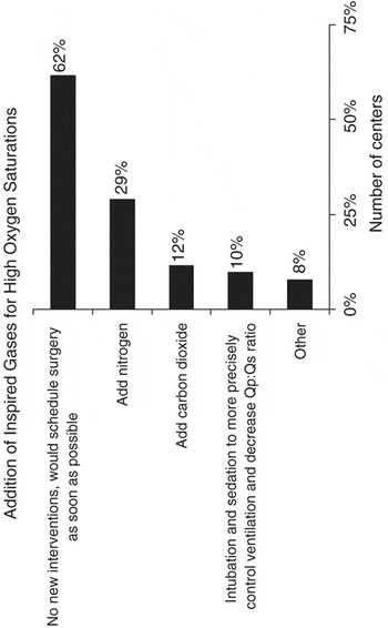
The typical dose and preferred site of administration of prostaglandin is shown in Figure 3. The preferred site of preoperative central venous access and arterial pressure monitoring are shown in Figures 4 and 5, respectively. A wide variety of responses were given regarding the frequency with which the hypothetical case described above would be given enteral feedings, either by mouth or nasogastric tube, as shown in Figure 6. The majority of centres prefer to operate within the first 5 days of life, as shown in Figure 7. The utilization of inspired gases and mechanical ventilation to manage pulmonary overcirculation is shown in Figure 8. For this question, more than one answer was allowed per respondent.
Responses to the questions: “Our USUAL dose of prostaglandin prior to surgery is:” and “Prostaglandin E1 is usually given:” IV = intravenous.

Response to the question: “Central venous access is PREFERENTIALLY provided:”

Response to the question: “Arterial access is PREFERENTIALLY provided:”

Response to the question: “In this hypothetical scenario, we would give enteral feedings (via tube or bottle) PREOPERATIVELY:”

Responses to the question: “The baby remains clinically well and is now 24 h old. The operating room schedule is – surprisingly – wide open. We would plan on surgery in this scenario, at what age:”

Response to the following question: “The baby is clinically well, with a measured oxygen saturation of 92–94%, mild tachypnea and tachycardia, without a metabolic acidosis. Echocardiography reveals a widely patent arterial duct. No other monitoring has been employed. In this scenario, we would TYPICALLY:” More than one response per respondent was allowed. Qp:Qs = the ratio of pulmonary to systemic flow.

Intraoperative and surgical strategies
Airway management and monitoring
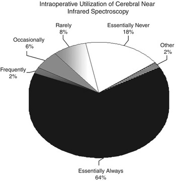
Neonates undergoing the first stage of reconstruction are intubated nasally in 54% of the centres, compared to orally in 36% of the centres. At 5 centres, the intubation site is based upon the preference of the physician. Near infrared spectroscopy of the brain is utilized in nearly two-thirds of the centres surveyed (Fig. 9).
Response to the question: “In the operating room, we use cerebral near infrared spectroscopy:”

Surgical and cardiopulmonary bypass techniques
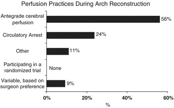
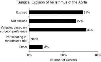
Most centres preferentially used antegrade cerebral perfusion in contrast to deep hypothermia with circulatory arrest, albeit that 11% of centres used a combination of these techniques (Fig. 10). Surgical management of the isthmus of the aorta was highly variable, based upon the practice of the surgeon and the centre (Fig. 11). Reconstruction of the aortic arch was typically performed with a homograft patch at 76.5% of the centres, and using heterograft material at 5.9%. Reconstruction with a patch was “rarely” used at 5.9%, and the reconstruction varied upon surgical preference in 9.8%.
Response to the question: “During the Norwood procedure during arch reconstruction, our routine practice is to use:”

Surgical strategy regarding the isthmus of the aorta.

The source of pulmonary flow following the first stage of reconstruction was highly variable. Of the 51 centres that responded to the question, 13 (25.5%) were participating in a multi-centric randomized clinical trial comparing the modified Blalock-Taussig shunt to the conduit placed from the right ventricle to the pulmonary arteries, the so-called “Sano” modification. Of the remaining 38 centres, 18 (47.4%) “usually” placed a conduit from the right ventricle to the pulmonary arteries, 14 (26.8%) “usually” used a modified Blalock-Taussig shunt, and at six centres, the decision was made “based upon the preference of the surgeon and/or the cardiologist”. Of the centres that stated that they utilized a conduit, 63% stated that they placed the conduit to the left of the neo-aorta, and 37% placed the conduit to the right of the neo-aorta.
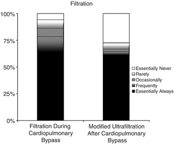
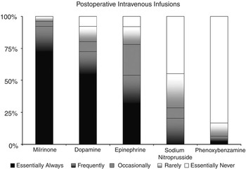
Conventional ultrafiltration and modified ultrafiltration were commonly used during and after cardiopulmonary bypass (Fig. 12), although many more centres responded with “essentially never” for modified ultrafiltration (27.5%), compared to “essentially never” for conventional ultrafiltration (5.9%). No centre was conducting or participating in a clinical trial of techniques for ultrafiltration. Medical support that was “essentially always or frequently” used following separation from cardiopulmonary bypass “in the usual case with adequate haemodynamics and good haemostasis” included milrinone (92%), dopamine (72%), epinephrine (54%), sodium nitroprusside (10.6%), phenoxybenzamine (8.4%), dobutamine (6.4%), vasopressin (4.3%), norepinephrine (2.1%) and thyroid supplementation (2.1%).
The use of filtration during and modified filtration after cardiopulmonary bypass.

Postoperative Management
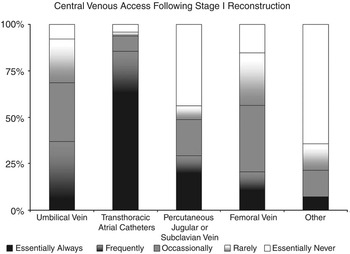
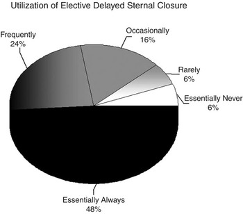
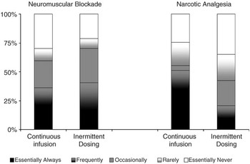
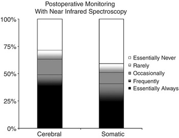
The types of central venous access used following surgery are shown Figure 13. Respondents were again asked to describe their “usual” practice for a patient with “adequate haemodynamics and good haemostasis”. Elective delayed sternal closure under these conditions was reported as essentially always or frequently in nearly three-quarters of the centres (Fig. 14). Under these conditions, the “typical” intravenous infusions of vasoactive agents are used in the intensive care unit as shown in Figure 15, and the use of neuromuscular blockade and analgesia is shown in Figure 16. The use of near infrared spectroscopy, both cerebral and somatic, is highly variable (Fig. 17). No centre was engaged in a randomized trial of this type of monitoring, although two centres, not included in Figure, listed ‘other’ and described an observational trial underway.
The reported use and location of central venous access at the completion of the first stage of reconstruction.

Response to the question: “Upon separation from cardiopulmonary bypass, in the USUAL case, with adequate haemodynamics and good haemostasis, we utilize delayed sternal closure:”

Use of vasoactive medications in the intensive care unit. Response to the question: “Upon separation from cardiopulmonary bypass, in the USUAL case, with adequate haemodynamics and good haemostasis, we utilize:”

The reported use and dosing strategy for neuromuscular blockade and analgesia following surgery “in the USUAL case, with adequate haemodynamics and good haemostasis”

Response to the question: “In the intensive care unit, we use CEREBRAL and SOMATIC (for example, on the abdomen) near infrared spectroscopy:”

Discharge Planning
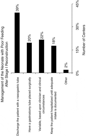
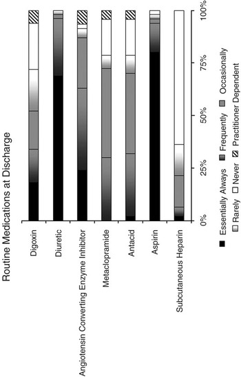
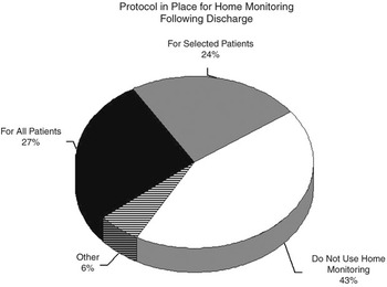
Respondents were given the following scene: “Feeding may be particularly problematic for many neonates after the first stage of reconstruction. (This does not pertain to patients with vocal cord paresis or frequent aspiration, but is meant for the neonate who is an otherwise healthy postoperative patient who is simply a poor feeder). If a neonate is not achieving adequate oral intake postoperatively, but is otherwise ready for hospital discharge, we…” The responses were quite variable across the centres, partly dependent upon preference of the practitioners, with nearly twice as many centres discharging the patient with a nasogastric tube when compared to those who place a surgical gastrostomy tube prior to discharge. The results are shown in Figure 18. Equally variable responses were given for “routine” medications at discharge (Fig. 19), and whether a ‘protocol’ for home monitoring was in place (Fig. 20). For a “stable patient with acceptable saturations of oxygen”, the elective timing of a superior cavopulmonary shunt was between 3 and 4 months in 17 of the 51 (33.3%) centres, 5 and 6 months in 29 (56.9%) centres, 6 and 8 months of age in 2 (3.9%), and a combination of these responses were given from 3 centres. One centre did not respond to this question.
The variable strategies employed for a neonate with feeding difficulties following stage I reconstruction. See text for details.

Response to the question: “For the TYPICAL neonate who is doing well following the first stage of the Norwood sequence and ready for discharge, the following medications are used:”

Response to the statement: “We have a PROTOCOL for home monitoring”

Discussion
Based upon the estimated number of Norwood operations performed by the centres involved in the survey, the results represent the management used in approximately 1000 neonates annually undergoing a Norwood operation, by over 90 different surgeons, and a larger number of cardiologists, intensivists, anaesthesiologists and nurses, at 52 different institutions around the world.
Inspection of the results shows interesting patterns. Some questions resulted in more-or-less a “consensus” opinion, where one answer predominated. Others could be considered somewhat “equivocal”, where the response options appeared to be relatively equal in frequency between three or more options across a continuum, while others could best be described as “controversial”, where a relatively equal response was seen to essentially opposite answers in a continuum, or a relatively equal choice between one of two mutually exclusive options. Of course, some of the answers to the survey could be considered to show an “in-between” type of response, but in general, our current strategies show a combination of some consensus, but predominantly significant variability in most instances.
In general, most centres, but certainly not all, use umbilical venous and arterial access prior to surgery, use a homograft patch to reconstruct the neo-aorta, monitor the patient in the operating room with cerebral near infrared spectroscopy, use conventional filtration during surgery, place transthoracic right atrial lines, do not close the sternum at the completion of the operation, and manage the patient early after surgery with dopamine and milrinone. At discharge, most of centres utilize diuretics and aspirin, and recommend construction of a cavopulmonary connection between the ages of 3 and 6 months. To our knowledge, none of these strategies have been shown to be superior based on randomized clinical trials, but instead have evolved over time, based upon the experience of many practitioners from many disciplines. Of these standard practices, only milrinone has been studied in a placebo-controlled trial,Reference Hoffman, Wernovsky and Atz2 but this trial did not include neonates with hypoplastic left heart syndrome. The use of milrinone, like so many other medications, has only been extrapolated to this population.Reference Zuppa, Nicolson and Adamson3
In contrast, nearly every other strategy for preoperative, intraoperative, and postoperative management shows considerable variability across centres, and occasionally across practitioners within centres. Areas which show the most equivocal responses tend to be those in the intensive care unit, and include variable strategies for management of high saturations of oxygen, route of delivery of prostaglandin, timing of surgery, choices of medication at all points of time from admission to discharge, issues of feeding, and the indications and use of home monitoring.Reference Ghanayem, Hoffman and Mussatto4 Areas of continued controversy tend to cluster in the operating room, and include the use of deep hypothermic circulatory arrest compared to antegrade cerebral perfusion,Reference Goldberg, Bove and Devaney5 management of the aortic isthmus, location of the conduit placed from the right ventricle to the pulmonary arteries, the use of modified ultrafiltration,Reference Gaynor, Kuypers and van Rossem6 and the postoperative use of near infrared spectroscopy for monitoring and management.Reference Ghanayem, Mitchell, Tweddell and Hoffman7, Reference Hoffman, Stuth and Jaquiss8
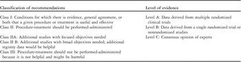
Just as reflected by this survey, review of the literature concerning management of patients with hypoplastic left heart syndrome reveals marked variation in practice and recommendations. Methodologies for assessment of evidence and clinical recommendations have been published by the Task Force of Practice of the American College of Cardiology and the American Heart Association. The strength of evidence for these recommendations is graded according to the type of studies available. Level A is derived from multiple randomized clinical trials, level B is derived for a single randomized trial or nonrandomized studies, and level C is based upon expert consensus. Most of the evidence of management of hypoplastic left heart syndrome, or most types of congenital cardiac disease, is at best level B, and often level C. Similarly, for hypoplastic left heart syndrome most of the clinical recommendations would be class IIA or IIB (see Table 1).Reference Eagle, Guyton and Davidoff9

Adapted from Eagle KA et al., ACC/AHA 2004 guideline update for coronary artery bypass surgery: Summary articleReference Eagle, Guyton and Davidoff9
As the data contained in this survey could not be externally validated, combined with our inability to correlate these practices with outcomes over the short and longer term, we are unable to make recommendations on ‘best practice’ at this time. The variability seen across the centres involved in this survey, nonetheless, provides a background for designing future research studies to improve outcomes for these complicated patients.
Given the variability in patterns of practice outlined by the centres involved in this internet-based survey, it might be difficult to perform randomized clinical trials for many of the issues raised in this survey. Randomized clinical trials are expensive, and time consuming to design and conduct. Bias of practitioners and lack of consent from parents may limit participation.Reference Hoehn, Wernovsky and Rychik10, Reference Hoehn, Wernovsky and Rychik11 And, while randomized clinical trials may be perfectly suited to answer some questions, for example, the use of deep hypothermic circulatory arrest compared to antegrade cerebral perfusion,Reference Goldberg, Bove and Devaney5 the multiple variations in patterns of practice may produce so much “background noise” that an impractical number of patients may be necessary to obtain a statistically significant difference in a proposed primary outcome. Strategies to reduce these confounding effects include stratifying by centre prior to randomization or the institution across centres of standardized management protocols. Standardized management protocols may not be embraced due to lack of equipoise, even if non-evidence based, and thus may limit the participation of centres in multicentric trials. It is also difficult to control, even in large trials,Reference Gaynor, Wernovsky and Jarvik12 patient-specific variations in response to similar therapies, for example, capillary leak after cardiopulmonary bypass, threshold for seizures, and so on. These all add additional ‘noise’ that may affect the primary outcome. Randomized multicentric clinical trials, nonetheless, are an essential part of clinical investigation, and many of the logistical, financial and ‘human-factor’ difficulties can be overcome, as has been recently demonstrated in the PRIMACORP study,Reference Hoffman, Wernovsky and Atz2 and the ongoing reconstruction trial for patients with functionally single ventricles sponsored by the Pediatric Heart Network of the National Heart, Lung and Blood Institute of the National Institutes of Health (www.pediatricheartnetwork.org).
An additional strategy for future investigation is to enroll all patients at a large number of centres in a prospective observational trial, beginning the collection of data at admission as a neonate, and making measurements of outcome throughout infancy and beyond. Without mandating change in the strategies for management across multiple centres and practitioners, outcomes could be measured prospectively, and could include hospital survival, growth of the pulmonary arteries, survival to one year of age, frequency of transplantation, neurodevelopmental outcome, parental satisfaction and stress, and long term quality of life, to name just a few.
There are many aspects of delivery of care that are difficult to measure even in tightly controlled trials, but may have an impact on important outcomes. For example, in the 27 centres who provided postoperative care in a dedicated unit for paediatric cardiac intensive care, 24 (89%) admitted the babies to that unit preoperatively as well, allowing for minimal transition of the care team before and after surgery, but most likely with variable involvement of specialists in neonatal medicine. In contrast, of the 24 centres who provided postoperative care in a multidisciplinary paediatric intensive care unit, only 7 (29%) provided preoperative care for those babies. Only one centre provided both pre- and postoperative care in a neonatal intensive care unit. It is not clear if these differences in the model for delivery of care, and composition of the team, have any effect on outcomes, length of stay in the hospital, postgraduate medical education, participation in clinical research, or parental stress and satisfaction. The variability in practice, nonetheless, allows for collaborative observational studies to be undertaken to answer these questions.
As mentioned previously, the results of our survey have not been independently validated at the centres themselves for accuracy, and although the participants were encouraged to seek the opinions of multiple practitioners in the program, this could not be confirmed. Geographical and other biases most certainly exist, as the majority of the participants were located in the United States of America, not all centres who manage neonates with hypoplastic left heart syndrome were contacted, as there was no registry to obtain such data, and management in Europe, Asia, Africa and South America were most likely not adequately represented. We did not address the composition of the team caring for the neonate, only the location of neonate before and after surgery. The location of care may be determined by the policy of the centre, bed availability, and other factors not specifically addressed in this survey. The frequency of prenatal diagnosis and the mode of admission, whether born in the same hospital or transported in, were not investigated, and thus none of these factors could be correlated with management practices of the “straight forward” neonate with hypoplastic left heart syndrome. The data obtained does, however, provide a ‘snapshot’ of current practices, and provides baseline data to plan future research efforts.
In conclusion, there is marked variability in practice across and within centres managing neonates with hypoplastic left heart syndrome. Despite, and perhaps because of, these variations, survival and outcomes in the midterm have continued to improve for children with hypoplastic left heart syndrome over the 25 years since the first successful report of staged reconstruction. Our challenge is to determine which of our current practices can be further improved with modifications of technique, medical therapies, management philosophy, and follow-up strategies, which can only be accomplished if specific practices can be unequivocally linked to outcomes in both the short and long term.
Appendix A – Survey Respondents
United States of America
Arnold Palmer Hospital for Children, Orlando
C. S. Mott Children’s Hospital, Ann Arbor
Children’s Hospital of the West Virginia University Hospitals, Morgantown
Children’s Heart Centre at Children’s Hospital and Regional Medical Centre, Seattle
Children’s Heart Centre at Phoenix Children’s Hospital, Phoenix
Children’s Heart Centre of the Schneider Children’s Hospital, New Hyde Park
Children’s Hospital of Omaha, Omaha
Children’s Hospital of Pittsburgh, Pittsburgh
Children’s Memorial Heart Centre, Chicago
Children’s National Medical Centre, District of Columbia
Congenital Heart Institute at Miami Children’s Hospital, Miami
Duke Children’s Hospital, Durham
Heart Institute at Children’s Hospital Los Angeles, Los Angeles
Heart Institute at Children’s Hospital of Orange County, Orange
Helen B. Taussig Children’s Cardiac Centre at Johns Hopkins, Baltimore
Herma Heart Centre at Children’s Hospital of Wisconsin, Milwaukee
Kosair Children’s Hospital, Louisville
Loma Linda International Heart Institute, Loma Linda
Medical University of South Carolina, Charleston
Monroe Carell Jr. Children’s Hospital at Vanderbilt, Nashville
Morgan Stanley Children’s Hospital of New York-Presbyterian, New York
Nemours Cardiac Centre at Alfred I. duPont Hospital for Children, Wilmington
Oregon Health and Science University, Portland
Primary Children’s Medical Centre, Salt Lake City
St. Christopher’s Hospital for Children, Philadelphia
Texas Children’s Heart Centre, Houston
The Cardiac Centre at The Children’s Hospital of Philadelphia, Philadelphia
The Cardiovascular Program at Children’s Hospital Boston, Boston
The Children’s Hospital Heart Institute, Denver
The Children’s Sibley Heart Centre, Atlanta
The Combined Cardiovascular Surgical Program at Stanford, Palo Alto
The Comprehensive Centre for Heart Care at Children’s Medical Centre, Dallas
The Congenital Heart Institute of Florida, Tampa and St. Petersburg
The Heart Centre at Cincinnati Children’s Hospital Medical Centre, Cincinnati
The Heart Centre at Columbus Children’s Hospital, Columbus
The Heart Centre at St. Louis Children’s Hospital, St. Louis
The Mount Sinai Medical Centre, New York
University of California at San Francisco Children’s Hospital, San Francisco
University of Virginia Children’s Hospital Heart Centre, Charlottesville
International
Asklepios Clinic, Sankt Augustin, Germany
Birmingham Children’s Hospital, Birmingham, England
Children’s Heart Centre, Bratislava, Slovak Republic
CHU Sainte-Justine, Montreal, Canada
Hospital for Sick Children at Great Ormond Street, London, England
L’Ospedale Pediatrico Bambino Gesù, Rome, Italy
Okayama University Hospital, Okayama City, Japan
Royal Brompton Hospital, London, England
Stollery Children’s Hospital, Edmonton, Canada
The Children’s Hospital at Westmead, Sydney, Australia
The Hospital for Sick Children, Toronto, Canada
The Montreal Children’s Hospital, Montreal, Canada
The Royal Children’s Hospital, Melbourne, Australia