Introduction
Schwannomas are benign peripheral nerve sheath tumours. Approximately 50 per cent of these tumours occur in the head and neck region, and less than 4 per cent arise in the paranasal sinuses. The distribution of schwannomas is ubiquitous, but in the paranasal sinuses they can be easily confused with malignant tumours. This is because of the radiological appearance of cartilaginous or bony erosion, but may also be due to their histological appearance whereby they lack encapsulation and are predominantly hypercellular. Although rare, the otolaryngologist, oncologist and pathologist should be aware of the existence of paranasal schwannomas and of their inherent diagnostic difficulties.
To our knowledge, there have been only two previously reported case series of paranasal schwannoma in the literature, and fewer cases of the cellular variant.
This retrospective case series from our tertiary referral centre presents our experience of five cases of paranasal schwannoma, and emphasises the difficulty in correctly diagnosing this rare, benign condition.
Materials and methods
Five cases of schwannoma in the sinonasal region were retrieved from the archives of the histopathology department at Christie Hospital between 1990 and 2001. All of the tumours had been referred from other hospitals, and the initial reporting pathologist had raised the possibility of malignancy in one case.
The specimens were fixed with formalin, embedded in paraffin and stained with haematoxylin and eosin. Immunohistochemical analysis was performed on all cases using the DAKO ChemMateTM EnVision detection system with 3-3′-diaminobenzidene as chromogen and using the following antibodies: S100 protein, HMB-45, GFAP (an intermediate filament protein found in glial cells such as astrocytes and ependymal cells and serves as a well recognised immunomarker), CD34, SMA (smooth muscle actin) desmin, smooth muscle antigen and melan A. Appropriate positive and negative controls were used in all the cases. All histological slides were reviewed by the same pathologist (SSB).
After obtaining approval from the local research ethics committee, clinical information and follow-up data were obtained from patients' case notes, both at the referring hospital and at Christie Hospital.
Results
Clinical features
This series represents one single institution's experience of five benign schwannomas in the paranasal region (see Table I). The male:female ratio was 1:1.5, and patients' ages at presentation ranged from 34 to 66 years (median 55 years). Four of the lesions were centred in the ethmoid space and one in the maxillary sinus. The most common presenting clinical symptom was nasal obstruction, but other symptoms included proptosis, epiphora, headaches, facial anaesthesia and epistaxis.
Main Clinical Features of 5 Cases of Paranasal Schwannoma

* The number in parenthesis refers to the time to recurrence from diagnosis in months. Pt no = patient number; yrs = years; mths = months; F = female; M = male; XRT = radiotherapy; LR = local recurrence
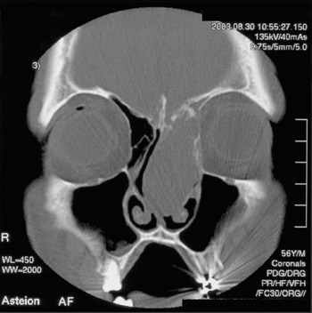
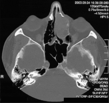
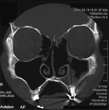
Pre-operative computed tomography (CT) scanning showed that two cases had evidence of bone destruction and that one schwannoma had invaded the cartilage of the nasal septum (see radiographs 1–3). Another case showed microscopic evidence of bone invasion.
Coronal CT image demonstrating extensive bilateral soft tissue mass obliterating left ethmoidal space and anterior frontal sinuses with erosion of left medial orbital wall.

Axial CT image showing invasion of bony architecture in left paranasal sinus with destruction of the midline vomer and nasal septum. Invasion of bony medial canthus and transosseous spread into subcutaneous tissue.

Coronal CT image after endoscopic debulking sinus surgery.

At surgery, two patients had evidence of invasion into the orbit, one had bony erosion of the orbital wall only, and one patient was found to have a small cerebrospinal fluid leak.
Four of the five cases received surgery as their primary, definitive treatment, and one was treated by primary radiotherapy. One patient was given radiotherapy adjuvantly and suffered late radiotherapy toxicity leading to blindness due to sacrifice of the retina and optic nerves. Follow up of patients ranged from 56 to 137 months (median 97 months). Two patients (both of whom received primary surgical treatment) relapsed during follow up and were surgically salvaged. At the time of writing, only one patient had died, from an intercurrent cause; the others remained alive without evidence of residual disease.
Pathology
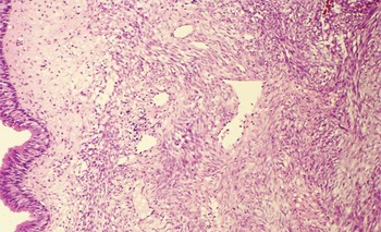
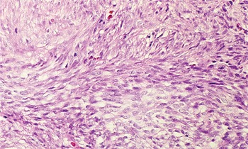
All histological tissue was received in fragments, and there was no discernible capsule to any of the tumour fragments. Three cases had features of cellular schwannoma (see Figures 1–3), and were composed of a compact proliferation of spindle-shaped cells arranged in interlacing fascicles with predominantly hypercellular Antoni A areas. The cells contained pale, fibrillar cytoplasm and bland, elongated or oval, plump nuclei. The other two cases had alternating hypercellular Antoni A and hypocellular Antoni B areas, with occasional Verocay bodies and foci of nuclear palisading, typical of the classical variant. None of the cases showed atypical mitoses, and there was no evidence of necrosis. The cells in one case contained melanin pigment, both within and outside the neoplastic cells. In three cases, there were occasional cells exhibiting degenerative nuclear atypia.
Photomicrograph showing tissue biopsy of cellular schwannoma at low magnification (H&E; ×40).

Plump, bland cells seen at higher power magnification (H&E;×100).

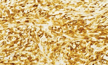
S-100 immunostaining seen at higher magnification (×100).

All cases showed strong and diffuse positivity for S100 immunostaining and patchy positivity for GFAP immunostaining. Immunostaining for HMB-45, melan A, CD34, SMA, smooth muscle antigen and desmin was negative.
Discussion
We describe five patients diagnosed with paranasal schwannoma, two with the classical form and three with the cellular variant, who all posed diagnostic problems both radiologically and histologically. Cartilage and bony erosion are more likely to be noted on CT scans and can give the mistaken impression of invasive, malignant disease. It has been postulated that erosion into osseous structures is a result of a pressure effect from a slowly growing mass.
The lack of encapsulation of these tumours and occasionally highly cellular appearance can make the histological diagnosis difficult. Moreover, it is not possible to comment on the status of the resection margins, as specimens are often fragmented as a result of piecemeal resection. The presence of tumour necrosis, cytological atypia and high mitotic activity (≥3 mitotic figures per 10 high power fields) are the main features predicting malignant potential and differentiating this lesion from a malignant peripheral nerve sheath tumour. The benign cellular variant can sometimes exhibit mitotic activity and nuclear pleomorphism. The decreased expression of S100 protein has been used as a pointer towards malignancy. Although rare, sinonasal malignant melanoma should be excluded; these tumours tend to show marked cytological atypia and a high mitotic index, with positive immunostaining with HMB-45 and melan A.
• Schwannomas are benign peripheral nerve sheath tumours
• The distribution of schwannomas is ubiquitous, but in the paranasal sinuses they can be easily confused with malignant tumours
• Cartilage and bony erosion is more likely to be noted on computed tomography scans and can give the mistaken impression of invasive, malignant disease
• Local excision without the need to attempt wide margins is usually sufficient as primary treatment
If a case of benign schwannoma is mistakenly diagnosed as malignant, then unnecessary over-treatment with external beam radiotherapy could lead to late effects, particularly to the neural tissue and the optic pathway, causing blindness, cataract, temporal lobe syndrome or epilepsy. Unnecessary chemotherapy could also be detrimental. We propose that radiotherapy should not be advocated as adjuvant treatment, due to the potential for late toxicity in this anatomical region and because results for surgery in recurrent cases are also good. However, there is a suggestion that these tumours might be radiosensitive, as evidenced by patient one, who received primary, definitive radiotherapy treatment and did not relapse up to her death 11 years later, and by patient five, who received adjuvant radiotherapy and had not relapsed 5.5 years later. If surgery is not possible then radiotherapy is an alternative approach and, with modern techniques such as intensity-modulated radiotherapy (IMRT), critical structures such as the optic pathway could be spared.
This series follows on from two previous case series reported in the literature. Hasegawa et al. Reference Hasegawa, Mentzel and Fletcher1 reported a series of five cases of sinonasal schwannoma, four of which were the cellular variant. There were no local recurrences, but the median follow up was only 27 months. Buob et al. Reference Buob, Wacrenier, Chevalier, Aubert, Quinchon and Gosselin2 reported five cases, but no distinction was made between the classical form and the cellular variant. These authors had a median follow up of six years, without any local recurrences. Both of these series reported similar diagnostic difficulties to those described above; however, neither study encountered any local recurrences. The present study reports results from a longer follow-up period (median 8.1 years) and exemplifies the possibility of local recurrence. There have been a number of other individual case reports of paranasal sinus schwannoma.Reference Donnelly, Al-Sader and Blayney3–Reference Perzin, Panyu and Wechter7
Typically, paranasal schwannoma does not metastasise but it can recur locally, particularly after incomplete surgery. Local excision without the need to attempt wide margins is usually sufficient as primary treatment, with further surgery undertaken for those cases with local recurrence. In patients deemed medically unfit or technically unsuitable for surgery, radiotherapy is an alternative.
Acknowledgements
We would like to thank the following: the Medical Records Departments at Christie Hospital, Wrexor Maelor Hospital, Huddersfield General Hospital, Tameside Hospital, Beardwood Hospital, Royal Preston Hospital and Royal Bolton Hospital; the Medical Illustration Department at Christie Hospital; and the Local Research and Ethics Committee of Manchester Metropolitan University (Academic Division), who gave approval to this research project.