Introduction
The patellogastropod limpets (hereafter referred to as ‘limpets’) are a monophyletic group of highly specialized marine gastropods with worldwide distribution and are usually found on tidal and subtidal rocky shores (Orton, Reference Orton1929; Blackmore, Reference Blackmore1969; Branch, Reference Branch1981; Nakano & Ozawa, Reference Nakano and Ozawa2007; Nakano & Sasaki, Reference Nakano and Sasaki2011; Espinosa & Rivera-Ingraham, Reference Espinosa, Rivera-Ingraham and Curry2017; Sousa et al., Reference Sousa, Vasconcelos, Riera, Pinto, Delgado and Henriques2019b). Limpets have been exploited as a fishery resource for more than 10,000 years (Klein, Reference Klein1979; Erlandson et al., Reference Erlandson, Braje, Rick, Jew, Kennett, Dwyer, Ainis, Vellanoweth and Watts2011), either as food or bait (Pombo & Escofet, Reference Pombo and Escofet1996; Harada et al., Reference Harada, Miyasaki, Maeda and Satoh1997; Guallart et al., Reference Guallart, Luque, Acevedo and Calvo2013; Espinosa & Rivera-Ingraham, Reference Espinosa, Rivera-Ingraham and Curry2017; Henriques et al., Reference Henriques, Delgado, Sousa and Ray2017). Currently, several limpet species of commercial interest around the world are threatened by increasing harvesting pressure, which leads to smaller specimens and reduced population sizes (Branch & Odendaal, Reference Branch and Odendaal2003; Espinosa et al., Reference Espinosa, Rivera-Ingraham, Fa and García-Gómez2009; Ramírez et al., Reference Ramírez, Tuya and Haroun2009; Erlandson et al., Reference Erlandson, Braje, Rick, Jew, Kennett, Dwyer, Ainis, Vellanoweth and Watts2011; Espinosa & Rivera-Ingraham, Reference Espinosa, Rivera-Ingraham and Curry2017; Martins et al., Reference Martins, Borges, Vale, Ribeiro, Ferraz, Martins, Santos and Hawkins2017), thus contributing to the status of vulnerable or endangered limpet populations (Espinosa et al., Reference Espinosa, Rivera-Ingraham, Maestre, González, Bazairi and García-Gómez2014; Espinosa & Rivera-Ingraham, Reference Espinosa, Rivera-Ingraham and Curry2017; Henriques et al., Reference Henriques, Delgado, Sousa and Ray2017).
The limpet Patella aspera Röding, 1798, is a native species from the Macaronesian region, which includes the Azores, Canary and Madeira archipelagos (Sousa et al., Reference Sousa, Delgado, Pinto and Henriques2017). The specific status of P. aspera was controversial until molecular studies differentiated the island populations of P. aspera from the continental populations of Patella ulyssiponensis Gmelin, 1791 (Côrte-Real, Reference Côrte-Real1992; Weber & Hawkins, Reference Weber and Hawkins2005). Patella aspera is a living resource of cultural and economic importance (Ferraz et al., Reference Ferraz, Menezes and Santos2001; Navarro et al., Reference Navarro, Ramírez, Tuya, Fernandez-Gil, Sanchez-Jerez and Haroun2005; Martins et al., Reference Martins, Borges, Vale, Ribeiro, Ferraz, Martins, Santos and Hawkins2017; Fernandes et al., Reference Fernandes, Fernandes and Cordeiro2019; Sousa et al., Reference Sousa, Vasconcelos, Henriques, Pinto, Delgado and Riera2019a, Reference Sousa, Vasconcelos, Riera, Pinto, Delgado and Henriques2019b). In the Azores archipelago, the harvesting pressure caused the collapse of the P. aspera fishery in the late 1980s and contributed to decreasing animal size (Ferraz et al., Reference Ferraz, Menezes and Santos2001; Martins et al., Reference Martins, Borges, Vale, Ribeiro, Ferraz, Martins, Santos and Hawkins2017). In Madeira, the harvested populations show a reduction in average body size, a higher male ratio, and less energetic investment in reproduction (Sousa et al., Reference Sousa, Vasconcelos, Henriques, Pinto, Delgado and Riera2019a, Reference Sousa, Vasconcelos, Riera, Pinto, Delgado and Henriques2019b, Reference Sousa, Pinto, Vasconcelos and Riera2020). Moreover, harvesting pressure could be behind the high inbreeding coefficients observed in P. aspera populations from all Macaronesia (Faria et al., Reference Faria, Pita, Martins, Ribeiro, Hawkins, Presa and Neto2018). The overfishing led to the implementation of regulation measures to promote the sustainable harvesting of this limpet species by the different regional governments (Ferraz et al., Reference Ferraz, Menezes and Santos2001; Martins et al., Reference Martins, Borges, Vale, Ribeiro, Ferraz, Martins, Santos and Hawkins2017; Sousa et al., Reference Sousa, Vasconcelos, Henriques, Pinto, Delgado and Riera2019a, Reference Sousa, Vasconcelos, Riera, Pinto, Delgado and Henriques2019b).
Fisheries management requires a good understanding of the life history and recruitment of the target species (Szuwalski et al., Reference Szuwalski, Vert-Pre, Punt, Branch and Hilborn2015). Major efforts have focused on limpet larval production, either by studying the factors that promote spawning induction, with variable success (Kay & Emlet, Reference Kay and Emlet2002; Ferranti et al., Reference Ferranti, Monteggia, Asnaghi and Chiantore2018, Reference Ferranti, Guallart, Fanciulli, Panzalis and Chiantore2021; Mau & Jha, Reference Mau and Jha2018; Mau et al., Reference Mau, Bingham, Soller and Jha2018; Nakano et al., Reference Nakano, Nakayama and Takahashi2020), or by using gametes obtained by dissection and artificially matured using alkalinized seawater baths, with consistent larval production (Dodd, Reference Dodd1957; Gould et al., Reference Gould, Stephano, Ortíz-Barrón and Pérez-Quezada2001; Ribeiro, Reference Ribeiro2008; Aquino De Souza et al., Reference Aquino De Souza, Tyler and Hawkins2009; Guallart et al., Reference Guallart, Peña, Pérez-Larruscaín, Luque and Templado2020; Cañizares et al., Reference Cañizares, Castejón, Haroun, Nogueira and Andrade2021; Castejón et al., Reference Castejón, Cañizares, Nogueira and Andrade2021). Development is brief, since in less than 24 h post-fertilization, limpet larvae can be observed as free swimming trochophores, which soon develop to competent pediveligers ready to settle and metamorphose to juveniles (Kay & Emlet, Reference Kay and Emlet2002; Nhan & Ako, Reference Nhan and Ako2019; Guallart et al., Reference Guallart, Peña, Pérez-Larruscaín, Luque and Templado2020; Nakano et al., Reference Nakano, Nakayama and Takahashi2020; Ferranti et al., Reference Ferranti, Guallart, Fanciulli, Panzalis and Chiantore2021). However, the settlement requirements for limpet larvae are mostly unknown, and feasible methods to obtain juveniles have yet to be explored (Mau & Jha, Reference Mau and Jha2018; Ferranti et al., Reference Ferranti, Guallart, Fanciulli, Panzalis and Chiantore2021).
Until now, the identification of adequate settlement inducers for limpets has been a difficult task (Mau & Jha, Reference Mau and Jha2018; Seabra et al., Reference Seabra, Cruz, Fernandes, Silva and Hawkins2019). Field studies have shown that crustose coralline algae (CCA) play a fundamental role as grazing substrates for several limpet species and other intertidal herbivores (Steneck, Reference Steneck1982; Steneck et al., Reference Steneck, Hacker and Dethier1991; Pueschel & Miller, Reference Pueschel and Miller1996; Maneveldt et al., Reference Maneveldt, Wilby, Potgieter and Hendricks2006). CCA are among the most widespread and abundant groups of marine algae dominating rocky benthic environments (Steneck, Reference Steneck1986; McCoy & Kamenos, Reference McCoy and Kamenos2015), in which different CCA species compete for space and try to overgrow other CCA species (Steneck, Reference Steneck1986; Steneck et al., Reference Steneck, Hacker and Dethier1991; McCoy & Pfister, Reference McCoy and Pfister2014). Experimental studies have shown that larval settlement in marine invertebrates, such as corals (Kenkel et al., Reference Kenkel, Traylor, Wiedenmann, Salih and Matz2011; Tebben et al., Reference Tebben, Motti, Siboni, Tapiolas, Negri, Schupp, Kitamura, Hatta, Steinberg and Harder2015), echinoderms (Keesing et al., Reference Keesing, Halford, Hall and Cartwright1997; Lambert & Harris, Reference Lambert and Harris2000), chitons (Barnes & Gonor, Reference Barnes and Gonor1973) and gastropods (Roberts et al., Reference Roberts, Kaspar and Barker2004; Spotorno-Oliveira et al., Reference Spotorno-Oliveira, Figueiredo and Tâmega2015), can be triggered by the presence of CCA. Ribeiro (Reference Ribeiro2008) reported an enhanced settlement rate in different patellid species in the presence of CCA. Moreover, the juveniles of several limpet species also occur naturally in environments associated with CCA, i.e. Patella depressa (Seabra et al., Reference Seabra, Hawkins, Espírito-Santo, Castro and Cruz2020), Patella pellucida (McGrath, Reference McGrath1992; McGrath & Foley, Reference McGrath, Foley and Wilson2005), P. ulyssiponensis (Seabra et al., Reference Seabra, Cruz, Fernandes, Silva and Hawkins2019, Reference Seabra, Hawkins, Espírito-Santo, Castro and Cruz2020), Patelloida mufria (Fletcher, Reference Fletcher1988) and Testudinalia testudinalis (as Acmaea testudinalis) (Steneck, Reference Steneck1982), thus suggesting that CCA could be involved in limpet settlement and recruitment (Kooistra et al., Reference Kooistra, Joosten and Van den Hoek1989; Mau & Jha, Reference Mau and Jha2018). Regarding other economically important marine gastropods, such as abalone, CCA was also shown to be an effective settlement inducer (Roberts & Nicholson, Reference Roberts and Nicholson1997; Daume et al., Reference Daume, Brand-Gardner and Woelkerling1999; De Viçose et al., Reference De Viçose, Viera, Bilbao and Izquierdo2010, Reference De Viçose, Viera, Huchette and Izquierdo2012; Roberts et al., Reference Roberts, Barker and Mladenov2010). Nevertheless, the true role and importance of CCA in limpet settlement and recruitment is under discussion. Alternatively, Nhan & Ako (Reference Nhan and Ako2019) also reported that a combination of different microalgae (i.e. the diatom Amphora and haptophycean Pavlova) promoted the settlement of the larvae of the Hawaiian opihi Cellana sandwicensis.
The main goal of this study is to evaluate different CCA communities and cultured microalgae strains added to the culture media as substrates to induce the settlement of the limpet P. aspera. Additionally, for the first time, a description of P. aspera pediveliger larvae, early postlarvae and juveniles is provided based on developmental and behavioural observations.
Materials and methods
Acquisition and maintenance of the adult specimens
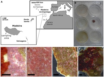
Adult specimens of the limpet P. aspera were collected by snorkel diving on the SW Madeira island coast (Figure 1A) between November 2020 and March 2021, coinciding with the peak of the annual reproductive period (Sousa et al., Reference Sousa, Delgado, Pinto and Henriques2017). The specimens were transported to the facilities of the Mariculture Centre of Calheta (CMC, Madeira, Portugal) and placed in 200-litre culture tanks at a maximum stocking density of 30 specimens per tank. The specimens were maintained under the following conditions: open system (water renewal set at 1–2 l min−1), high aeration (O2 saturation higher than 95%), natural temperature (20 ± 2 °C) and salinity (36 ± 1 g l−1). The adult specimens were dissected between 1 and 5 days after capture to obtain gametes.
(A) Map showing the location of the sampling area. (B) Culture cell plates used for the settlement assays. (C) Example of ‘mosaic-like’ CCA community (MSC) used in Assay 1. (D) Example of ‘homogeneous pink’ CCA community (HMG) used in Assay 2. (E) Example of ‘non-dominant CCA’ community (NDC) used in Assay 2. (F) Example of ‘dominant dark red’ CCA community used in Assay 3. Scale bar = 2 mm.

Larval production
The larvae of P. aspera were obtained using the methodology designed by Castejón et al. (Reference Castejón, Cañizares, Nogueira and Andrade2021) for this species, which employs oocytes obtained by dissection and matured artificially using alkalinized seawater (see Table 1 for details). Briefly, the adult specimens were characterized using the shell length, total mass and female gonadosomatic index (GSI) following Sousa et al. (Reference Sousa, Delgado, Pinto and Henriques2017). The gametes were obtained from dissected gonads and pooled in filtered sterilized seawater (FSS). The oocytes were matured artificially using NaOH alkalinized seawater (pH 8.4 ± 0.1) for 3 h. Then, the oocytes were carefully washed in a 55 μm mesh and placed in 600 ml glass beakers for fecundation, adding 105 sperm cells ml−1, following Guallart et al. (Reference Guallart, Peña, Pérez-Larruscaín, Luque and Templado2020) for the limpet Patella ferruginea. The incubation was static inside a controlled temperature room at 17 ± 1 °C for 24 h.
Patella aspera. General information of the Assays

GSI, gonadosomatic index; n, size of each adult pool; OD, oocyte density; ILD, initial larval density; TM, total mass; SL, shell length.
Date of realization, male and female general morphometry, oocyte density during the incubation period (24 h), initial larval density during the larval culture period (48 h), and the initial larval density at the start of the assays. Values are shown as average ± SD.
Larval culture methodology
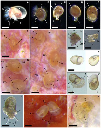
The larvae obtained after the incubation period (24 h) were free-swimming trochophores, which were generally found swarming near the water surface of the glass beakers. The trochophores were gathered by siphoning the upper two-thirds of the water column of the beakers over a 55 μm mesh to collect the larvae. This step was key to collecting a majority of trochophores with normal morphology while avoiding collecting the debris accumulated on the bottom of the beakers (e.g. non-fertilized oocytes, abnormal larvae and tissue remains), which could interfere negatively with the larval culture. The trochophores were immediately redistributed to glass beakers filled with 500 ml clean FSS for their culture for an additional 48 h at 17 ± 1 °C (larval density is presented in Table 1). Feeding was not provided at any time since preliminary tests showed that P. aspera larvae can develop without feeding. After this additional culture period (48 h), the larvae reached the pediveliger stage characterized by the development of the eyes, velum, protoconch, foot and operculum (Figure 2A–E). The pediveligers were gathered by siphoning the upper three-quarters of the water column of the glass beakers and using a 55 μm mesh to collect the larvae. As before, this step was fundamental to collect a majority of viable larvae while avoiding the content of the bottom of the beakers (e.g. debris and non-viable and dead specimens).
Patella aspera. Identification and behaviour of different early life stages. Pediveliger larvae (A–E; at least 72 h post-fertilization, scale bar = 100 μm): (A) key characters to identify the pediveliger larva; (B) swimming behaviour; (C) withdrawal behaviour; (D) crawling behaviour; (E) resting behaviour. Comparison between the pediveliger larvae and the early postlarvae (F–H; at least 5–6 days post-fertilization, scale bar = 100 μm): (F) early postlarva, note the lack of velum; (G) and (H) pediveliger larvae and early postlarvae, note the lack of velum in the early postlarvae. Juveniles (I–K, scale bar = 100 μm): (I) juvenile, crawling, 15 days post-fertilization; (J) juveniles, resting, 8 days post-fertilization; (K) juvenile, crawling, 17 days post-fertilization. Deformed specimens (L–M; scale bar = 100 μm): (L) pediveliger larvae; (M) early postlarvae. (N) Empty protoconchs (larval shells) from pediveliger larvae and early postlarvae (scale bar = 100 μm). (O) Empty juvenile shells (scale bar = 100 μm). CT, cephalic tentacle; E, eyespots; EPL, early postlarvae; F, foot; O, operculum; P, protoconch (larval shell); PVL; pediveliger larvae; T, teleoconch; V, velum; VM, visceral mass.

Preparation of the substrates
This study used two types of substrates as settlement inducers: cultured microalgae strains and CCA. The microalgae strains consisted of two benthic diatom species able to form biofilms: Halamphora coffeaeformis and Navicula incerta, which were provided by the Universidad de Las Palmas de Gran Canaria (Canary Islands, Spain), and the haptophycean Pavlova sp., which was obtained from the Roscoff Culture Collection (RCC3458 Pavlova_sp; Sorbonne Université, France). The algal strains were cultured at CMC facilities.
The shells of P. aspera are usually colonized by epiphytic encrusting coralline algae (Martins et al., Reference Martins, Faria, Furtado and Neto2014). On Madeira Island, epiphytic CCA communities colonize the shells of two sympatric and congeneric limpet species, Patella candei d'Orbigny, 1839 and P. aspera (Figure 1C–F; Supplementary Files 1 and 2). Limpet shells were used as CCA-covered substrata due to its ease of manipulation when compared with other substrata (e.g. boulders). Although CCA are a taxonomically difficult group whose biodiversity has yet to be determined in Macaronesia (Sangil et al., Reference Sangil, Martins, Hernández, Alves, Neto, Ribeiro, León-Cisneros, Canning-Clode, Rosas-Alquicira, Mendoza, Tittley, Wallenstein, Couto and Kaufmann2018), in the present study, the authors followed previous studies performed in other marine gastropods, and taxonomic identification of CCA was not verified (Roberts & Nicholson, Reference Roberts and Nicholson1997; Ribeiro, Reference Ribeiro2008; De Viçose et al., Reference De Viçose, Viera, Bilbao and Izquierdo2010; Seabra et al., Reference Seabra, Cruz, Fernandes, Silva and Hawkins2019). This study used a morphology-based classification of the different CCA communities. The epiphytic algal communities from the limpet shells were grouped into four categories based on the presence of pink and dark red coralline algal crusts: (1) ‘mosaic-like’, the shell surface was covered by pink and dark red crusts (MSC; Figure 1C; Supplementary File 1a); (2) ‘homogeneous pink’, the shell surface was only covered by pink crusts (HMG; Figure 1D; Supplementary File 1d); (3) ‘non-dominant CCA’, the shell surface was dominated by non-encrusting algae, albeit numerous pink crusts were present (NDC; Figure 1E; Supplementary File 1c); and (4) ‘homogeneous red’, the shell surface was dominated by dark red crusts (Figure 1F; Supplementary File 2).
Nevertheless, prior to use, limpet shells were prepared to minimize the influence of any potential adult cue on the settlement and to obtain pieces able to fit in the culture cells. First, the soft body of the animal was removed using a scalpel. Then, the shells were placed inside a culture tank under the same conditions as adults for 1–2 days to soften the tissue remains attached to the shell aperture; these remains were removed using paper and water. Finally, the shells were wrapped in paper and broken using a hammer to obtain pieces with a size suitable depending on the purpose of the assays (see Assays description). The shell pieces were washed again and placed in a culture tank until required.
Design of the settlement assays
The assays were performed in cell culture plates (Sigma® cell culture plate, size 6 wells, sterile, treated tissue culture), in which each individual culture cell corresponded to a different treatment (Figure 1B). The initial density of pediveliger larvae is indicated in Table 1. The placement of the larvae in the culture cells marked the start of the assays. The final volume in each culture cell was adjusted to 8 ml FSS. The assays were performed at 17 ± 1 °C.
Assay 1: test of different substrates used as settlement inducers (14 days)
Six treatments with five replicates each were employed: (1) control (CNT) treatment without substrate; (2) ‘mosaic-like’ community obtained from P. candei shell pieces of 68 ± 13 mm2 (MSC; Figure 1C; Supplementary File 1a); (3) H. coffeaeformis (HAL; 8.9 × 10⁴ cell aggregations ml−1); (4) N. incerta (NAV; 8.6 × 10⁴ cells ml−1); (5) Pavlova sp. (PAV; 9.4 × 10⁴ cells ml−1); and (6) combined H. coffeaeformis and Pavlova sp. (HAL + PAV) at the same densities as specified before. The cultured microalgae treatments followed Nhan & Ako (Reference Nhan and Ako2019) for the settlement of C. sandwicencis.
Assay 2: test of different CCA communities (14 days)
Five treatments with six replicates each were employed: (1) control (CNT), treatment without substrate; (2) ‘mosaic-like’ community obtained from P. candei shells (MSC, Figure 1C; Supplementary Figure 1a); (3) ‘homogeneous pink’ community obtained from P. aspera shells (HMG; Figure 1D; Supplementary Figure 1b); (4) ‘non-dominant CCA’ community from P. aspera and P. candei shells (NDC; Figure 1E; Supplementary File 1c); and (5) ‘mosaic-like’ community air-dried for one month to test its use for long-term conservation (ADD; Supplementary File 1d). This assay used limpet shell pieces 240 ± 67 mm2 in area.
Assay 3: test of CCA coverage influence on settlement (12 days)
This assay used a CCA community dominated by dark red crusts (Figure 1F; Supplementary File 2), as this was the single community available on P. aspera and P. candei shells collected in March 2021. Four treatments with six replicates were employed: (1) control (CNT), treatment without substrate; (2) small-sized pieces of shells 3 ± 1 mm2 in area representing 0.35 ± 0.07% coverage (CCs; Supplementary File 2b); (3) medium-sized pieces of shell 40 ± 10 mm2 in area representing 4.25 ± 1.05% coverage (CCm; Supplementary File 2c); and (4) large-sized pieces of shell 316 ± 78 mm2 in area representing 33.21 ± 8.15% coverage (CCl; Supplementary File 2d).
Daily monitoring
The assays were monitored daily using a dissecting microscope (Leica M165, Leica Microsystems, Wetzlar, Germany) connected to a camera and image analysis software (LAS V4.12; Leica Microsystems, Wetzlar, Germany) to count the total number of daily settled specimens (total early postlarvae + total juveniles) in each treatment. In Assay 3, the daily number of swimming pediveliger larvae was counted to calculate the daily ratio of swimming pediveligers (RSP = total swimming pediveligers [in a given day] × total specimens−1 [end of the assay]).
Ratios calculated at the end of the assays
All specimens of each culture cell were identified at the end of each assay. Live specimens were identified as pediveliger larvae (Figure 2A–E, G, H), early postlarvae (Figure 2F–H; Supplementary File 3), juveniles (Figure 2I–K; Supplementary File 4) and deformed specimens (Figure 2L, M). Empty protoconch shells (Figure 2N) and empty juvenile shells (Figure 2O) were also identified. If the treatment was a shell piece, the identification process was performed as follows: (1) the shell piece was placed on a Petri dish with FSS to identify the specimens located on their upper and lower surfaces; (2) next, the FSS from the culture cell was collected using a Pasteur pipette, and gathered specimens were identified on a Petri dish; and (3) finally, the bottom and wall of the culture cell were examined carefully to identify the remaining specimens. If the treatment was a control or any cultured algal strain, only steps 2 and 3 were applied.
The total number of specimens was calculated as the sum of all specimens excluding the deformed specimens (originated empty shells already included in the sum). The following ratios were calculated: ratio of juveniles (RJ = (total alive juveniles + total empty juvenile shells) × total specimens−1), ratio of deformed specimens (RD = total deformed specimens × total specimens−1), and ratio of living pediveligers (RAP = total alive pediveligers × total specimens−1).
Statistical analyses
R software version 3.6.3 (R Development Core Team, 2020) was used to perform all statistical analyses. The ANOVA assumption of homogeneity of the variances was tested using Levene's test (‘car 3.0–7’, Fox & Weisberg Reference Fox and Weisberg2019), and the normality of the residuals was tested using the Shapiro–Wilk test, with a critical level (α) of 0.05 to reject the null hypothesis. The ratio of juveniles (RJ), ratio of deformed specimens (RD) and ratio of alive pediveligers (RAP) were arcsine square root transformed when they did not meet the ANOVA assumptions. A one-way design (one-way ANOVA, type II) was used to analyse the RJ (all assays) and RAP (assays 2 and 3), considering the substrate treatments as factors and a critical level (α) of 0.05 to reject the null hypothesis. The post hoc Tukey's HSD test was applied when differences were significant. The Kruskal–Wallis test and post hoc Dunn's test with Bonferroni correction were employed when the data did not meet the ANOVA assumptions after the transformation, i.e. RD (all assays) and RAP (assay 1). For this purpose, the R package ‘PMCMRplus 1.9.0’ (Pohlert, Reference Pohlert2014; Pohlert & Pohlert, Reference Pohlert2018) was applied.
Results
Identification and behaviour of the larval and postlarval stages
The pediveliger larvae, early postlarvae and juveniles were characterized by their major features (see Table 2 for a summary). The pediveliger larvae had a rounded velum forming a cephalic shield with cilia and a pair of eye spots; the shell was a globular protoconch, and the foot had a dorsal operculum (Figure 2A–E). The pediveliger larvae showed four different behaviours: (1) ‘swimming’, characterized by quick sprints or spins propelled by the velum cilia (Figure 2B); (2) ‘withdrawal’, in which the specimen remained withdrawn inside the shell (Figure 2C); (3) ‘crawling’, characterized by the ability to crawl on the substrate using the foot (Figure 2D); and (4) ‘resting’, identified as a withdrawal in which the animal remained in vertical position inside the shell and supported by the foot (Figure 2E). The early postlarval stage showed key differences from the previous pediveliger stage: the velum was absent, and the ability to swim was lost (Figure 2F–H; Supplementary File 3). Later, specimens showed the beginnings of the teleoconch formation. The most common behaviour of the early postlarvae was ‘resting’ with occasional lateral turns without forward displacement (Supplementary Video 1). ‘Crawling’ was rarely observed. The early postlarvae were usually observed over the CCA, forming aggregations (Supplementary File 3; Supplementary Video 1). The duration of the early postlarval stage was estimated between 2–3 days after settlement. The juveniles were identified by two key differences from the previous early postlarval stage: the well development of the crescent moon-shaped teleoconch (Figure 2I–K; Supplementary File 4), and the active grazing behaviour (Supplementary Videos 2 and 3), with frequent deposition of faeces. The faeces were occasionally propelled towards the shell covering it (Supplementary Figure 4a–c). The digestive system of the juveniles was visible through the protoconch (Supplementary Figure 4b–d). Two types of deformed specimens, characterized by detachment of the shell, were observed: (1) pediveliger larvae (Figure 2L) and (2) early postlarvae (Figure 2M).
Suggested nomenclature to identify pre-settlement and post-settlement stages of true limpets

P, protoconch shell; T, teleoconch shell.
Distinctive characteristics and behaviours are indicated.
Assay 1: test of different substrates used as settlement inducers
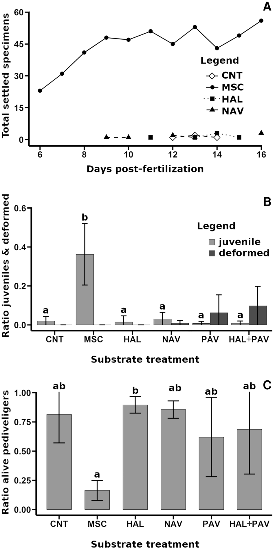
The earliest settlement (early postlarval) was observed in the ‘mosaic-like’ communities (MSC) (Figure 3A), with a total of 23 settled specimens at Day 6 post-fertilization. In the N. incerta, H. coffeaeformis and control treatments, one specimen settled on Day 9 post-fertilization, Day 11 post-fertilization, and Day 12 post-fertilization, respectively. Settled specimens were not observed during monitoring in Pavlova sp. and H. coffeaeformis + Pavlova sp. treatments (Figure 3A). The MSC showed signs of slow alteration, such as whitening and growing of fungi and algal biofilms (Supplementary Figure 5a, b).
Patella aspera. Assay 1: test of different substrates used as settlement inducers. (A) Total number of identifiable settled specimens during daily monitoring, zero values were not showed. (B) Ratio (mean ± SD) of juveniles (grey) and deformed specimens (dark grey). Different letters indicate significant differences (P < 0.05) among treatments in the ratio of juveniles. (C) Ratio of alive pediveliger larvae at the end of the assay (mean ± SD). Different letters indicate significant differences (P < 0.05) among treatments. CNT, control; HAL, diatom H. coffeaeformis; MSC, ‘mosaic-like’ CCA community; NAV, diatom N. incerta; PAV, haptophyte Pavlova sp.; HAL + PAV, combination of H. coffeaeformis and Pavlova sp.

One-way ANOVA and post hoc tests showed that the ratio of juveniles (RJ) was significantly greater in the MSC treatment (mean ± SD: 0.36 ± 0.16; F 5,24 = 18.81, P < 0.001; Figure 3B). In contrast, the RJ in the cultured algal strain treatments was similar to that in the control (average RJ = 0.02 ± 0.02; Figure 3B). Regarding the ratio of deformed specimens (RD), the Kruskal–Wallis test showed barely significant differences (χ2 = 11.64, P = 0.04), which were not supported by post hoc Dunn's test. Higher RD occurred in Pavlova sp. and H. coffeaeformis + Pavlova sp. treatments (RD = 0.06 ± 0.09 and 0.10 ± 0.10, respectively), while RD was almost zero in the other treatments (Figure 3B). The ratio of live pediveligers (RAP) was lower in MSC treatment (χ2 = 13.11, P = 0.022; Figure 3C).
Assay 2: test of different CCA communities
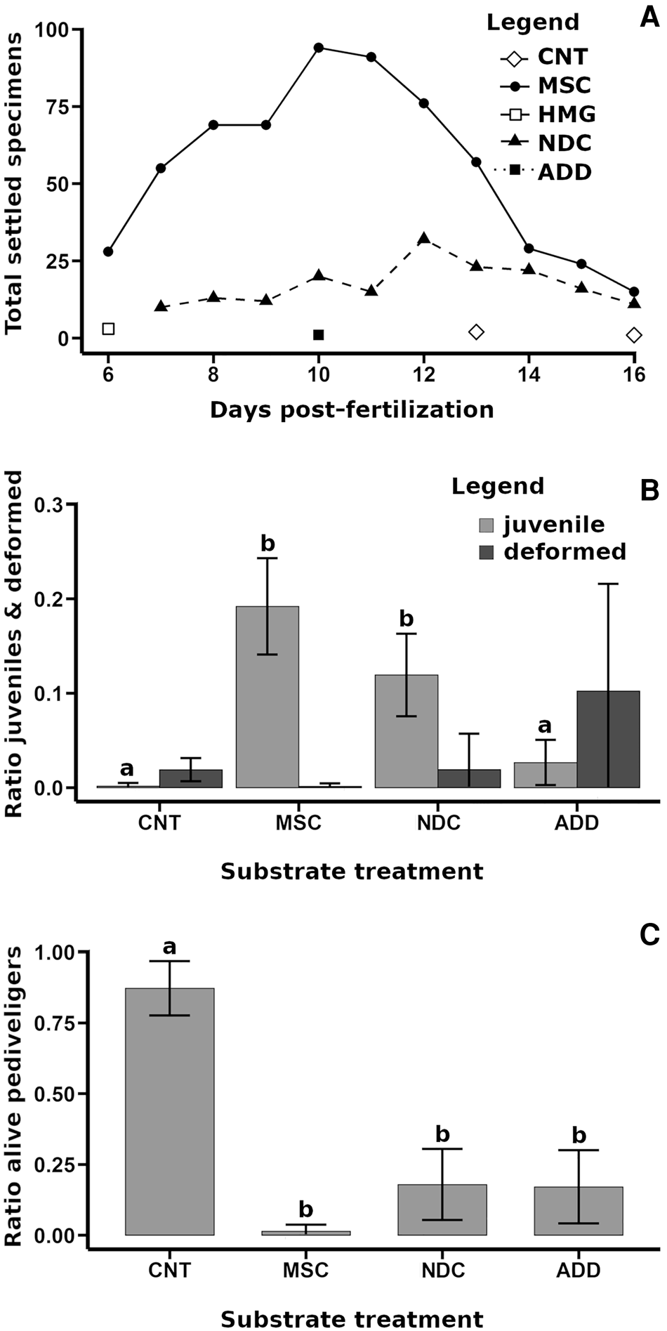
The earliest settlement (early postlarval) was observed at Day 6 post-fertilization in the MSC with a total of 28 settled specimens and ‘homogeneous pink’ communities (HMG) with three settled specimens (Figure 4A). Next, settlement occurred in ‘non-dominant CCA’ communities (NDC) with 10 total settled specimens at Day 7 post-fertilization. Settled specimens were observed continuously in MSC and SHL, but their abundance decreased after Days 11–12 post-fertilization (Figure 4A) in favour of empty (dead) juvenile shells. The observation of settled specimens was marginal in the control (CNT) and air-dried treatments (ADD) (Figure 4A). HMG treatments were not analysed statistically since they were removed at Day 5 of the assay (equivalent to Day 8 post-fertilization) because they showed important signs of a fast alteration that could have compromised the entire assay, such as decolouration (Supplementary File 5c), water turbidity, proliferation of microorganisms and bad smell. HMG showed high mortality, ranging from 0.37 ± 0.18 (Day 1 of the assay) to 0.94 ± 0.08 (day of the removal; Supplementary File 5d). The ADD treatment did not show clear alterations but high proliferation of microorganisms.
Patella aspera. Assay 2: Test of the influence of different CCA communities. (A) Total number of identifiable settled specimens during daily monitoring, zero values were not showed. (B) Ratio (mean ± SD) of juveniles (grey) and deformed specimens (dark grey). Different letters indicate significant differences (P < 0.05) among treatments in the ratio of juveniles. (C) Ratio of alive pediveliger larvae at the end of the assay (mean ± SD). Different letters indicate significant differences (P < 0.05) among treatments. CNT, control; ADD, ‘air dried’ CCA community; HMG, ‘homogeneous pink’ CCA community; MSC, ‘mosaic-like’ CCA community; NDC, ‘non-dominant’ CCA community.

The RJ was significantly greater in the MSC and NDC treatments (F 3,20 = 37.78, P < 0.001; Figure 4B). The RD was greater in ADD, but the differences were not statistically significant (χ2 = 7.17, P = 0.067). The RAP was significantly higher in the control than in the shell piece treatments (F 3,20 = 83.51, P < 0.001; Figure 4C).
Assay 3: test of CCA coverage influence on settlement
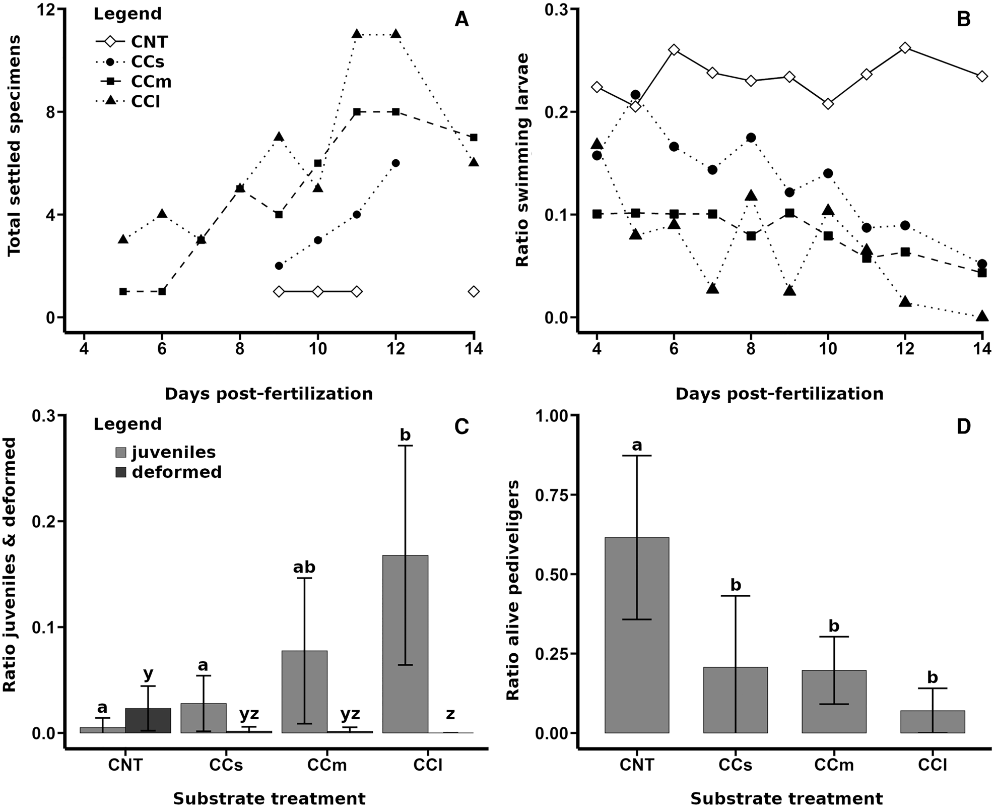
The first settled specimens (early postlarvae) were observed at Day 5 post-fertilization (second experimental day) in the medium (CCm) and large shell pieces (CCl), with 1 and 3 settled specimens, respectively (Figure 5A). In the control (CNT) and small shell pieces (CCs), the earliest settled specimens occurred at Day 9 post-fertilization (Figure 5A). The swimming pediveliger ratio was greater and constant in the control, while it tended to decrease in all shell piece treatments (Figure 5B). The CCl treatment was characterized by a notable abundance of metazoans, including nematodes (Supplementary File 6a), polychaetes (Supplementary File 6b), crustaceans (copepods and tanaids, Supplementary File 6c and d) and a midget larva (Supplementary File 6e and f). The CCl also showed alterations over time, including high metazoan activity (reproduction of copepods, accumulation of faeces, and elaboration of tubes as refuges), proliferation of microorganisms and fungal growth (Supplementary File 5e).
Patella aspera. Assay 3: test of CCA coverage influence on settlement. (A) Total number of identifiable settled specimens during daily monitoring, zero values were not showed. (B) Ratio of swimming pediveliger larvae observed during daily monitoring, standard deviation bars were not showed due to high overlapping. (C) Ratio (mean ± SD) of juveniles (grey) and deformed specimens (dark grey). Different letters indicate significant differences (P < 0.05) among treatments in the ratio of juveniles (a, b) and in the ratio of deformed specimens (y, z). (D) Ratio of alive pediveliger larvae at the end of the assay (mean ± SD). Different letters indicate significant differences (P < 0.05) among treatments. CCl, large shell piece; CCm, medium shell piece; CCs, small shell piece; CNT, control.

The RJ increased significantly with the size of the shell (Figure 5C; F 3,20 = 6.43, P < 0.01). The RD was greater and barely significant in the control (Figure 5C; χ2 = 9.30, P = 0.026). The RAP was greater in the control than in the shell piece treatments (F 3,20 = 10.10, P < 0.001; Figure 5D).
Discussion
The results of the current study proved the suitability of the Castejón et al. (Reference Castejón, Cañizares, Nogueira and Andrade2021) methodology to obtain viable trochophores of P. aspera and to observe their development, settlement and metamorphosis when reaching the juvenile stage. Similarly, Ribeiro (Reference Ribeiro2008), studying different limpet species from the UK, employed dissection and maturation in vitro to obtain viable trochophores able to settle in laboratory conditions, e.g. P. depressa, P. ulyssiponensis and P. vulgata. Trochophores of P. aspera did not show any special requirements to develop into competent pediveliger larvae and could be maintained in glass beakers during development. The exclusive gathering of swimming larvae (either trochophores or pediveligers) from the water column and their posterior placement in clean containers probably contributed to an increase in the number of viable larvae able to reach posterior stages of development. A similar methodology was used successfully for the larval culture of P. ferruginea (Ferranti et al., Reference Ferranti, Guallart, Fanciulli, Panzalis and Chiantore2021). Previous studies maintained limpet larvae at low density to study their development, ranging from less than 1 larvae ml−1 (Ribeiro, Reference Ribeiro2008) to 3–7 larvae ml−1 (Seabra et al., Reference Seabra, Cruz, Fernandes, Silva and Hawkins2019), 10 larvae ml−1 (Mau et al., Reference Mau, Bingham, Soller and Jha2018) and 10–15 larvae ml−1 (Ferranti et al., Reference Ferranti, Guallart, Fanciulli, Panzalis and Chiantore2021). For precautionary principles, we also recommend low density (less than 10 larvae ml−1) until further studies are performed. The larvicultural protocol used in this study could facilitate future studies focused on the early life and settlement requirements of different limpet species.
A clear nomenclature is fundamental to avoid misinterpretations among different life stages of limpets and to promote future studies on their biology and ecology. The crawling pediveliger larvae have been previously called ‘settled larvae’ or simply ‘settlers’, referring to these specimens as indicators of settlement (Nhan, Reference Nhan2014; Nhan & Ako, Reference Nhan and Ako2019; Seabra et al., Reference Seabra, Cruz, Fernandes, Silva and Hawkins2019), while ‘crawling larva’ was a term used to define the juveniles (Nakano et al., Reference Nakano, Nakayama and Takahashi2020). The results of this study showed three distinct life stages related to the settlement process: pediveliger larvae, early postlarvae and juveniles. The competent (or crawling) pediveliger larvae cannot be considered ‘settled larvae’ or ‘settlers’, as they represent an intermediate planktonic-benthic stage able to shift between crawling and swimming (Table 2; Figure 6). Therefore, the term ‘settled’ or ‘settlers’ should only be considered after the shedding of the velum with the consequent loss of the ability to swim, as occurred at the early postlarval stage. Velum shedding is one of the characteristics used to describe the onset of metamorphosis in abalone (Searcy-Bernal et al., Reference Searcy-Bernal, Salas-Garza, Flores-Aguilar and Hinojosa-Rivera1992; Roberts & Nicholson, Reference Roberts and Nicholson1997; De Viçose et al., Reference De Viçose, Viera, Bilbao and Izquierdo2010) and other marine gastropods (Zhao & Qian, Reference Zhao and Qian2002; Salas-Garza et al., Reference Salas-Garza, Parés-Sierra, Gómez-Rigalt and Carpizo-Ituarte2009; Ferranti et al., Reference Ferranti, Guallart, Fanciulli, Panzalis and Chiantore2021). Limpets in the early postlarval stage develop into juveniles, characterized by a well-developed crescent-moon shaped teleoconch and active grazing behaviour.
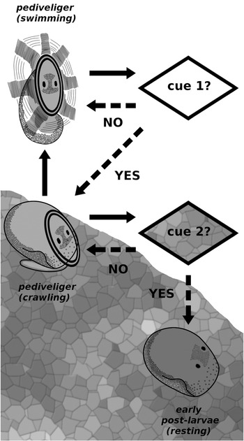
Hypothetical two-step model for the settlement of larvae of the true limpets. This model considers two settlement cues: the cue 1 is soluble and induces the sinking of the larvae to facilitate the contact with the substrate; the cue 2 is physically attached to the substrate and induces the metamorphosis to early postlarval stage.

The larval development of P. aspera is short, ranging from 5–6 days post-fertilization for the earliest settlers (early postlarvae) to 10–14 days, when the peak of settled specimens (early postlarvae and juveniles) occurred. This timing is within the range of other limpet taxa, including species of the same genus with 4–15 days (Ribeiro, Reference Ribeiro2008; Guallart et al., Reference Guallart, Peña, Pérez-Larruscaín, Luque and Templado2020; Ferranti et al., Reference Ferranti, Guallart, Fanciulli, Panzalis and Chiantore2021), as well as the genera Cellana with 4–6 days (Mau et al., Reference Mau, Bingham, Soller and Jha2018) and Lottia with 6–12 days (Kay & Emlet, Reference Kay and Emlet2002; Nakano et al., Reference Nakano, Nakayama and Takahashi2020). Although the timing for development in limpets is specific and temperature dependent (Ribeiro, Reference Ribeiro2008; Ferranti et al., Reference Ferranti, Guallart, Fanciulli, Panzalis and Chiantore2021), the short duration of the larval stages implies a limited dispersal phase, explaining the genetic and morphometric differences observed between the Macaronesian and continental populations (Côrte-Real et al., Reference Côrte-Real, Hawkins and Thorpe1996a, Reference Côrte-Real, Hawkins and Thorpe1996b; Weber & Hawkins, Reference Weber and Hawkins2005; Carreira et al., Reference Carreira, Shaw, Gonçalves and McKeown2017).
Previous studies (Ferranti et al., Reference Ferranti, Monteggia, Asnaghi and Chiantore2018; Mau & Jha, Reference Mau and Jha2018) have recommended the use of microalgae diets to increase the survival and settlement success of limpet larvae. In contrast, Kay (Reference Kay2002) did not observe an improvement in the settlement rate of the limpet Lottia digitalis when using microalgae. Our results support the observations of Kay (Reference Kay2002), i.e. microalgae did not enhance the survival and/or settlement success of the pediveligers of P. aspera, suggesting that limpet larvae are similar to those of abalone (genus Haliotis), which are lecithotrophic animals able to survive several weeks in the absence of food, relying on their yolk reserves (Takami et al., Reference Takami, Kawamura and Yamashita2000; Roberts & Lapworth, Reference Roberts and Lapworth2001; Moran & Manahan, Reference Moran and Manahan2003; Takami & Kawamura, Reference Takami and Kawamura2003). Several observations support the lecithotrophic hypothesis for the larvae of P. aspera. First, the larvae can develop into competent pediveliger larvae without any food source. Second, the larvae are able to swim for 2 weeks in the absence of food, hypothetically at the expenditure of energy from rich yolk reserves. Third, the larvae did not show any clear feeding activity in the presence of substrates, either microalgae or CCA. Fourth, the juveniles showed a shift to active grazing and excretory activity (Supplementary Videos 2 and 3), similarly to the abalone species (Takami et al., Reference Takami, Kawamura and Yamashita2000; Roberts & Lapworth, Reference Roberts and Lapworth2001; Moran & Manahan, Reference Moran and Manahan2003; Takami & Kawamura, Reference Takami and Kawamura2003). Finally, the digestive system was more conspicuous after metamorphosis to juveniles (Supplementary File 4).
The aforementioned observations suggest that the P. aspera digestive system at the early postlarval stage undergoes dramatic changes, involving the development of structures required to ingest and digest particulate food, thus placing the ‘early postlarval’ stage as a differentiated and metamorphic stage. Nevertheless, the hypothesis requires confirmation by histological observations. After the early postlarval stage, the juveniles of P. aspera showed remarkable grazing activity (Supplementary Videos 2 and 3), indicating that juveniles need to grow quickly at the expense of energetically costly crawling behaviour (Davies et al., Reference Davies, Hawkins and Jones1990; Niu et al., Reference Niu, Nakao and Goshima1998). The epithallial shedding of the CCA (and its associated epibionts) could be a primary food source for the juvenile limpets, as they were observed grazing upon epithelial shedding, giving their resultant faeces a similar white colouration (Supplementary File 4, Supplementary Video 2). This food resource is consistent with the limpet-feeding strategy as non-selective grazers and opportunistic consumers (Della Santina et al., Reference Della Santina, Sonni, Sartoni and Chelazzi1993; Burgos-Rubio et al., Reference Burgos-Rubio, De la Rosa, Altamirano and Espinosa2015; Schaal & Grall, Reference Schaal and Grall2015).
Our study suggests that CCA communities are the natural settlement substrate for P. aspera larvae. The CCA shortened the timing for settlement and increased the ratio of successful juvenile metamorphosis in comparison to the control treatments. These results are also supported by field observations where juveniles of several patellid limpet species were observed to be associated with environments dominated by CCA (McGrath & Foley, Reference McGrath, Foley and Wilson2005; Seabra et al., Reference Seabra, Cruz, Fernandes, Silva and Hawkins2019). Also, Ribeiro (Reference Ribeiro2008) observed increased settlement ratio in Patella larvae when exposed to CCA. In contrast, Kay (Reference Kay2002) did not observe a clear influence on the settlement ratio in Lottia larvae in the presence of CCA. These different observations can be explained by differential settlement requirements in each limpet taxa, as well by differential settlement inductors in different CCA communities. In Assay 2, different CCA communities presented dissimilar settlement success for P. aspera, in accordance with the reduced settlement rate in Assay 3 compared with Assay 1, i.e. ‘mosaic-like’ communities were preferred by the larvae of P. aspera over communities dominated by dark red crusts. Similarly, the competent larvae of the abalone Haliotis iris also presented dissimilar settlement success in the presence of different CCA species (Roberts et al., Reference Roberts, Kaspar and Barker2004).
Following the results obtained in this study, we propose that settlement and metamorphosis in limpets are triggered by a two-step mechanism (Figure 6), as suggested for other marine gastropods (Chia & Koss, Reference Chia and Koss1988; Zhao & Qian, Reference Zhao and Qian2002). The first step consists of soluble cues that inhibit swimming and induce crawling behaviour (Figure 6). This is suggested by the lower swimming activity of larvae when CCA were included, opposed to constant swimming larvae without CCA. It is accepted that CCA contain GABA-mimetic peptides and that abalone larvae react to GABA and CCA extracts, ceasing swimming behaviour and promoting sinking and crawling (Barlow, Reference Barlow1990). The second step of this model requires a different cue, which is responsible for triggering metamorphosis (Figure 6). If this second cue accumulates in certain areas of the substrate, it would explain the common small aggregations of early postlarvae over the substrate (Supplementary File 3), suggesting that P. aspera larvae may discriminate settlement cues at microscale levels.
Supplementary material
The supplementary material for this article can be found at https://doi.org/10.1017/S0025315421000916
Acknowledgements
The sampling of limpets for scientific purposes during closed season was authorized by the Fisheries Directorate, Secretary for the Sea and Fisheries, Regional Government of Madeira. The authors would like to thank Gercende Courtois de Vicose for the algal strains donations and valuable advice on marine gastropods culture. The authors also would like to thank the technicians at Mariculture Center of Calheta for their assistance and local fishermen of Calheta for helping provide the specimens. The authors have no conflict of interest to declare.
Funding
Financial support provided by the project ‘AQUAINVERT - Development of sustainable, integrated and innovative aquaculture in Macaronesia. Research and Development to promote the production of marine invertebrates of commercial interest (MAC/1. 1ª/282)’ founded by FEDER under the INTERREG MAC 2014-2020 program. D.C. was supported by a grant under the project AQUAINVERT.
Conflicts of interest/Competing interests
The authors declare no conflicts of interest.
Ethics approval
The authors confirm that the ethical policies of the journal, as noted on the journal's author guidelines page, have been adhered to. All applicable international, national, and/or institutional guidelines for the care and use of animals were followed.
Availability of data and material
The data that support the findings of this study are available from the corresponding author upon reasonable request.
Authors' contributions
Castejón, D.: animal management, experimental design, experiment realization, animal observation and description, sampling and samples analyses, statistical analyses, drafted paper. Nogueira, N.: drafted paper revision, project elaboration. Andrade, C.A.P.: drafted paper revision, project elaboration, coordination and direction.