INTRODUCTION
The phylum Myxozoa contains over 2180 species, almost all of which are considered to be obligate parasites of aquatic animals (Lom and Dyková, 2006). It is composed of 2 classes, the Myxosporea and the Malacosporea, members of which are important pathogens responsible for severe economic losses in cultured and wild fisheries (Lom and Dyková, 2006). While the life-cycles of a number of myxosporeans have been elucidated and involve an alternation of hosts between an annelid and fish, to date no malacosporean life-cycle has been completed (Tops and Okamura, 2005).
Tetracapsuloides bryosalmonae is the malacosporean parasite that causes proliferative kidney disease (PKD) in salmonid fish. In Europe and North America, this disease has been a major constraint on the cultivation of rainbow trout Oncorhynchus mykiss and other salmonids for over a quarter of a century. It primarily affects fingerling fish (i.e. those in their first year of contact) and usually results in 100% morbidity on an affected farm with up to 20% mortality (Hedrick et al. 1993). Impacts on wild populations are generally unknown but are potentially substantial (Feist et al. 2002; Whali et al. 2002).
PKD is characterized by a chronic inflammation of the anterior and posterior kidney which is caused by a massive accumulation of lymphocytes and extensive granuloma formation (Chilmonczyk and Monge, 1999). The inflammatory response is centred on the interstitial pre-sporogonic stages of T. bryosalmonae that undergo extensive proliferation in the fish host (Ferguson and Needham, 1978). Initial studies on these stages could not adequately speciate the parasite leading it to be referred to simply as PKX (Seagrave et al. 1980). Kent and Hedrick (1985, 1986) described renal intra-tubular sporogonic stages, some of which possessed polar capsules and identified the parasite as a member of the phylum Myxozoa. While polar capsules are a distinguishing feature of myxozoan spores, the spores of T. bryosalmonae lacked hard valves and were therefore thought to be immature. Together with the severe inflammation caused by the parasite this led to speculation that salmonids were an aberrant host (Seagrave et al. 1980; Kent and Hedrick, 1986, 1987). However, extensive studies on wild fish populations consistently failed to identify any other potential fish host (Hedrick et al. 1986; Feist, 1988; MacConnel and Smith, 1990; Feist and Bucke, 1993; Kent et al. 1993; Kent et al. 1998; Morris et al. 2000a).
Two papers by Okamura (1996) and Canning et al. (1996) respectively described an unusual Myxozoan parasite Buddenbrockia plumatellae (syn. Tetracapsula bryozoides) infecting the freshwater bryozoan Cristatella mucedo. This parasite formed sacs within the host filled with spores that lacked hard valve cells. Subsequent 18ssu rDNA analysis and microscopical studies determined that T. bryosalmonae also infected a range of freshwater bryozoans (Anderson et al. 1999; Longshaw et al. 1999). The unusual sporogenesis and cytoplasmic nature of the spores led to the class Malacosporea being erected in the phylum Myxozoa to accommodate these parasites (Canning et al. 2000). Transmission experiments confirmed that spores of T. bryosalmonae released from bryozoans could infect salmonids resulting in PKD (Feist et al. 2001). While bryozoan to bryozoan transmission is not known to occur (Tops et al. 2004), the role of salmonids remains in question with T. bryosalmonae being reported from bryozoans in lakes with no known salmonid populations (Anderson et al. 1999; Okamura et al. 2001). Furthermore, a series of experiments to transmit T. bryosalmonae from infected salmonids to naïve bryozoans failed, leading to the suggestion that fish are either facultative or aberrant hosts and are not essential for the spread of the disease (Tops et al. 2004; Tops and Okamura, 2005).
Here we provide evidence that transmission of T. bryosalmonae from a salmonid to a bryozoan does occur, thus completing the first life-cycle of a malacosporean parasite and indicating that native salmonids are an important part of this parasite's life-cycle.
MATERIALS AND METHODS
Collection, culture and verification of naïve bryozoans
To reduce the possibility of collecting parasite-resistant bryozoans (Tops and Okamura, 2003), colonies of Fredericella sultana were collected from a river site in Southern England where T. bryosalmonae-infected bryozoans had been previously obtained (50 °47′22″N, 02 °28′15″W) during March 2005. Portions of the colonies or their attached substrata were glued onto 9 cm diameter Petri dishes using a cyanacrylate adhesive and macroinvertebrates were removed using a dissecting microscope and fine forceps. The colonies were initially maintained for 1 month at 18–19 °C in a custom-built, closed re-circulating culture system and fed a mixture of laboratory-cultured algae Cryptomonas ovata, Cholorgonium capillatum, Rhodomonas sp. and Synura sp. (McGurk, 2005). The culture system was comprised of a backlit 25 L aquarium filled with Jarwoski's algal medium pumped into a circular bucket. The bucket had a central stand to clip the Petri dishes onto and an airlift to aerate the media and generate a gentle current. The outflow of the bucket returned to the aquarium.
Due to the transparent nature of the cystid associated with cultured bryozoans, overt T. bryosalmonae infection could be assessed by regular examination using an inverted microscope. Eight distinct colonies were selected which appeared to be parasite free. Portions of these colonies comprising of 6 zooids each were glued onto a new Petri dish using cyanacrylate adhesive and transferred to a fresh culture system. These colonies were maintained in this system for a further 4 months before the start of the transmission experiment. During this time the growing colonies were regularly examined for signs of T. bryosalmonae infection using an inverted microscope. At approximately monthly intervals, 5 zooids were removed from different areas of each of the colonies, DNA was extracted using a magnetic mini-prep kit (ABGene, UK), quantified using a nanodrop spectrophotometer (Nanodrop Technologies, USA), and examined for the presence of T. bryosalmonae using PCR and the primers 5F and 6R (Kent et al. 1998) following the recommended cycling conditions at both 35 and 40 thermocycles. Portions of collected bryozoans exhibiting overt T. bryosalmonae infection were used as positive controls. Immediately prior to the exposure experiments, further control colonies were obtained by removing relatively substantial portions of the naïve colonies (>20 zooids) from 4 discrete areas of the Petri dish. To encourage the adherence and growth of these pieces, netting (0·75 mm diameter mesh) was glued over the Petri dishes. The removed colony fragments were introduced into the space between the netting and the dishes, which were subsequently inverted and placed into the original culture system.
Infection of brown trout
Brown trout Salmo trutta eggs, obtained from a commercial hatchery, were transferred to the Aquatic Research Facility at the University of Stirling and hatched. This facility is specific pathogen-free, and uses temperature-controlled, dechlorinated, mains tap water to feed experimental 25 L flow-through tanks at a rate of 0·5 L min−1. The trout were fed a proprietary diet and grown to ~7 g. Prior to exposure, 12 fish were killed by an overdose of benzocaine followed by severing of the spinal cord just behind the head, kidney samples were taken and tested using PCR and primers 5F, 6R. To further validate the T. bryosalmonae naïve status of the fish, a portion of the posterior kidney of these fish was fixed in formalin and immunohistochemically stained using the T. bryosalmonae-specific monoclonal antibody mAb P01 (Aquatic Diagnostics, Stirling, UK) following the manufacturer's protocol.
Using the procedure of McGurk et al. (2006) a total of 110 brown trout were exposed twice on consecutive days to water containing T. bryosalmonae spores released from infected bryozoan colonies collected during the above sampling. Fifty of these trout were transferred to a separate tank for the transmission experiment, the other 60 being used to monitor the progression of spore development within the fish. Monitoring was started 2 weeks post-exposure and all fish maintained at 18 °C. Six of these fish were killed on a weekly basis, kidneys removed and fixed in neutral buffered formalin, embedded in paraffin wax, sectioned and stained using immunohistochemistry with mAb P01 and mAb B4. The mAb B4 was previously developed against the sporogonic stage of the parasite (Morris et al. 1997). Immunohistochemistry using mAb B4 was conducted as for mAb P01 except that the mAb was used as neat hybridoma supernatant rather than reconstituted.
Transmission of Tetracapsuloides bryosalmonae to naïve Fredericella sultana
At the time when the brown trout were first noted to possess multicellular sporogonic stages and spores as verified by immunohistochemistry, water from the tank of 50 infected fish was introduced into the system containing naïve F. sultana continuously for 6 weeks (0·5 l h−1) using a peristaltic pump while aquarium water was introduced into the system containing the control colonies. During this period all of the bryozoans were routinely examined on a weekly basis for overt T. bryosalmonae infection using a dissecting microscope.
Confirmation of T. bryosalmonae infection in exposed colonies
A portion of F. sultana colony, comprising 3 zooids with putative T. bryosalmonae spore sacs within it was excised and the DNA isolated using a magnetic mini-prep kit (ABGene, UK). The DNA was then examined using PCR, as above, using specific primers 5F and 6R. The entire DNA of the control colonies was also extracted and tested using PCR and these primers.
A further portion of the infected colony, containing spores and mature spore sacs was dissected apart in a separate 5 cm Petri dish. The contents of this dish were then washed into a 25 l flow-through aquarium containing 6 T. bryosalmonae naïve rainbow trout (~10 g) obtained from eggs grown in the facility. The fish were maintained for 6 weeks at 18 °C before examining for T. bryosalmonae infection using immunohistochemistry with mAb P01 as described above. Aquaria containing unexposed rainbow trout were used as a control.
RESULTS
Collection of naïve bryozoans
Large numbers of F. sultana were found encrusting the roots of a willow tree, situated on a riverbank. Small sections of these roots were transported to the lab for culture. The entwining and fragmentary nature of this bryozoan species, coupled with an opaque cystid meant that enumerating absolute numbers of individuals within a colony was not possible.
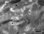
Evidence of infection of the F. sultana was first noted 1 week after collection, within the metacoel of several individuals, in the form of recognizable malacosporean spherical spore sacs. One month after collection the majority of bryozoans appeared to be infected. The infection continued in the colonies with increasing numbers of spore sacs being observed within their metacoels. After 2 months the severity of the infections, meant that for many infected individuals, the metacoel was packed with maturing sacs with the zooids either dead or misshapen with stunted lophophores and guts (Fig. 1). Statoblasts (specialized bryozoan propagules) were not produced by overtly infected colonies in contrast to those that were not infected in which they continued to develop. At the end of 3 months only bryozoans that never demonstrated parasitic infection, including those that were sampled for the transmission experiment, remained alive in the culture. These colonies continued to grow and appeared normal throughout the culture period.

Fig. 1. Dead Fredericella sultana zooids packed with developing spore sacs, indicated with *. A healthy zooid can be observed at the top of the figure, indicated with an arrow.
From the 8 naive colonies selected for the transmission experiment only 2 survived the transfer and subsequent gluing to the fresh Petri dish. These 2 colonies subsequently colonized the dish and did not show any sign of T. bryosalmonae infection, with PCR results repeatedly failing to demonstrate the presence of parasite DNA.
Transmission of T. bryosalmonae to brown trout
After exposure, pre-sporogonic parasites were observed in all of the fish sampled. Initially, these were invariably noted adjacent to tubules but as the infection progressed increasing numbers of interstitial parasites were observed, together with associated inflammatory changes in the tissues surrounding them, as described in previous studies (Ellis et al. 1985; Kent and Hedrick, 1986). Intra-luminal sporogonic stages were first noted 21 days post-exposure and appeared as single cells within the tubules. Spores and multi-cellular sporogonic stages were noted 9 weeks post-exposure and it is at this time that the bryozoans were exposed.
Exposure of naïve Fredericella sultana to water containing brown trout
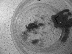
At the time of exposure the bryozoans had colonized the majority of the Petri dish (Fig. 2). Natural fragmentation by these colonies resulted in the formation of numerous clonal individuals connected together by a common cystid.

Fig. 2. Growth of experimental Fredericella sultana colonies over a 9 cm Petri dish immediately prior to exposure experiment.
Forty-two days post-initial exposure to water supplied from the tank of brown trout, numerous single cells of about 10 μm in length were observed, swirling within the metacoel of a single bryozoan comprising of 7 zooids, followed by observable spore sacs 2 days later. Spore sacs within this individual were initially only intermittently produced, being present in the coelom for around 48 h before they disappeared. However, the incidence of developing sacs gradually increased so that by the second to third week of the overt infection sacs of varying sizes were being continuously observed. During this time T. bryosalmonae infection was noted within other areas of the colonized Petri dish. Due to the bushy nature of the cultured F. sultana colonies it was difficult to precisely follow development in the majority of the infected individuals and we could not accurately determine the level of infection. However, some branches of bryozoan had adhered and grown along the plastic, aiding in the repeated examination of the infection.
Spore sac development during the overt infection progressed as follows. From the beginning of the overt infection single cells were always observed in the metacoel of an infected individual. These cells formed aggregations that floated within the bryozoan's metacoel. The smallest of these was composed of only 2 or 3 cells, but much larger aggregates were observed containing numerous cells. These aggregates gradually resolved themselves into spherical bodies (spore sacs) onto which masses of cells remained attached. As the spore sacs matured, increasing in size, so the attached cells disappeared from their surface resulting in the sacs appearing uniformly spherical. The sacs then ruptured releasing mature spores into the metacoel where they were presumably released by the host into the environment. Statoblast production ceased in those individuals displaying the overt infection, while in apparently healthy parts of the colony their development continued.
During the experiment, the infection only affected a minority of individual bryozoans and occurred in parts of the colonies that were still attached to non-infected individuals through a common cystid. Those colonies grown in the separate culture system did not show any signs of infection with subsequent PCR examination of these colonies failing to amplify T. bryosalmonae DNA. PCR of the experimentally infected colonies produced a product of the expected size. In addition, the rainbow trout exposed to the dissected colonies developed clinical signs of PKD, with moderate kidney swelling and pre-sporogonic parasites within their kidneys in contrast to the control fish.
DISCUSSION
This study has demonstrated that Tetracapsuloides bryosalmonae can be transmitted to a bryozoan host from infected brown trout, thus resolving speculation regarding the role of a salmonid in this parasite's life-cycle. Furthermore, fish to bryozoan to fish transmission was achieved, indicating that the parasite can cycle between these hosts indefinitely without the need for an additional host.
For the transmission experiment it was important that the hosts were known to be free of infection prior to the study. Although specific pathogen-free brown trout eggs were used, the bryozoans obtained for the trial were collected from a T. bryosalmonae enzootic river so there is a possibility that they were infected prior to the experimental exposure. The presaccular development of T. bryosalmonae in Fredericella sultana involves proliferating, amoeboid stages that are distributed throughout an infected colony (Morris and Adams, 2006a). If these stages were present within the original bryozoan fragments used in the transmission experiment it would be expected that all of the subsequent colonies derived from these would also be infected. Therefore, we believe that the steps taken to verify the naïve status of these colonies, such as an extended quarantine period (5 months), regular PCR analysis of zooids and co-culture of the original colony and substantive colony fragments were adequate to demonstrate the absence of any covert or latent infection. PCR analysis of the control bryozoans also did not demonstrate infection, as expected if the original colonies were naïve. Further more, only a proportion of the exposed colonies became overtly infected, suggesting that the bryozoans became infected after natural colony metacoelomic fragmentation had occurred on the dish.
While identification of the precise stage infecting bryozoans was not determined, it is reasonable to assume that this was a spore derived from an infected fish. Spores were present within the tubules of the brown trout used to monitor the infection at the time of the experimental exposure, and recognizable malacosporean spores have been previously reported from the urine of T. bryosalmonae-infected fish (Hedrick et al. 2004). Earlier studies to naturally transmit T. bryosalmonae from fish to bryozoans have failed. However, none of these transmission trials precisely timed the experimental exposures to when fish could be expected to shed mature spores i.e. when spores were observed in kidney tubules. The trials being conducted when only sporogonic stages were recognized (Morris et al. 2002a,b; Tops et al. 2004). This suggests that failure to elicit infection was the result of infective spores not being present within the salmonids at the time of exposure. Numbers of mature spores in the urine increase towards the end of the clinical disease (Hedrick et al. 2004), correlating to their visualization in the brown trout used to monitor disease progression for the transmission experiment. However, only sporogonic stages, not spores, have been regularly reported as persisting in fish after the clinical disease has subsided (Kent and Hedrick, 1986; Morris et al. 2000a). This suggests that infective spores are only produced periodically during infection and therefore timing is an important factor to achieve successful transmission. This may also explain why a previous experiment to infect bryozoans by exposure to macerated infected kidney produced a positive PCR result (Morris et al. 2002a) while subsequent attempts to infect bryozoans by this method failed (Morris et al. 2002b; Tops et al. 2004). In theory, while feeding macerated kidney which contained spores could transmit the infection to bryozoans, nothing is known about the conditions needed for the spores to become or remain infectious, therefore such transmission methods need to be further validated.
It is notable that although infection was induced in the exposed colonies the incidence within them appeared to be relatively low. This may have been, in part, due to the use of algal media within the culture system rather than freshwater that may have adversely affected spore infectivity. Little is known about spore development and release, although studies on rainbow trout in the USA suggest it may be a rare event in diseased and recovering fish (Hedrick et al. 2004). However, the observation that Arctic charr produce large numbers of spores suggests that the quantity of spores produced in one salmonid species are not indicative for all species (Kent et al. 2000). Sporogonic stages are known to persist in fish long after they have recovered from the clinical disease and it is likely that intra or extra-piscine environmental cues may induce further sporogenesis (Kent and Hedrick, 1986; Hedrick et al. 2004). Until more is known about sporogony in the fish host, transmission to bryozoans will be difficult to optimize.
The transmission of T. bryosalmonae from brown trout to a bryozoan suggests that all species of salmonid that produce mature T. bryosalmonae spores are capable of transmitting the infection to susceptible bryozoan species. Spores have been reported from Arctic charr Salvelinus alpinus, Chinook salmon O. tshawytscha, Coho salmon O. kisutch, cutthroat trout O. clarki, Atlantic salmon S. salar and brown trout S. trutta (Hedrick et al. 1993). However, for rainbow trout O. mykiss, reports of spore production have been conflicting, with spores being observed in North America but not Europe (Bucke et al. 1991). This led to the suggestion that there is a European strain of the parasite and a North American one, adapted to species of Salmo and Oncorhynchus respectively (Bucke et al. 1991; Morris et al. 1997). Distinct genetic differences have been found to exist between T. bryosalmonae isolates obtained from the two continents (Henderson and Okamura, 2004) and this further supports the hypothesis of T. bryosalmonae strains adapted to indigenous salmonids. It is important that the ability of a range of salmonids to transmit the infection to bryozoans is further examined; both to verify the results of the brown trout transmission experiment and to determine precisely which species are involved in the life-cycle of this parasite.
The report of T. bryosalmonae infecting bryozoans from Cowan Lake, in Ohio where salmonids have not been found suggests that either infected bryozoans can persist and spread independently of salmonids or there is another host capable of completing the life-cycle (Okamura et al. 2001). The observation that high temperatures exacerbate the infection in F. sultana suggests that the warm waters of this water body may be detrimental to infected bryozoans and therefore to maintain the infection another host may be required. While salmonids are well documented as hosts for the parasite, there are reports of the Northern pike Esox lucuis being infected with an organism reminiscent of T. bryosalmonae (Seagrave et al. 1981; Bucke et al. 1991). This parasite was identified in the kidney and was ultrastructurally identical to T. bryosalmonae. In addition, both the extrasporogonic and intra-tubular sporogonic stages of this parasite stained with immunohistochemistry and in situ hybridization techniques developed for the detection of T. bryosalmonae further confirming its identity (Morris et al. 2000a,b). Esociform fish are believed to be closely related to salmonids which could help to explain how T. bryosalmonae infect members of both groups (Canning and Okamura, 2004). While E. lucuis does not inhabit Cowan lake, a closely related species, E. masquinongy, is stocked and therefore has the potential to act as the vertebrate host. However, data regarding T. bryosalmonae infections of Esox spp. is at present very limited and is in need of additional research.
The development of the infection in the exposed colonies correlated with that observed by McGurk et al. (2006). These authors reported that T. bryosalmonae was detrimental to the host with much of an infected colony dying during the overt infection. In the present study, all of the infected bryozoans eventually died of the infection. The development of T. bryosalmonae in fish and bryozoan hosts is closely linked to temperature, with lower temperatures reducing the impact of the disease in rainbow trout (Ferguson, 1981; Clifton-Hadley et al. 1987; Gay et al. 2001; de Kinkelin and Loriot, 2002). This is thought to be due to elevated temperatures influencing the rate of parasite proliferation within the host, with the parasite continuing to replicate until the host is either able to mount an effective immune response or the temperature is lowered (Morris et al. 2005). The relatively high constant temperature at which the collected bryozoans were cultured during the quarantine period appeared to permit unregulated proliferation of the parasite, eventually resulting in the death of the infected hosts. This suggests that in the wild, during long, hot summers overtly infected bryozoans would regress and die while those infected later in the season, when temperatures were beginning to fall, would survive either remaining infected until the following year or producing infected over-wintering statoblasts. This correlates with the known epidemiology of the parasite both in brown trout populations, where fish are infected in early summer (June) and spores are detected 2–3 months later (Ellis et al. 1985; Clifton-Hadley and Feist, 1989; Morris and Adams, present study) and in bryozoans, where infected F. sultana populations are present throughout the winter but, in hot summers, the prevalence of infected colonies appears to be reduced, making them difficult to identify from some PKD enzootic sites (Gay et al. 2001; Okamura et al. 2001).
While the bryozoan F. sultana overwinters as a living colony, other species of freshwater bryozoans regress and survive as statoblasts. It has been postulated that these asexual propagules may harbour the infection, allowing temporal survival of the parasite over-winter and enhance the dissemination of the parasite when they are released from infected colonies (Okamura, 1996; Okamura et al. 2001; Henderson and Okamura, 2004; Tops et al. 2004). Dispersal of statoblasts through waterfowl has been suggested as the original route of introduction of T. bryosalmonae from North America into Europe (Henderson and Okamura, 2004). Such dispersal also correlates with the diverse nature of habitats that T. bryosalmonae-infected bryozoans have been identified from, including water bodies with no known salmonids present (Okamura and Wood, 2002). However, in all malacosporean infections of bryozoans examined to date, statoblast production is either markedly reduced or ceases, while attempts to hatch those statoblasts obtained from infected colonies have failed (Okamura, 1996; Canning et al. 2002; Morris et al. 2002b; McGurk, 2005). However, this does not preclude successful infection of statoblasts which form prior to the overt infection as suggested by Tops and Okamura (2005). As such while the role of statoblasts from infected bryozoans cannot be discounted, it appears that a principal route for the spread and maintenance of the parasite in bryozoans is through exposure to spores released from native salmonids. Therefore, the farming of native salmonids and restocking of salmonid fisheries are likely to increase the levels of parasite in endemic areas.
In addition to waterfowl transporting infected statoblasts, translocation of T. bryosalmonae to new water bodies could occur through the introduction of infected salmonids either through anadromous fish that have migrated off course or stocking activities. The stocking of infected fish has been suggested as a mechanism to account for the spread of PKD in Swiss brown trout populations (Wahli et al. 2002). The original intercontinental translocation of the parasite pre-dates fisheries activities (Henderson and Okamura, 2004) and may have occurred through species of susceptible bryozoans and salmonids such as Artic charr that have natural distributions spanning the northern hemisphere. The remarkable ecological plasticity of some salmonids such as brown trout would further facilitate the distribution of the parasite to a diverse range of habitats while intra-clonal propagation through fragmentation of bryozoan colonies may assist the establishment of the parasite into new water bodies by increasing the spread of infected bryozoans within a body. This in turn would increase the spatial pattern of spore release thus increasing the likelihood of more fish becoming infected. Intraclonal propagation may also allow the parasite to be maintained and proliferate for extended periods within a lake or river without the need for another host (Morris and Adams, 2006b). However, this would rely on environmental conditions that would permit the infection to persist whilst not killing the bryozoan host.
While the number of malacosporean species reported from bryozoan hosts is increasing (Tops et al. 2005), malacosporean-like parasites have also been reported from non-salmonid fish such as cyprinids (Voronin, 1993). Tetracapsuloides bryosalmonae is the first malacosporean for which an alternation of host has been demonstrated indicating that the life-cycles of other malacosporeans may also require a bryozoan and fish host. This cycling between an invertebrate and vertebrate host is consistent with the other myxozoan class Myxosporea where at least some of the species alternate between an annelid and fish.
The authors would like to thank Niall Auchinachie for fish husbandry. This research was funded by a grant from the Biotechnology and Biological Sciences Research Council (grant no: BBC5050241) awarded to D.J.M. and A.A.