Introduction
The separate developments of the first and second polar bodies are fundamental biological events in the physiology of oocyte–zygote transition, and evaluation of the morphology of these structures is a crucial step in the assisted reproduction laboratory routine.
The first polar body is extruded after the first meiosis has been completed, i.e. during the maturation of the oocyte. It contains cellular components such as the nucleus and other organelles, but its volume is extremely small compared with that of the ovum. Unlike the oocyte, the first polar body always undergoes degeneration, i.e. it is programmed to die even when fertilization occurs. The decay of the first polar body is usually complete by approximately 20 h after its extrusion (Ortiz et al., Reference Ortiz, Lucero and Coxatto1983). Although it is known that this degradation is an active process that can be reversed [in Mos-deficient mouse oocytes–Mos: upstream activator of mitogen-activated protein kinase–the first polar body persists instead of degrading and sometimes undergoes an additional cleavage (Choi et al., Reference Choi, Fukasawa, Zhou, Tessarollo, Borror, Resau and Vande Woude1996)], its mechanism has so far not been clearly elucidated. The presence of fragmented DNA in the nuclear area of the first polar body in unfertilized mouse (Takase et al., Reference Takase, Ishikawa and Hoshiai1995; Fujino et al., Reference Fujino, Ozaki, Yamamasu, Ito, Matsuoka, Hayashi, Nakamura, Ogita, Sato and Inoue1996) and human oocytes (Van Blerkom & Davis, Reference Van Blerkom and Davis1998; Corn et al., Reference Corn, Hauser-Kronberger, Moser, Tews and Ebner2005) leads to the suggestion that first polar body elimination occurs by apoptosis. However, no other apoptotic features were observed in previous studies (Zakeri et al., Reference Zakeri, Lockshin, Criado-Rodríguez and Martínez2005). As DNA degradation visualized by TUNEL assay can also be connected with necrotic cell death (Darzynkiewicz et al., Reference Darzynkiewicz, Bedner and Traganos2001), there is as yet still no definitive proof for apoptotic way of death.
The morphology and the cellular content of the second polar body are very similar to the first polar body. Matured oocytes release the second polar body immediately after fertilization. It can be present in the perivitelline space of the developing embryo for several days; however, in the end, it is eliminated too. The decay of the second polar body is usually complete before the embryo reaches the blastocyst stage, and there is much more evidence for the involvement of apoptosis in its death. Beside DNA fragmentation, which was detected in the second polar bodies of rat (Szołtys et al., Reference Szołtys, Tabarowski and Pawlik2000) and pig preimplantation embryos (Tatemoto et al., Reference Tatemoto, Sakurai and Muto2000; Hao et al., Reference Hao, Lai, Mao, Im, Bonk and Prather2004), mouse embryos from the 2-cell stage also showed exteriorization of phosphatidylserine on the outer plasma membrane leaflet (Zakeri et al., Reference Zakeri, Lockshin, Criado-Rodríguez and Martínez2005) and the presence of caspase activity (Martinez et al., Reference Martinez, Rienzi, Iacobelli, Ubaldi, Mendoza, Greco and Tesarik2002). Caspase activity was also observed in the second polar bodies of healthy human embryos (Exley et al., Reference Exley, Tang, McElhinny and Warner1999).
The aim of this study was to compile a physiological overview of elimination of mouse first and second polar bodies during early preimplantation development–i.e. from the zygotic to the 16-cell embryonic stage. Although some partial data on extrusion and degradation of unnecessary gametic/embryonic material were documented previously, so far no conclusive study has been performed. Information on the physiological basics of this elimination process would help to select appropriate and distinctive morphological features in order to distinguish the first from the second polar body during the assisted reproduction laboratory routine. Furthermore, the present study should present data on possible differences in polar body degradation between healthy embryos developed in vivo and in vitro. As embryo culture accompanies the majority of biotechnological techniques used at the present time, information on any effects of artificial conditions on eliminative mechanisms, characterizing biochemical facilities of specific embryonic structures, would be beneficial.
Materials and methods
Embryo recovery and culture
Female mice (ICR strain, Velaz, Prague, Czech Republic; 4- to 5-week-old) underwent synchronization treatment with pregnant mare's serum gonadotropin (eCG 4 UI ip; Foligon, Intervet International, Boxmeer, Holland), followed 47 h later by administration of human chorionic gonadotropin (hCG 6 UI i.p.; Pregnyl, Organon, Oss, Holland). Females were mated with males of the same strain overnight and mating was confirmed by identification of a vaginal plug.
In vivo developed zygotes, then 2-cell, 4-cell and 8- to 16-cell stage embryos were flushed from the oviduct of animals killed by cervical dislocation using a flushing-holding medium [FHM (Lawits & Biggers, Reference Lawits, Biggers, Wassarman and DePhamphilis1993)] at 28 h, 36 h, 48 h and 58 h after administration of hCG, respectively. In vitro-derived embryos were obtained from freshly isolated zygotes, which were pooled, washed in KSOM culture medium (Specialty Media Group, Phillipsburg, USA), transferred to culture drops (1 embryo/1μL KSOM) and cultured under standard conditions (5 % CO2 and 37°C) to the 2-cell, 4-cell and the 8 to 16-cell stage.
All animal experiments were reviewed and approved by the Ethical Committees for animal experimentation of the Institute of Animal Physiology approved by the State Veterinary and Food Administration of the Slovak Republic and were performed in accordance with Slovak legislation based on Directive 86/609/EEC on the protection of animals used for experimental and other scientific purposes.
Cell death assays
Immediately after isolation or culture, mouse embryos were randomly divided into three groups, subjected to three different staining protocols (Fabian et al., Reference Fabian, Makarevich, Chrenek, Bukovská and Koppel2007) and evaluated under a fluorescence microscope (BX 51 Olympus, Japan) for the presence of specific features of apoptotic or necrotic cell death.
AV/PI (annexin V/propidium iodide/Hoechst 33342) fluorescence staining
A staining kit (Annexin-V-FLUOS; Roche Diagnostics, Penzberg, Germany) was used according to the manufacturer's instructions. Vital embryos were washed in phosphate-buffered saline (PBS) supplemented with bovine serum albumin (BSA; Sigma-Aldrich; 1 mg/ml), incubated in a mixture of binding buffer, fluorescein isothiocyanate (FITC)-conjugated annexin V (AV, stains redistributed phosphatidylserine in plasma membrane) and propidium iodide (PI; stains DNA of dead cells only) for 10 min at room temperature, and again washed in PBS (with BSA). Afterwards, embryos were stained with Hoechst 33342 (bisbenzimide 20 μg/ml in PBS; stains DNA of all nuclei, Sigma-Aldrich) for 5 min at room temperature, washed, and mounted on slides.
Staining enabled morphological assessment of nuclear material (M; blue), assessment of nuclear membrane stability (PI±; red) and detection of phosphatidylserine redistribution in the plasma membrane of polar bodies (AV; green).
T/PI (TUNEL/propidium iodide/Hoechst 33342) fluorescence staining
Vital embryos were first stained with PI (10 μg/ml; Sigma–Aldrich, St. Louis, USA), then washed in PBS (with BSA), fixed in 4% paraformaldehyde (Merck, Darmstadt, Germany) in PBS at room temperature for 1 h and optionally stored in 1% paraformaldehyde in PBS at 4°C (up to 1 week). Fixed embryos were washed, permeabilized for 1 h in PBS with 0.5% Triton X-100 (Sigma–Aldrich) and again washed in PBS. Then they were incubated in 10 μl of terminal deoxynucleotidyl transferase and 90 μl of fluorescein-conjugated dUTP (TUNEL, In Situ Cell Death Detection Kit; Roche, Penzberg, Germany) for 1 h at 37°C in the dark. After the TUNEL reaction, all embryos were stained with Hoechst 33342 (20 μg/ml) for 5 min at 37°C, washed, and mounted on slides.
Staining enabled morphological assessment of nuclear material (M; blue), assessment of nuclear membrane stability (PI±; red) and detection of specific DNA degradation in polar bodies (T±; green).
T/C3 (TUNEL/caspase-3/Hoechst 33342) fluorescence staining
Embryos were washed in PBS (with BSA), fixed in 4% paraformaldehyde, and optionally stored in 1% paraformaldehyde at 4°C. Fixed embryos were washed in 0.3 % Triton X-100, permeabilized in 1% Triton X-100 at room temperature for 2 h and subjected to TUNEL reaction (see above). TUNEL-reacted embryos were then blocked for 4 h in a buffer consisting of PBS with 5% normal goat serum (Invitrogen) and 0.1% Triton X-100, and incubated overnight with a primary anti-active-caspase-3 antibody (polyclonal rabbit cleaved caspase-3 (Asp175) antibody; Cell Signaling Technology, Danvers, MA, USA). Then they were washed and incubated for 90 min with a Texas Red-conjugated secondary antibody (goat anti-rabbit IgG; Jackson ImmunoResearch, West Grove, PA, USA). Finally, embryos were extensively washed in 0.3 % Triton X-100 and PBS, stained with Hoechst 33342 (20 μg/ml) and mounted on slides. Immuno-cytochemical negative control staining was performed on embryos by either: (1) pre-absorption of the primary antibody with specific antigen (cleaved caspase-3 (Asp175) blocking peptide; Cell Signaling Technology); or (2) omission of secondary antibody.
Staining enabled morphological assessment of nuclei (M; blue), detection of specific DNA degradation (T±; green) and detection of caspase-3 activity in polar bodies (C3±; red).
Statistical analysis
The percentage of embryos with specific signs of cell death represented the number of embryos that contain such features in at least one polar body relative to the total number of embryos evaluated by respective fluorescence staining. Standard chi-squared tests were used to detect differences in the incidence of cell death features in polar bodies between embryos at various stages of preimplantation development. Values of P < 0.05 were considered significant.
Results
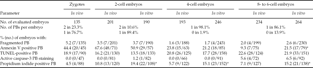
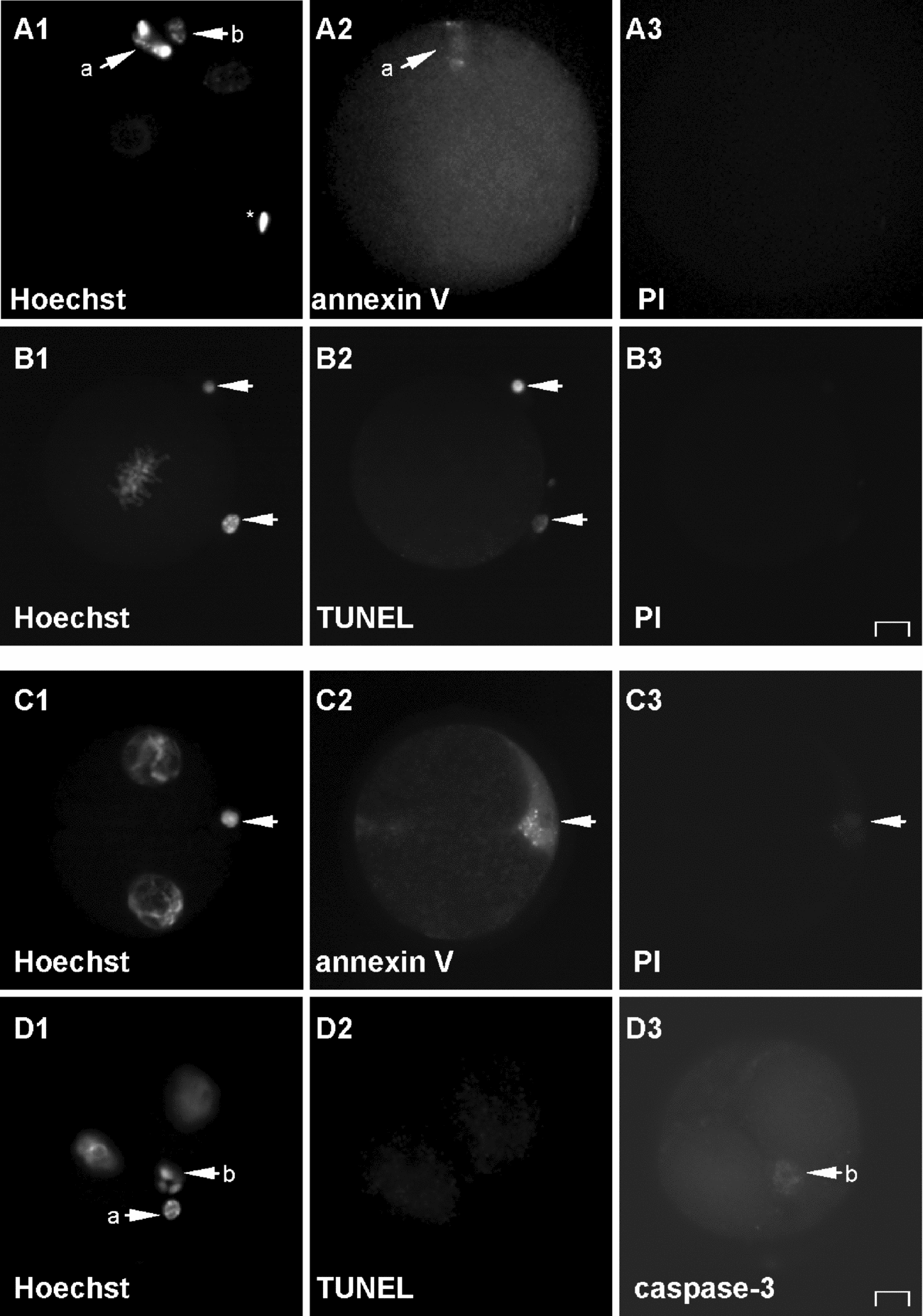
Approximately one-quarter of freshly isolated zygotes contained two polar bodies, and the remainder of them contained one polar body (Table 1 and Fig. 1A,B). The majority of these polar bodies showed condensed nuclear morphology (Fig. 1B1). Some of them (around 5%) showed nuclear decay into fragments (Fig. 1A1). The redistribution of phosphatidylserine on the outer plasma membrane leaflet of at least one polar body (AV+) was recorded in almost half of the zygotes (44%; Fig. 1A2) and the presence of specific DNA degradation in the polar body nuclear area (T+) was observed in almost 19% of them (Fig. 1B2). These apoptotic features were only occasionally accompanied with damage to the nuclear membrane (PI+). However, in zygotic polar bodies no positive IHC signal for active caspase-3 was recorded.
Polar body (PB) distribution and incidence of various features of apoptosis and secondary necrosis at particular stages of preimplantation development of mouse embryos developed in vivo and in vitro

The difference in the incidence of observed features between in vivo- and in vitro-derived embryos was evaluated by chi-squared test (*P < 0.05).
Illustrative fluorescence micrographs of mouse embryos at the zygotic stage (A,B) and the 2-cell stage (C,D). Original magnification: x400. Scale bar: 10 μm. Nuclear morphology is visualized by chromatin staining with Hoechst 33342 (A1–D1); redistribution of phosphatidylserine on the outer plasma membrane leaflet is visualized by annexin V staining (A2,C2); specific DNA degradation is visualized by TUNEL labeling (B2,D2); loss of embryo viability is visualized by vital propidium iodide (PI) DNA staining (A3–C3); and the presence of active caspase-3 is visualized by IHC labelling (D3). (A) Zygote containing two polar bodies (arrowheads) and spermhead (asterisk)–first polar body (a) shows partially fragmented morphology and positive annexin V staining, second polar body (b) does not show any features of cell death. (B) Zygote containing two polar bodies (arrowheads)–both with condensed nuclear morphology, TUNEL-positive, still viable (PI-negative). (C) Two-cell embryo with one (second) polar body– condensed, annexin V positive, dead (PI-positive). (D) Two-cell embryo with two polar bodies–the first one (a) condensed, TUNEL-negative, showing no IHC signal; the second one (b) with fragmented nuclear morphology, TUNEL-negative, showing clear signal for active caspase-3.
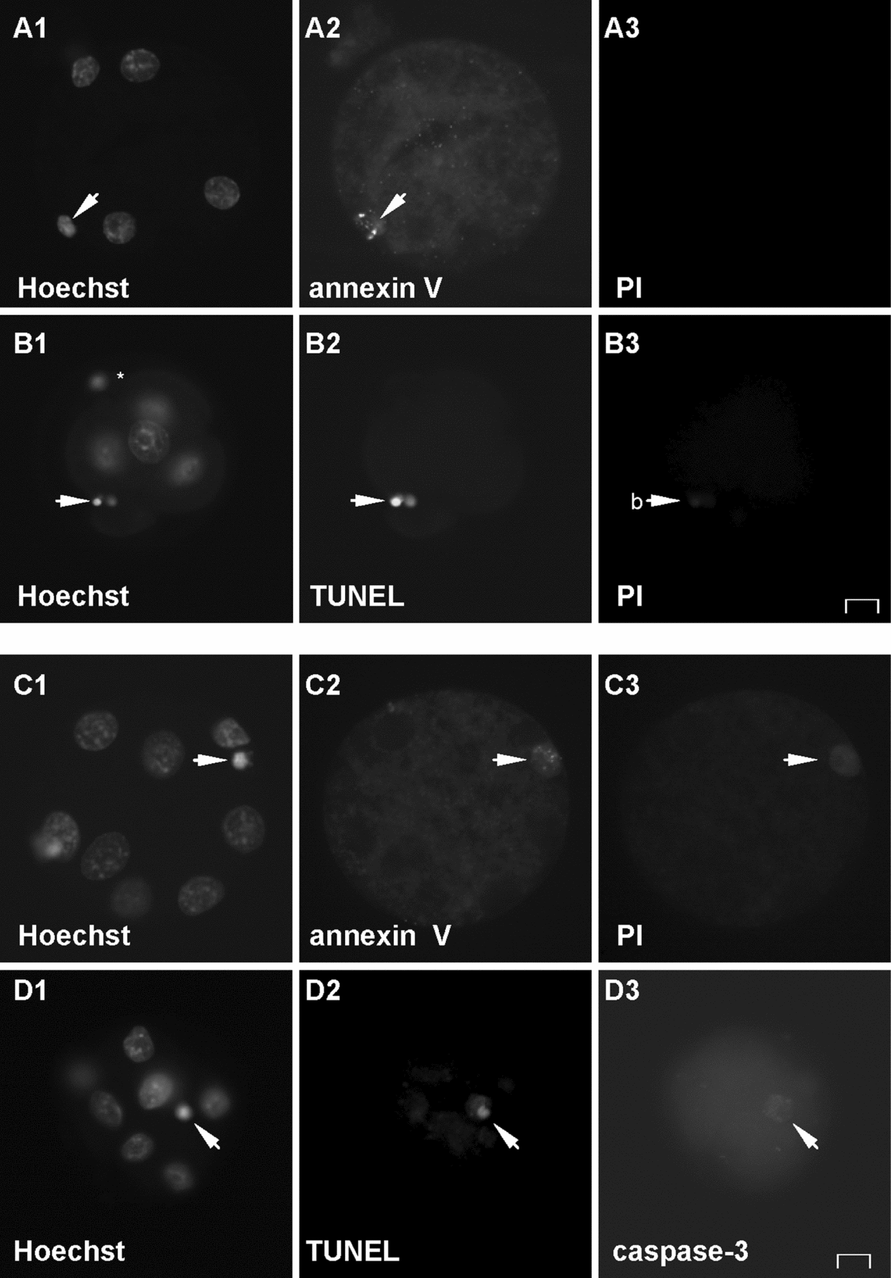
After reaching the 2-cell stage (Fig. 1C,D), two polar bodies were present only in 10% of embryos. In almost all 4-cell embryos (Fig. 2A,B) only one polar body was observed. Finally, after reaching the 8-cell or higher stage of development (Fig. 2C,D), in almost 15% of embryos both polar bodies were lost.
Illustrative fluorescence micrographs of mouse embryos at the 4-cell (A,B) and the 8-cell stage (C,D). Original magnification: x400. Scale bar: 10 μm. Nuclear morphology is visualized by chromatin staining with Hoechst 33342 (A1–D1); redistribution of phosphatidylserine on the outer plasma membrane leaflet is visualized by annexin V staining (A2,C2); specific DNA degradation is visualized by TUNEL labeling (B2,D2); loss of embryo viability is visualized by vital propidium iodide (PI) DNA staining (A3–C3); and the presence of active caspase-3 is visualized by IHC labeling (D3). A: Four-cell embryo with one (second) polar body (arrowhead) showing positive annexin V staining. (B) Four-cell embryo with spermhead (asterisk, out of focus) and one (second) polar body (arrowhead) –fragmented, TUNEL-positive, dead (PI-positive). (C) Eight-cell embryo with one (second) polar body (arrowhead)–condensed, annexin V positive, dubious viability (slight PI staining). (D) Eight-cell embryo with one (second) polar body (arrowhead)–condensed, TUNEL-positive, showing clear signal for active caspase-3.
The nuclear morphology of polar bodies showed very similar character during both in vivo and in vitro embryonic development (Table 1). As in the zygotes, the majority of embryonic polar bodies showed condensed nuclear morphology and some of them (approx. 2–4%) showed nuclear decay into fragments (Fig. 1C1,D1; Fig. 2A1–D1).
After reaching the 2-cell stage, the proportion of embryos with polar bodies showing phosphatidylserine flip (Fig. 1C2) slightly increased (68% for in vivo, P<0.05; 51% for in vitro, P>0.05; when compared with zygotes). In contrast, during further embryonic development (Fig. 2A2,C2) the incidence of this apoptotic feature in polar bodies gradually decreased to 24% (P<0.001) and 21% (P<0.001) for 4-cell embryos in vivo and in vitro respectively; and to 9% (P<0.05) and 19% (P>0.05) for 8-cell and later embryos in vivo and in vitro respectively; when compared with the previous developmental stage).
The occurrence of other apoptotic features was different. The 2-cell stage embryos and the 4-cell stage embryos showed relatively similar incidence of specific DNA degradation in the polar body nuclear area to that in zygotes (around 15% in 2-cells, P > 0.05 when compared with zygotes; and around 19% in 4-cells, P > 0.05 when compared with zygotes or 2-cells; taken together for both in vivo- and in vitro-developed embryos; Fig. 2B2). A tendency of slight increase in the incidence of TUNEL labeling was recorded only in polar bodies of the 8-cell and later embryos (Fig. 2D2), but it was not significant either, however (P > 0.05 when compared with 4-cells). Positive IHC signal for active caspase-3 was recorded only in one 2-cell embryo developed in vitro (Fig. 1D3) and several 8-cell or later embryos (6% for in vivo and 7% for in vitro; Fig. 2D3).
In developing embryos–similarly to zygotes–both positive annexin V staining and TUNEL labelling in polar bodies were only rarely accompanied with damage to the nuclear membrane (PI+; Fig. 2B3,C3). The highest incidence of polar bodies which were not able to exclude PI from the nuclear area, was recorded in the 2-cell embryos (11% for in vivo and 19% for in vitro, Table 1). In all other cases it did not exceed 7.1 % (for in vivo) or 15.2 % (for in vitro). In all evaluated samples, polar bodies of in vitro-derived embryos showed significantly higher incidence of PI staining than polar bodies of in vivo derived embryos (P > 0.05).
Discussion
Polar bodies begin to die during the earliest preimplantation development, but spontaneous cell death in the embryo itself usually does not appear before the 8-cell stage, depending on the species (Zakeri et al., Reference Zakeri, Lockshin, Criado-Rodríguez and Martínez2005; Fabian et al., Reference Fabian, Makarevich, Chrenek, Bukovská and Koppel2007). Furthermore, it has been proved that early embryos (from the 2-cell to the morula stage) are usually quite resistant to cell death induction (Fabian et al., Reference Fabian, Makarevich, Chrenek, Bukovská and Koppel2007). Thus, the mechanism of polar bodies’ elimination represents very important physiological event.
As shown in our study, in mice three-quarters of the first polar bodies are lost even at the zygotic stage, which is in accordance with previous observations (Ortiz et al., Reference Ortiz, Lucero and Coxatto1983); the 4-cell stage embryos contain only one (second) polar body, and elimination of the second polar body proceeds continuously during later development (polar body is missing in 15% of the 8-cell and 16-cell embryos). Finally, in mouse blastocysts polar bodies can be found only occasionally (based on previous observations, data not shown).
Our results prove that the majority of polar bodies commit ‘endogenously programmed suicide’, i.e. they die by way of apoptotic cell death. Both the first and the second polar body show several typical features of apoptosis: (1) phosphatidylserine flip in the early stages of the apoptotic process (which overlaps predominantly with the zygotic and 2-cell stages of embryonic development); (2) specific DNA degradation and condensed (rarely fragmented–contrary to the differentiated embryonic cells (Fabian et al., Reference Fabian, Makarevich, Chrenek, Bukovská and Koppel2007)) nuclear morphology during the executive phase of the apoptotic process (found continuously throughout embryonic development at approximately the same incidence); and (3) the inability to exclude cationic dye (PI) from the nucleus during the terminal stage of the apoptotic process (so-called ‘secondary necrosis’–first wave during the 2-cell stage of development, probably overlapping with the final elimination of the first polar body, and second wave at the 16-cell stage and later, probably overlapping with the final elimination of the second polar body). As each polar body is usually short of adequate physical contact with neighboring cells, it cannot be phagocytosed, and secondary necrosis (accompanied with massive autolytic degradation of cellular membranes (Vitale et al., Reference Vitale, Zamai, Mazzotti, Cataldi and Falcieri1993)) is its natural fate–similarly as in normal embryonic cells, which are extruded to the blastocoelic cavity or perivitelline space (Fabian et al., Reference Fabian, Koppel and Maddox-Hyttel2005).
Although the frequency of IHC signal representing active caspase-3–the main enzyme of the apoptotic cascade (Papandile et al., Reference Papandile, Tyas, O'Malley and Warner2004)–was relatively low in polar bodies of preimplantation embryos, its presence at later stages of development definitely proved the apoptotic origin of the cell death process at least for the second polar body. At earlier phases of embryonic development (the 2-cell stage) only one caspase-3 positive polar body was observed (Fig. 1D3) and as it was a singular case, it probably represented the decay of the second polar body too.
These results are in accordance with previous observations by Martinez et al. (Reference Martinez, Rienzi, Iacobelli, Ubaldi, Mendoza, Greco and Tesarik2002) and Zakeri et al. (Reference Zakeri, Lockshin, Criado-Rodríguez and Martínez2005), who suggested that caspase activation does not occur until oocyte activation by the fertilizing spermatozoon. Thus, the death of the first polar body probably proceeds by a caspase-independent mechanism (e.g. by means of endonuclease G release from mitochondria and its translation to the nucleus), and the death of the second polar body can involve caspases. This theory is supported by the fact that inhibitors of caspases do not block the death of the first polar bodies (Zakeri et al., Reference Zakeri, Lockshin, Criado-Rodríguez and Martínez2005).
In our study, no significant differences between data obtained in vivo and in vitro were recorded in the majority of cases (Table 1). However, in polar bodies of in vitro-developed embryos a slightly earlier presence of one apoptotic feature (the presence of active caspase-3 during the 2-cell stage), higher incidence of another apoptotic feature (phosphatidylserine flip during the 8-cell stage), and significantly higher incidence of PI nuclear staining (during all stages) were observed. These findings suggest that stressing (or inadequate) developmental conditions might potentiate apoptotic processes not only in normal embryonic cells (reviewed in Fabian et al., Reference Fabian, Koppel and Maddox-Hyttel2005) but also in polar bodies. Besides, increased incidence of PI nuclear staining also suggests earlier entrance of eliminated structures into the secondary necrotic stage of cell death.
In conclusion, from the morphological point of view, mouse polar bodies act very similarly to damaged embryonic cells which have lost contact with their neighboring blastomeres. In spite of their relatively small volume and organelle content, they possess all the molecular equipment necessary for triggering and executing an active suicide process, i.e. apoptotic cell death. Furthermore, similarly as in dying embryonic cells, stressing external conditions (such us culture in vitro) might accelerate and increase the incidence of apoptotic elimination of the polar bodies in embryos.
Acknowledgements
We thank Dana Čigašová for her technical assistance and Andrew Billingham for his English proof-reading. This work was supported by the Slovak Academy of Sciences under contract VEGA 2/0049/11 and the Slovak Research and Development Agency under contract APVV-0620-07.