Introduction
Species interactions are a major factor structuring biological communities, and symbiotic interactions can be an important force driving evolution (Margulis & Fester Reference Margulis and Fester1991). In lichens, one of the ‘textbook examples’ of symbiosis, the local composition of symbiotic partnerships between lichen-forming fungi and their photobionts is a fascinating issue (Beck et al. Reference Beck, Friedl and Rambold1998, Reference Beck, Kasalicky and Rambold2002; Yahr et al. Reference Yahr, Vilgalys and DePriest2004; Ohmura et al. Reference Ohmura, Kawachi, Kasai, Watanabe and Takeshita2006). Progress has been made in recent years due to the development of molecular techniques that allow a straightforward identification of photobiont and fungal genetic strains and species (Scheidegger & Werth Reference Scheidegger and Werth2009; Werth Reference Werth2010). In this way, photobiont guild structure and one-to-one species, as well as haplotype interactions, have been analyzed in the lichen symbiosis.
The symbiotic mutualistic associations of lichen fungi and their photobionts can be described by their degree of specificity and selectivity. These terms characterize the taxonomic range of the partners with which an organism associates (Smith & Douglas Reference Smith and Douglas1987). Following the suggestion by Beck et al. (Reference Beck, Kasalicky and Rambold2002), I use the term specificity for the symbiotic association as a whole, that is, from the view of both photobionts and mycobionts. A narrow taxonomic range (e.g., a one-to one relationship) would be indicative of the high specificity of the association. I refer to selectivity as the taxonomic range of the interaction viewed from the perspective of only one biont (Beck et al. Reference Beck, Kasalicky and Rambold2002). Accordingly, a mycobiont would be highly selective in its partner choice if it were found in association with few, closely related photobiont species.
Previous studies have shown that lichen fungi may exhibit a varying degree of selectivity towards their photobionts. Some fungi show high selectivity, and they associate with a particular photobiont phylogenetic clade or species (Kroken & Taylor Reference Kroken and Taylor2000; Helms et al. Reference Helms, Friedl, Rambold and Mayrhofer2001; Beck et al. Reference Beck, Kasalicky and Rambold2002; Piercey-Normore Reference Piercey-Normore2004, Reference Piercey-Normore2006; Yahr et al. Reference Yahr, Vilgalys and DePriest2004; Stenroos et al. Reference Stenroos, Högnabba, Myllys, Hyvönen and Thell2006; Myllys et al. Reference Myllys, Stenroos, Thell and Kuusinen2007). Other fungal species are far less selective towards their photobiont partners (Wirtz et al. Reference Wirtz, Lumbsch, Green, Türk, Pintado, Sancho and Schroeter2003; Piercey-Normore Reference Piercey-Normore2004; Yahr et al. Reference Yahr, Vilgalys and DePriest2004; Myllys et al. Reference Myllys, Stenroos, Thell and Kuusinen2007); some may occasionally or always associate with two photobiont species (Piercey-Normore Reference Piercey-Normore2006; Casano et al. Reference Casano, del Campo, Garcia-Breijo, Reig-Arminana, Gasulla, del Hoyo, Guera and Barreno2011). Some fungal species are able to partner up with photobionts belonging to different kingdoms, and which photobiont partner gets involved in the symbiosis may depend on environmental conditions (Goffinet & Bayer Reference Goffinet and Bayer1997; Tønsberg & Goward Reference Tønsberg and Goward2001; Summerfield et al. Reference Summerfield, Galloway and Eaton-Rye2002).
Many open questions remain in the field of fungal-algal associations in lichens: for instance, are fungal-photobiont relationships entirely random or are they structured in a complex way, that is, nested or compartmentalized? In the case of random associations, no apparent pattern of association would be found in species interaction matrices (Bascompte et al. Reference Bascompte, Jordano, Melian and Olesen2003; Jordano et al. Reference Jordano, Bascompte and Olesen2003). In nested species interactions, specialists tend to interact with subsets of the species that interact with generalist species (Bascompte et al. Reference Bascompte, Jordano, Melian and Olesen2003). In terms of the lichen symbiosis, this would mean that the specialized fungal species are interacting almost exclusively with the photobiont species that the generalistic fungi are interacting with. This nested type of species interaction leads to a pattern in species interaction matrices that resembles an asymptotic curve (Bascompte et al. Reference Bascompte, Jordano, Melian and Olesen2003; Jordano et al. Reference Jordano, Bascompte and Olesen2003). Translated to the world of lichen fungi and their photobionts, that would mean that few generalist mycobiont species interact with many photobiont species, and few generalist photobiont species interact with many mycobiont species. Additionally, specialized mycobiont species would interact with a subset of the photobiont species used by the generalists, and vice versa, specialized photobionts would interact with a subset of the mycobiont species. Nestedness can not only be studied at the level of fungal and algal species, but also at the level of haplotypes of each symbiosis partner.
In the case of compartmentalized species interactions (Bascompte et al. Reference Bascompte, Jordano, Melian and Olesen2003; Jordano et al. Reference Jordano, Bascompte and Olesen2003), certain fungal species would interact with a defined subset of photobiont species, while other fungal species would interact with a non-overlapping subset of photobiont species; interaction matrices would thus exhibit independent subwebs. Compartmentalization and participation of comparatively few species has recently been documented from networks of mutualistic, symbiotic ant and plant species with a high degree of interaction intimacy, while networks with low interaction intimacy were generally species-rich and nested (Guimarães et al. Reference Guimarães, Rico-Gray, Oliveira, Izzo, dos Reis and Thompson2007). Results from previous studies indicate that mycobiont-photobiont interactions are not random, and they seem to be independent of the green algal species present in free-living populations at a site (Beck et al. Reference Beck, Friedl and Rambold1998; Rambold et al. Reference Rambold, Friedl and Beck1998).
Many lichen fungi disperse clonally with symbiotic propagules which contain photobiont cells, which leads to vertical transmission of the photobiont (Nash Reference Nash1996; Werth et al. Reference Werth, Wagner, Gugerli, Holderegger, Csencsics, Kalwij and Scheidegger2006a,Reference Werth, Wagner, Holderegger, Kalwij and Scheideggerb) unless the photobiont is exchanged during establishment (Ohmura et al. Reference Ohmura, Kawachi, Kasai, Watanabe and Takeshita2006; Wornik & Grube Reference Wornik and Grube2010; Werth & Scheidegger 2012). Also, lichen-forming ascomycetes reproduce sexually with ascospores, which are dispersed independently of their photobiont cells (i.e., horizontal transmission), though a few exceptions exist where both partners are co-dispersed (Honegger Reference Honegger and Nash2008). Germinating ascospores have to associate with new photobionts (relichenization) in order to form a thallus. This phenomenon is probably common, but has been reported from only a few species (Beck et al. Reference Beck, Kasalicky and Rambold2002; Romeike et al. Reference Romeike, Friedl, Helms and Ott2002; Sanders & Lücking Reference Sanders and Lücking2002; Werth & Sork Reference Werth and Sork2008). It has also been suggested that germinating spores could gain photobionts either from vegetative propagules produced by other lichen species, or from adult thalli of other species (Ott Reference Ott1987a; Ott et al. Reference Ott, Schröder and Jahns2000; Rikkinen Reference Rikkinen2003). Alternatively, they may retrieve cells from free-living Trebouxia populations (Beck et al. Reference Beck, Friedl and Rambold1998).
Ramalina menziesii (Ramalinaceae) is a common epiphytic lichen in coastal oak savannas of southern California. It is associated with the photobiont genus Trebouxia (Brodo et al. Reference Brodo, Sharnoff and Sharnoff2001), which contains some frequent and widely distributed photobiont species. Recently, the local genetic structure was investigated for this lichen fungus across four sites, and high gene flow was found among sites (Werth & Sork Reference Werth and Sork2008). Werth & Sork (Reference Werth and Sork2008) reported Trebouxia decolorans as photobiont of R. menziesii. The reproductive strategy of Ramalina menziesii in the study site was determined as almost exclusively sexual, involving relichenizations, whereas clonal reproduction was negligible (Werth & Sork Reference Werth and Sork2008). These results necessitated a more detailed study of fungal-algal relationships using juvenile thalli.
Here, I specifically asked the following questions: 1) What proportion of juveniles of R. menziesii had an opportunity to interact with other species (as evidenced by growing in close proximity)? 2) Is the lichen-forming fungus R. menziesii selective in its choice of photobiont strains? 3) With which other lichen-forming fungi does R. menziesii share a common pool of algal species or haplotypes? If pairs of juvenile R. menziesii and its nearest neighbour on a branch have identical photobiont haplotypes, photobiont exchange may have occurred between them. Take-over of algae has been observed in some lichens (Friedl Reference Friedl1987; Ott Reference Ott1987a, Reference Ottb; Ott et al. Reference Ott, Schröder and Jahns2000). 4) Are fungal species associations and algal species/haplotype associations random in R. menziesii and the other members of the lichen community, or are they structured in a more complex way (e.g. nested)?
Materials and Methods
Study site
The study area was located at UCSB Sedgwick Reserve in the Santa Ynez Valley of Santa Barbara County. This field station is administered by UC Santa Barbara as part of the University of California Natural Reserve System (California, United States, 34°42′N, 120°02′W; 2358 ha; elevation 290–790 m). The majority of the reserve is an oak savanna dominated by three tree species: valley oak (Quercus lobata; Fagaceae), blue oak (Q. douglasii), and coastal live oak (Q. agrifolia). Sork et al. (Reference Sork, Davis, Dyer, Smouse, Standiford and Purcell2002a, Reference Sork, Davis, Smouse, Apsit, Dyer, Fernandez and Kuhnb) provide details of the study site.
Data sampling
To study the potential for interaction with other lichen species, juvenile thalli (≤5 cm long) of Ramalina menziesii Tayl. (Ramalinaceae) were observed in the field, and it was recorded whether they were growing by themselves or within 2 mm from another lichen species, or from an adult conspecific. A cut-off level of 2 mm was chosen because preliminary observations of distance, between thalli indicated that within this distance, other species were often found. Also, it was assumed that for exchange of algae to happen, physical contact with the neighbouring thallus was required, which was why a rather short distance was used as cut-off level.
Juveniles were observed on multiple branches per tree on a total of 11 trees. The number of juveniles observed per tree varied between 2 and 22, depending on availability of juveniles on branches at an accessible height on the tree (≤2 m). The aim of these observations was to quantify how often R. menziesii juveniles had close contact with thalli of other lichen species. Also, these data yielded a qualitative overview of community composition.
The following design was used to study fungal-algal relationships on a total of five trees at the study site: within each tree, 2–3 branches were selected that were more than 1 m from each other; within each branch, 3–4 lichen thalli were collected, depending on availability: 1) a juvenile Ramalina menziesii (=0 cm); 2) a lichen species other than R. menziesii, situated within 2 mm from the juvenile (the ‘nearest neighbour’); 3) a lichen species other than R. menziesii, situated at a distance larger than 1 cm from the juvenile, range 1·6–12·3 cm (>1 cm); 4) an additional R. menziesii, if present on branch, juvenile or adult. In this way, a total of 45 samples were collected. This design allowed me to test whether pairs of juveniles of R. menziesii and other species shared algal DNA sequence haplotypes.
Molecular methods
DNA sequences were obtained from the ITS (Internal Transcribed Spacer) region of the photobiont, situated within the nuclear ribosomal gene cluster. ITS was used because the same locus was used in several other studies of lichen photobionts, including those where Trebouxia type cultures or strains from culture reference collections had been sequenced.
Owing to the photobiont specificity of the molecular assay, it was not necessary to obtain axenic cultures prior to sequencing. Instead, the photobiont ITS was amplified by Polymerase Chain Reaction (PCR) from thallus extracts, and sequenced directly.
Each algal-specific PCR contained 1×Multiplex PCR Master Mix (Qiagen, Hilden, Germany), 200 nM each of forward and reverse primer, 1 μl genomic DNA (0·05–2 ng), and H2O to a final volume of 30 μl. The Multiplex PCR Master Mix (Qiagen) consisted of an unspecified quantity of dNTPs, HotStarTaq DNA polymerase, and 3 mM MgCl2 (pH 8·7). PCR conditions were as follows: 15 min at 96°C to activate the hot-start polymerase of the multiplex kit, 35 cycles of 30 s at 95°C, 60 s at 56°C, 90 s at 72°C, and a final extension of 10 min at 72°C. The primers used for PCR and sequencing were ITS1T and ITS4T (Kroken & Taylor Reference Kroken and Taylor2000). PCR purification and sequencing were performed as described in Werth & Sork (Reference Werth and Sork2008). DNA sequences were edited with Sequence Scanner Software (Applied Biosystems, Foster City, California, USA) and aligned using ClustalW (Thompson et al. Reference Thompson, Higgins and Gibson1994) as implemented in MEGA version 4.1 (Kumar et al. Reference Kumar, Tamura and Nei2004). Alignments were adjusted manually after the automated alignment had been performed. All unique ITS sequences were deposited in GenBank (accessions EU717911-EU717936; Table 3).
Data analysis
ITS sequences of the photobionts were grouped by tree and branch, and the ITS haplotypes were compared within each hierarchical level (among trees, among branches within tree, within branch). Then, photobiont haplotypes were compared for R. menziesii juveniles and their nearest neighbouring lichen fungi. Matches of photobiont haplotypes among thalli indicated photobiont sharing among fungi.
Phylogenetic trees excluding gaps were constructed using MrBayes version 3.1.2 (Ronquist & Huelsenbeck Reference Ronquist and Huelsenbeck2003). I ran PAUP version 4.0 to generate model scores (Swofford Reference Swofford1998), and used MrModeltest version 2.2 (Posada & Crandall Reference Posada and Crandall1998; Nylander Reference Nylander2004) to infer the appropriate substitution model for the phylogenetic analyses. The best model according to the AIC criterion was selected for all analyses (GTR+G). Four chains were run in parallel in MrBayes with a generation number of 1 000 000, sampling trees every 100th generation. A total of 10 000 trees were sampled, and the first 1000 trees of these were discarded (burn in). Results from a second run of MrBayes were inspected, and tree topology remained consistent across runs (data not shown). In addition, a neighbour-joining analysis was run with 1000 bootstraps in Mega version 4 (Tamura et al. Reference Tamura, Dudley, Nei and Kumar2007) using Jukes-Cantor distance, which resulted in a similar tree topology as the Bayesian analysis (data not shown).
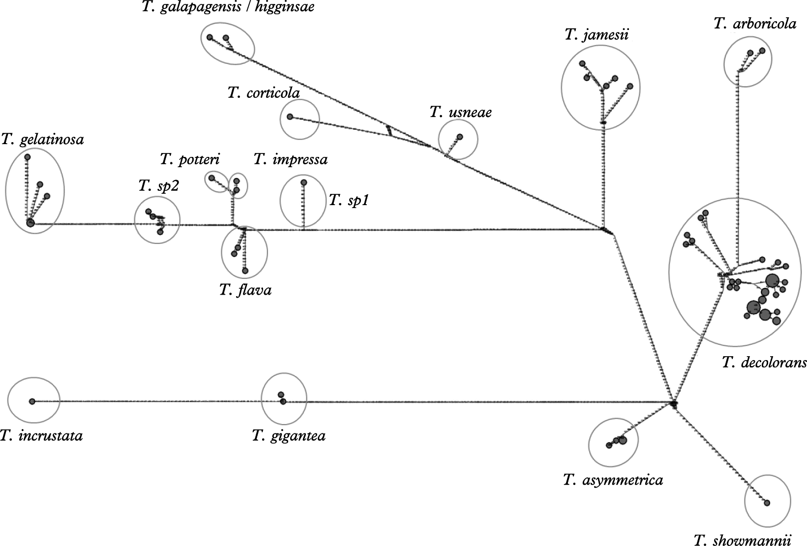
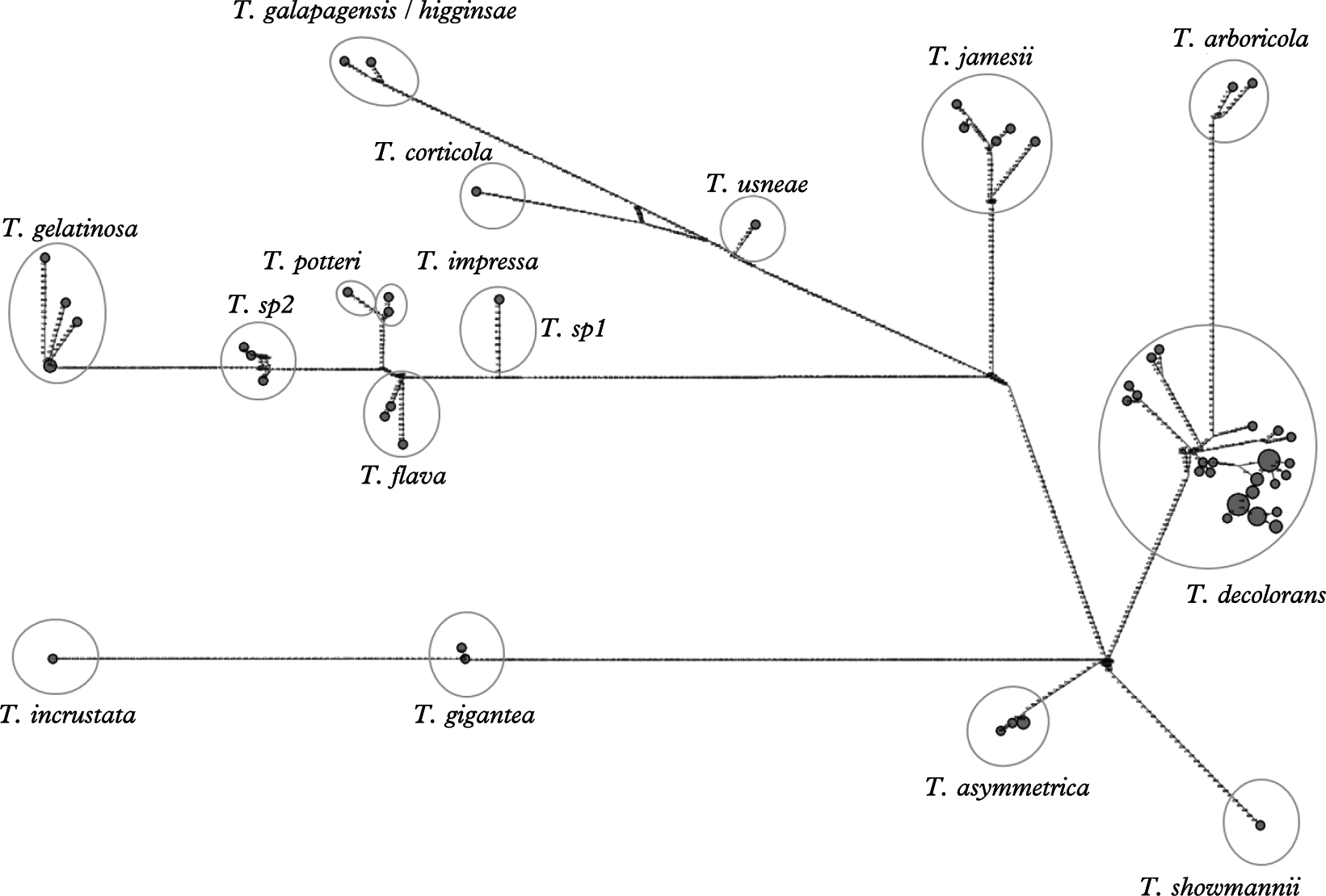
Moreover, median-joining haplotype networks as implemented in the software Network (Bandelt et al. Reference Bandelt, Forster, Sykes and Richards1995, Reference Bandelt, Forster and Röhl1999) were constructed. These networks allow a simple inspection of how well a group of sequences is separated from other groups by a plot of the mutated positions on the branches, and hence, to delineate species. Incongruences in the data, for example due to recombination, are shown as loops in the network.
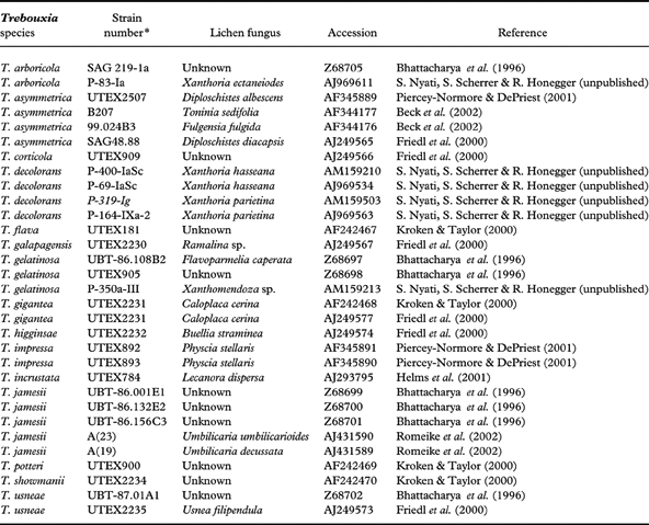
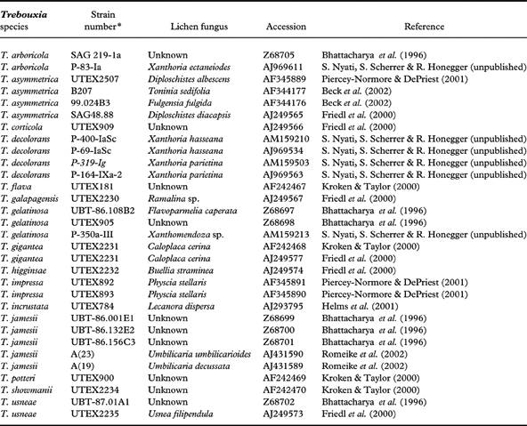
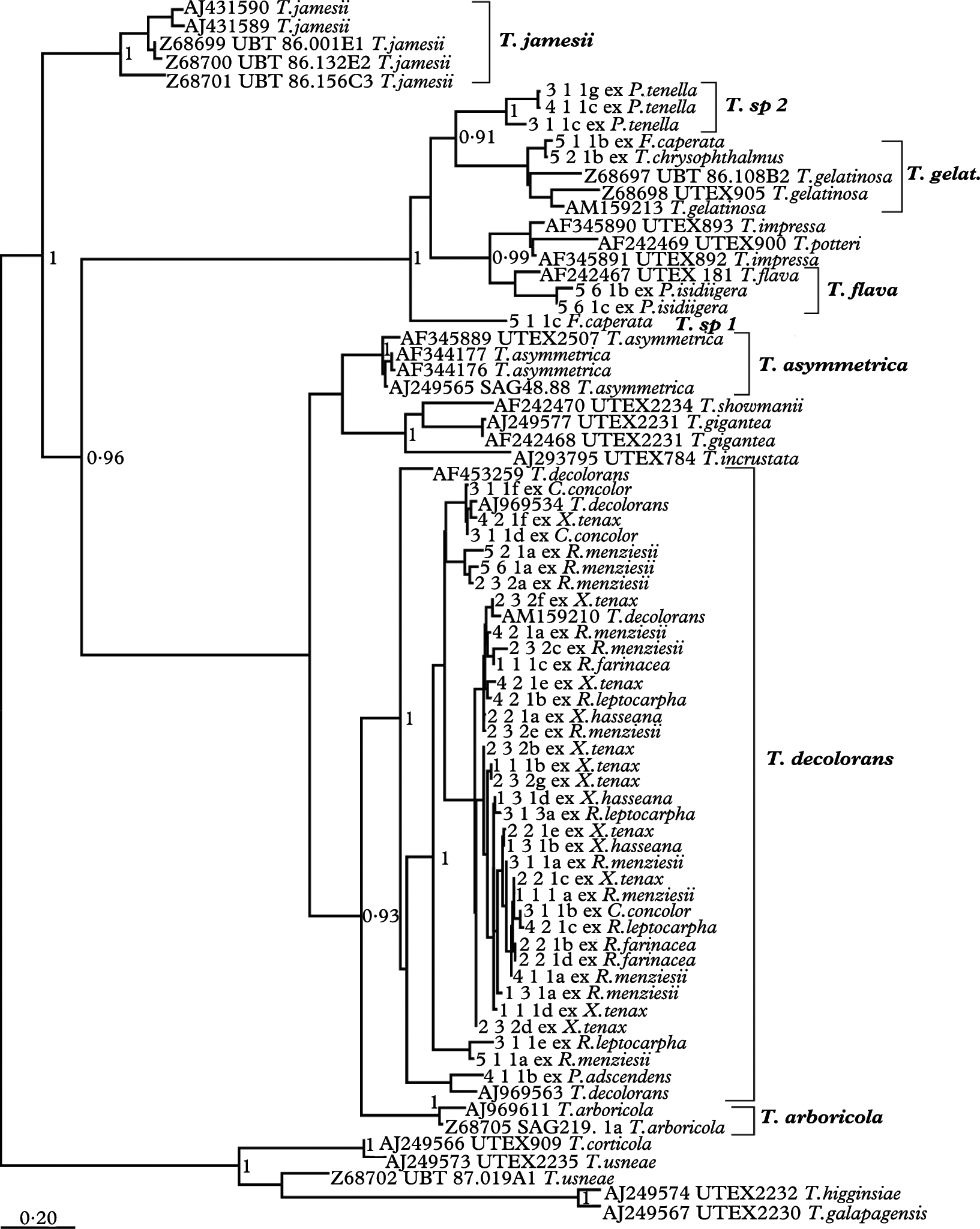
To identify photobiont species, BLAST searches (Altschul et al. Reference Altschul, Madden, Schäffer, Zhang, Zhang, Miller and Lipman1997) for the photobiont haplotypes were performed, ITS sequence data of known photobionts were obtained from reference cultures from GenBank, and included in the Bayesian analysis. Photobiont species were considered identical if they were in the same, supported clade as a known species in the median-joining haplotype network and the phylogenetic tree (posterior probability >0·9), or were considered a ‘new’ species when clades had a high bootstrap support and did not group within known species. These ‘new’ algal species may represent either known species for which sequences have not yet been deposited in GenBank, or species new to science. Table 1 gives an overview of the GenBank sequences used in the phylogenetic analysis.
Sequences from GenBank included within this study, showing species of Trebouxia analyzed, GenBank accession numbers, and references to ITS rDNA sequences
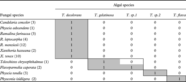
To test for nestedness of the interaction of algal species and their lichen-forming fungi (species-level interaction), a fungi-by-algae matrix was constructed that showed the presence/absence of associations between fungal and photobiont species (rows: fungal species; columns: photobiont species; entries: 1=algae found as a symbiotic partner of this fungal species, 0=algae not found as a partner of that fungus). To test for nestedness in the association of algal haplotypes (columns) and fungal species (rows), a second matrix was constructed. The program Aninhado version 2.03 (Guimarães & Guimarães Reference Guimarães and Guimarães2006) was run to determine the matrix temperature T, a measure of how a given presence/absence matrix differs from a perfectly nested matrix, with values ranging from 0 (perfect nestedness) to 100 (no nestedness) (Atmar & Patterson Reference Atmar and Patterson1993). Nestedness was defined as N=(100-T)/100, and ranged between 0 (no nestedness) and 1 (maximum nestedness) (Bascompte et al. Reference Bascompte, Jordano, Melian and Olesen2003; Guimarães & Guimarães Reference Guimarães and Guimarães2006). Using Aninhado, I tested if a respective matrix was significantly nested by comparing the observed matrix temperature with the temperatures of random replicate matrices. These random replicates were constructed using four null models implemented in Aninhado: 1) In the first null model (hereafter referred to as ‘ER’), presences were assigned randomly to any matrix cell, keeping the total number of presences constant. 2) The second null model (‘COL’) assigned presences randomly within columns, thereby keeping column sums constant. 3) The third null model (‘LI’) differed by assigning presences randomly within rows, thereby keeping row sums constant. 4) The fourth null model referred to as ‘CE’ kept both columns and rows constant, and was the most conservative null model because it controlled for the degree of nestedness expected by the heterogeneity of interactions across species (Bascompte et al. Reference Bascompte, Jordano, Melian and Olesen2003). For each null model, 1000 random matrices were generated, their matrix temperatures were calculated in Aninhado, and N was determined, and compared with the nestedness of the observed matrices. Following Bascompte et al. (Reference Bascompte, Jordano, Melian and Olesen2003), the probability P of a random matrix being equally or more nested than the observed matrix as a test statistic was used.
To compare nestedness among the two matrices (1, fungal species – algal species; 2, fungal species – algal haplotypes), I calculated their relative nestedness as N*=(N−NR)/ NR, where N was the observed nestedness, and NR was the averaged nestedness of 1000 random replicates. Positive values of relative nestedness indicate that the network shows asymmetrical specialization and a generalist core (i.e., nestedness), while negative values might be associated with symmetric interaction patterns and the existence of compartments (Guimarães et al. Reference Guimarães, Rico-Gray, Oliveira, Izzo, dos Reis and Thompson2007).
Nestedness analysis is not an adequate technique to test for non-nested patterns; if associations are not nested, they are either random or, alternatively, they may show a different type of organization (Guimarães et al. Reference Guimarães, Rico-Gray, Oliveira, Izzo, dos Reis and Thompson2007). For instance, compartments may exist, which are indicated by isolated subwebs. Therefore, columns and rows were sorted and visually inspected the interaction networks. Moreover, the number of such subwebs within the matrices were counted.
Results
Potential for interaction and diversity
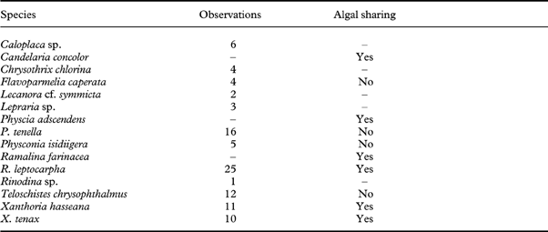
In total, 103 juveniles of R. menziesii were surveyed on 11 trees (Table 2). Only four out of the 103 juvenile lichens had no neighbour lichen within a 2 mm distance. At the tree level, the mean number of R. menziesii juveniles growing in close proximity to another lichen neighbour ( =9·0) was 24·8 times larger than that of juveniles growing by themselves (=0·364). Thus, many juveniles of R. menziesii may have had the potential to interact with other species of lichens during their development.
Species observed in a field survey as nearest neighbours of Ramalina menziesii, giving the number of observations and whether fungal species shared photobiont with R. menziesii as determined from a DNA sampling survey (specimens included in the DNA sampling are listed in Table 3).
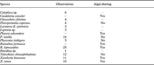
– no data.
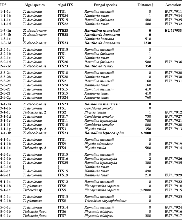
For the genetic analyses, ten lichen species were sampled from the trees containing R. menziesii juveniles (Table 3, Fig. 1). These included two species of Xanthoria, X. hasseana and X. tenax, Ramalina leptocarpha and R. farinacea, and two Physcia species. A total of 26 algal haplotypes were found among the juvenile thalli of R. menziesii and the 10 other species (Table 3).
Algal – fungal relationships in lichens associated with juvenile Ramalina menziesii. The most frequent ITS haplotype, ITS23, is marked in bold
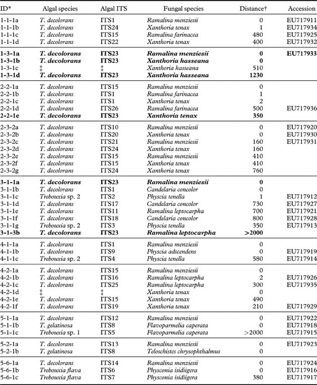
* sample ID consists of the tree number, branch number, twig number, and sample letter
† the distance from sample a, the juvenile R. menziesii [mm]
‡ sequencing failed.
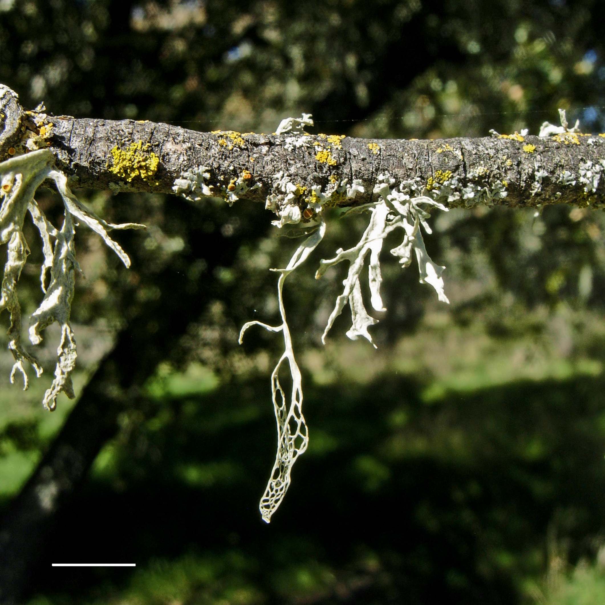
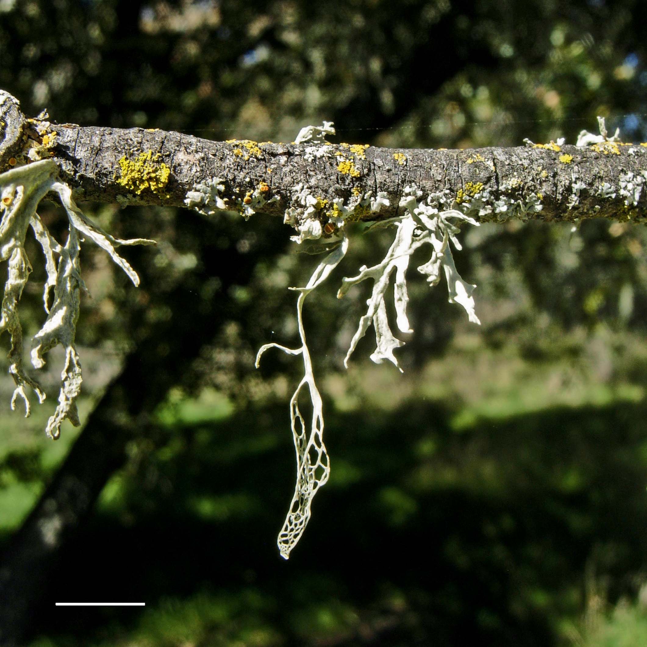
A typical juvenile, net-formed thallus of the model species Ramalina menziesii (lace lichen, Ramalinaceae), the dominant epiphytic lichen of a southern Californian oak savannah. The juvenile R. menziesii grows next to other members of the epiphytic community, Xanthoria tenax and Ramalina leptocarpha. Scale=1 cm. In colour online.
Algal sharing
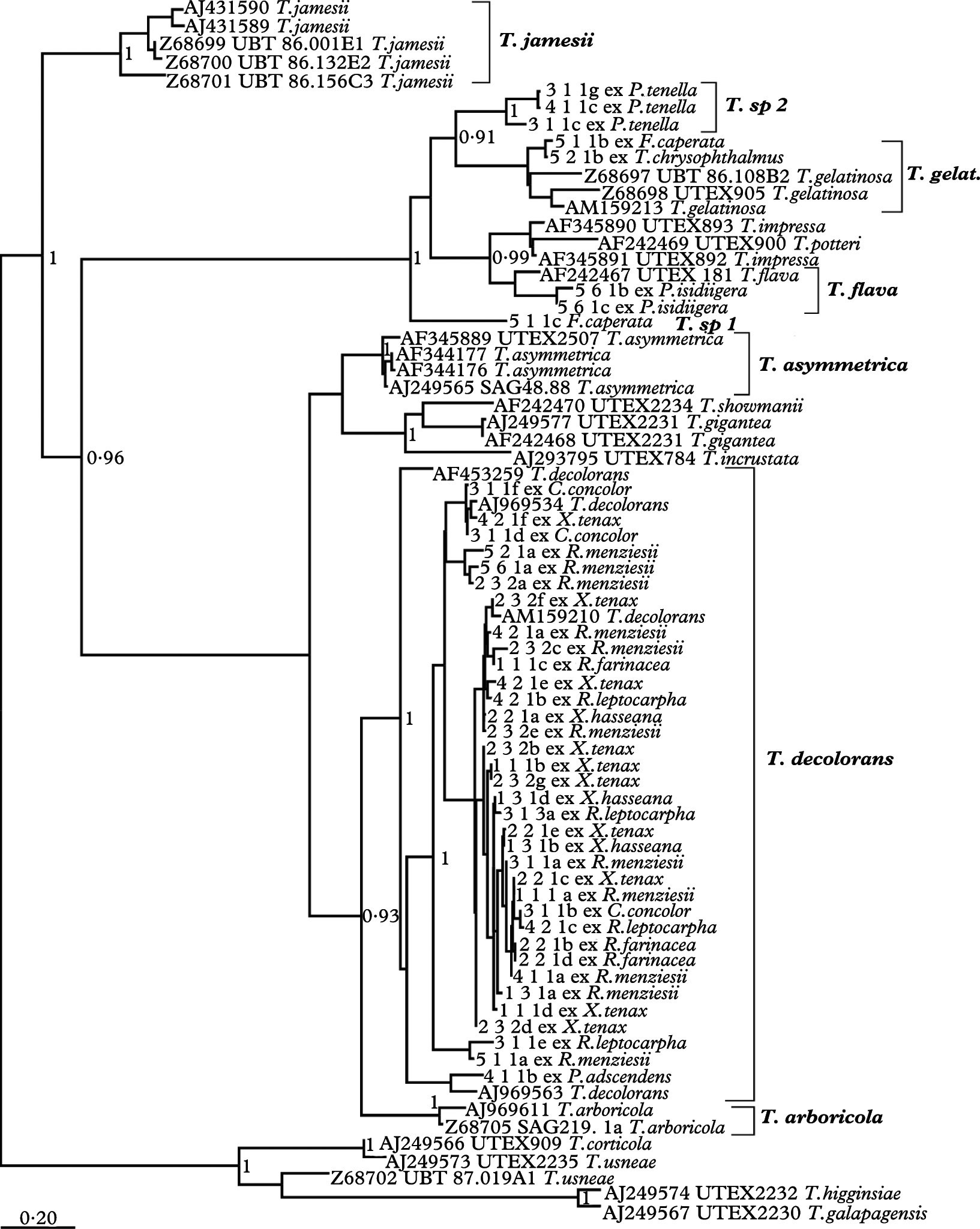
Four algal haplotypes were shared among the lichen fungi. Juveniles of R. menziesii utilized eight algal ITS haplotypes which belonged to a single algal species (Trebouxia decolorans) (Table 3, Figs 2 & 3). In 90% of the observations, the algal ITS haplotype of the nearest neighbour was different from that of the juvenile R. menziesii. On one twig, an algal haplotype was shared between a juvenile and its nearest neighbour Xanthoria hasseana (1-3-1b), as well as with a X. hasseana thallus growing at a distance of 12·3 cm (1-3-1d). The same haplotype (ITS23) occurred also on two other trees (2-2-1e, 3-1-1a). On another tree, X. tenax (2-3-2f) shared an algal haplotype with a R. menziesii (2-3-2e) growing at the same spot. Interestingly, the same algal haplotype occurred also on other trees (individuals 1-1-1c, 4-2-1a). Also, another algal haplotype occurred on several trees (ITS1; trees 1, 2, 3, and 4).
Phylogenetic tree from Bayesian analysis of the photobionts associated with Ramalina menziesii and other lichen fungi from a southern Californian oak savanna. See Table 1 for more information on the sequences from GenBank. The numbers show Bayesian posterior probabilities.
Median-joining haplotype network of the photobiont community associated with R. menziesii and other lichen fungi in a southern Californian oak savanna. Branch length is proportional to the number of mutated positions. Haplotypes are shown as dots, with dot size proportional to a haplotype's frequency. Conspecific individuals are marked with circles.
Fungal-algal associations
Five species of the genus Trebouxia were found to be associated with the lichen fungi studied. Trebouxia decolorans was found to be the photobiont of seven lichen-forming fungal species, Candelaria concolor, Physcia adscendens, Ramalina farinacea, R. leptocarpha, R. menziesii, Xanthoria tenax, and X. hasseana (Table 3, Fig. 2). In contrast, Physcia tenella and Flavoparmelia caperata (individual 5-1-1c) were each associated with a different, unknown (i.e., either not included in GenBank or new to science) species of the genus Trebouxia (Fig. 2). The second thallus of F. caperata (5-1-1b) and Teloschistes chrysophthalmus were found in association with T. gelatinosa (Fig. 2, Table 3). The only photobiont species of R. menziesii in the study area was T. decolorans. The photobiont of P. isidiigera was deemed conspecific with Trebouxia flava, and that of P. tenella (e.g. 3-1-1g) was distantly related to T. gelatinosa (Fig. 2), but represented an unknown species.
Random or structured associations
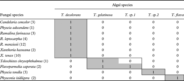
The species interaction matrix, algal species by fungal species, was not significantly nested (N=0·564, a value of 1 indicating perfect nestedness) (Guimarães & Guimarães Reference Guimarães and Guimarães2006). The matrix is available in Table 4. This matrix was not significantly nested for any of the null models employed (Table 5). For two null models, the values of relative nestedness, N*, were negative. These negative values may indicate the existence of subwebs (compartmentalization). Visual inspection of the network revealed four reciprocally isolated subwebs, which suggests that the species-level matrix was indeed organized in compartments. The largest subweb consisted of seven fungal and one algal species, and the remaining three represented one-by-one associations or two-by-two associations.
Interaction matrix of fungal and algal species, sorted to reveal potential subwebs. Numbers in parentheses indicate sample sizes.
Relative nestedness N* of fungal-algal associations for four null models.

* significant at P=0·05.
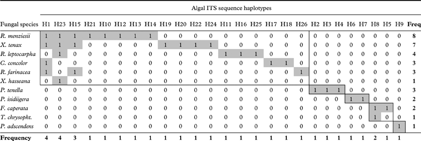
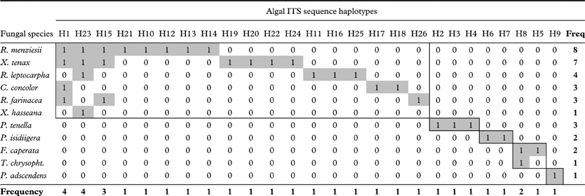
The nestedness of the matrix of fungal species and algal ITS sequence haplotypes was N=0·716. Relative nestedness of fungal species and algal haplotypes was higher than that of the fungal-algal species matrix (Table 5). Significant nestedness was found with the ER and COL null model, the LI model was near to significant, and the conservative CE model was not significant.
Overall, the tests above revealed some degree of nestedness in the fungal species-algal ITS haplotype matrix, while the species-by-species matrix was not significantly nested. However, when visually inspecting the haplotype-level network (Table 6), a few isolated subsets nevertheless existed, implying compartmentalization (e.g. for the fungal species F. caperata, T. chrysophthalmus and P. tenella), while the largest part of the network, a group of fungal species including R. menziesii and X. hasseana, appeared to be nested.
Interaction matrix of fungal species and algal ITS sequence haplotypes, sorted by their frequency in columns and rows to reveal potential subwebs; for names of fungal genera and species, see Table 4
Discussion
First, the potential for R. menziesii juveniles to interact with other species was studied using field observations, and I found that juveniles had indeed ample opportunities to interact. Then, I investigated photobiont sharing among fungal species in the epiphytic lichen community with molecular techniques, and found evidence of sharing of one photobiont species (Trebouxia decolorans) by several fungal species. There was also some evidence of haplotype sharing among the focal species of the study, R. menziesii, and another species growing as its nearest neighbour (e.g., X. hasseana), though in most observations (90%), the algal haplotypes of juvenile R. menziesii differed from those of their nearest neighbours. The fungal-algal species associations were concordant with compartmentalization, a pattern in the data where groups of fungal species share the same algal partners.
Diversity
The lichen-forming fungi in this study belonged to the orders Teloschistales and Lecanorales, their photobionts to Microthamniales (genus Trebouxia). The Bayesian phylogenetic tree based on ITS sequences of Trebouxia sp., as well as results from the median-joining haplotype networks, were in agreement with other published phylogenies of lichen photobionts (Beck et al. Reference Beck, Friedl and Rambold1998; Dahlkild et al. Reference Dahlkild, Kallersjo, Lohtander and Tehler2001; Helms et al. Reference Helms, Friedl, Rambold and Mayrhofer2001; Hauck et al. Reference Hauck, Helms and Friedl2007). The algal species T. decolorans associated with multiple fungal species, including the species of Ramalina and Xanthoria. Only Flavoparmelia caperata associated with different algal species (T. gelatinosa and T. sp1). These observations are consistent with some of these fungi sharing a common pool of algae; they may represent a photobiont-mediated fungal ecological guild. Photobiont-mediated guilds have been reported earlier for cyanobacterial lichens (Rikkinen et al. Reference Rikkinen, Oksanen and Lohtander2002; Rikkinen Reference Rikkinen2003). The photobiont community was dominated by the alga T. decolorans. This alga is a frequent lichen photobiont and is distributed worldwide. Trebouxia decolorans is also known as a photobiont of Xanthoria parietina, a common and widespread lichen species (S. Nyati, S. Scherrer & R. Honegger, unpublished data).
Factors influencing juvenile development in R. menziesii
Why do most R. menziesii juveniles grow close to other lichen thalli, even though they mostly do not seem to utilize the algae of other lichens? This is particularly intriguing as the branches I observed were not completely covered with lichens, seemingly leaving ample space for new colonizations.
Interestingly, this observed pattern is the opposite of what one would expect if interspecific competition played a major role. In this case, juveniles of R. menziesii should avoid growing close to adult lichen thalli that may outcompete them in the pursuit of light. Competition by other epiphytic species has been identified as an important mortality factor during juvenile development (Sillett et al. Reference Sillett, McCune, Peck and Rambo2000a; Zoller et al. Reference Zoller, Frey and Scheidegger2000). Allelopathic effects (Fahselt Reference Fahselt1994), phenolic compounds leached from other lichen species hindering the growth of juveniles, do not seem to play a major role in the development of R. menziesii in the study area. If allelopathic effects were important, I would expect to see juveniles of R. menziesii growing at a larger distance from other lichens on the branches, rather than within a few millimetres from their neighbours, as observed in most juvenile thalli included in this study. Juveniles require a particular microclimate for their development (Schuster Reference Schuster1985). Possibly, the area within 2 mm from an occupied spot on a branch may contain microclimatic conditions suitable for spore germination and juvenile development of R. menziesii, so that specimens nearby have a facilitation effect on young thalli. For instance, the presence of a nearest neighbour might favour shading of a germinating spore, and increase the water potential of the site relative to an ‘empty’ site. While the morphology of juvenile development and survival rates of diaspores have been studied in detail (Ott Reference Ott1987a; Scheidegger Reference Scheidegger1995; Ott et al. Reference Ott, Schröder and Jahns2000; Sillett et al. Reference Sillett, McCune, Peck and Rambo2000a, Reference Sillett, McCune, Peck, Rambo and Ruchtyb; Zoller et al. Reference Zoller, Frey and Scheidegger2000; Hilmo & Såstad Reference Hilmo and Såstad2001; Hilmo & Ott Reference Hilmo and Ott2002), the environmental conditions required for the development of lichens from symbiotic propagules and from ascospores are still poorly understood, though the latter are key to the population dynamics of sexual lichen fungi.
Algal sharing
The many juvenile thalli of R. menziesii that were found growing close to neighbours highlight the potential of R. menziesii juveniles to have interacted with other species in the course of their development, and it cannot be ruled out that presence of neighbouring thalli leads to a facilitation effect, that is, increased establishment success. While the observational data show that R. menziesii juveniles grow most often in proximity with other species, this is obviously no indication that an interaction has indeed occurred with the neighbouring lichen species. I therefore used genetic data to investigate haplotype sharing between juvenile R. menziesii and neighbouring thalli.
Juveniles of R. menziesii used 31% of the algal haplotypes found in this study. In only one case was there evidence for algal sharing with a nearest neighbour (i.e. X. hasseana, 1-3-1a and 1-3-1b). In branch 1-3, the same photobiont was also found within the same branch at a larger distance, suggesting that the juvenile R. menziesii may have associated with a locally abundant, free-living algal haplotype, rather than a true algal exchange having taken place. In all other cases, there was no evidence of algal exchange among the juvenile and its nearest neighbour (Table 3), despite an apparently ample opportunity for interactions.
Several factors could explain these results. Firstly, the alga of the nearest neighbour may or may not be compatible (Yahr et al. Reference Yahr, Vilgalys and DePriest2004) with a developing R. menziesii sporeling. For instance, all nearest neighbouring algae found on tree number five were apparently not photobionts of R. menziesii. Secondly, R. menziesii may have retrieved its algae either from free-living algal populations or from vegetative, symbiotic lichen propagules. There is evidence that some species of Trebouxia can occur in free-living populations (Bubrick et al. Reference Bubrick, Galun and Frensdorff1984; Mukhtar et al. Reference Mukhtar, Garty and Galun1994). Thirdly, the algal layer of another lichen thallus may not be easily accessed by a R. menziesii sporeling. Many lichens, including the foliose and fruticose species investigated, have a dense upper cortex of fungal hyphae (Honegger Reference Honegger and Nash2008). Algal cells are usually situated in the algal layer below the cortex, and are often surrounded by fungal hyphae, and haustoria connect to the algal cells (Honegger Reference Honegger and Nash2008). Therefore, few algal cells may be available from an adult lichen thallus, unless for some reason the upper cortex is lacking. This could be the case in sorediate lichens, where the upper cortex breaks up to release soredia. Also, as a thallus gets damaged, for example by herbivores, the algal layer might become exposed leading to algal release. Indeed, it has been shown that in some cases Trebouxia cells can be released from damaged lichen thalli, given that sufficient moisture is available (Richardson Reference Richardson1999). However, for a sporeling or a juvenile lichen thallus, algae might also be available from propagules, free-living algae, or thallus fragments (Bubrick et al. Reference Bubrick, Galun and Frensdorff1984; Ahmadjian Reference Ahmadjian1988; Mukhtar et al. Reference Mukhtar, Garty and Galun1994; Rikkinen Reference Rikkinen2003). Fourthly, algal sharing due to clonal propagation is not a likely explanation of the pattern found, as juveniles were investigated in the species-level study, and R. menziesii does not have any particular structures facilitating vegetative dispersal (e.g. soredia, isidia). Juveniles therefore have to result from sexual propagation, which is in accordance with the findings of a previous study (Werth & Sork Reference Werth and Sork2008). Fifthly, the juvenile thalli studied were between 0·5 cm and 5 cm long; these size classes may represent different ages. A species which was the nearest neighbour during the time of sampling, may not actually have been present on the branch when an ascospore was germinating and incorporating its alga. Nevertheless, if algal ‘takeover’ by R. menziesii was a common phenomenon, one would expect more observations of algal sharing; at least algal sharing among neighbouring thalli containing the compatible algal species, T. decolorans.
Fungal-algal associations
The fungal species included in this study utilized one algal species, with the notable exception of Flavoparmelia caperata, which used two algal species. A pool of seven fungal species belonging to the orders Teloschistales (e.g., Xanthoria hasseana) and Lecanorales (e.g., Ramalina menziesii) each utilized the same algal species, T. decolorans. A second group of fungal species of the same orders used several Trebouxia species of an algal clade including T. gelatinosa. Such high fungal selectivity for particular algal species has also been reported in other studies (Beck Reference Beck1999; Beck et al. Reference Beck, Kasalicky and Rambold2002; Piercey-Normore Reference Piercey-Normore2004, Reference Piercey-Normore2006; Yahr et al. Reference Yahr, Vilgalys and DePriest2004). Doering & Piercey-Normore (Reference Doering and Piercey-Normore2009) documented the photobionts of a lichen community on Jack pine, and found that fungal species were associated with at least five algal species. Similar to findings in the present study, taxonomically different fungal species (e.g. those belonging to Physciaceae and Parmeliaceae) associated with the same algal species.
Random or structured associations
The results demonstrate that Ramalina menziesii interacts with a specific subset of the photobiont species present in the lichen community. The interaction between mycobionts and photobionts is specific at the level of individual species, thus corroborating the results of other studies (Beck et al. Reference Beck, Kasalicky and Rambold2002; Piercey-Normore Reference Piercey-Normore2004, Reference Piercey-Normore2006; Yahr et al. Reference Yahr, Vilgalys and DePriest2004; Ohmura et al. Reference Ohmura, Kawachi, Kasai, Watanabe and Takeshita2006; Hauck et al. Reference Hauck, Helms and Friedl2007).
Most interestingly, the structure of fungal-algal associations changed with decreasing scale. The organization of fungal and algal species was consistent with compartmentalization, a pattern where several independent subsets of species interact with one another. This pattern was different from what has been observed in less intimate mutualistic interactions, such as plant-pollinator or plant-herbivore interactions, in which the observed species associations were significantly nested, that is with few generalist species interacting with many species, and many specialists interacting with subsets of the species interacting with the generalists (Bascompte et al. Reference Bascompte, Jordano, Melian and Olesen2003; Jordano et al. Reference Jordano, Bascompte and Olesen2003; Bascompte & Jordano Reference Bascompte and Jordano2007). However, symbiotic mutualisms of ant and plant species exhibited compartmentalized interactions as well (Guimarães et al. Reference Guimarães, Rico-Gray, Oliveira, Izzo, dos Reis and Thompson2007). In the present study, fungal and algal species showed evidence of compartmentalization. However, it has to be considered that the sample sizes were relatively small and the communities were certainly not completely sampled. Further studies that employ a more intensive sampling scheme are necessary to investigate whether the patterns reported here hold with increased sample sizes, and in different lichen communities.
The test for nestedness was significant at the smaller spatial scale, that is for the network of fungal species and algal haplotypes, probably because a large part of the network was comprised of the fungal species associating with haplotypes of T. decolorans, and, at this scale, some evidence for nestedness appeared to be present in the data (i.e. T. decolorans haplotypes interacting with many fungal species). The nestedness within the T. decolorans-mediated subweb may have masked the compartmentalized structure of the small remaining part of the network, making the statistical test of nestedness significant in 50% of the permutation tests performed. To conclude, also at the scale of haplotypes, the network showed significant organization, which differed slightly from the organization at the species level.
For the analyses of fungal-algal interactions, I investigated only part of a community (i.e. the lichens that were found on the same branches as juvenile R. menziesii). An increased sampling effort would have revealed more associations among species, which might have made it more likely to observe significant nesting at the haplotype level. The statistical significance of nestedness, despite the limited sample size, implies that some nested structure is indeed present in the haplotype-level data. Increased sampling could also have revealed more subwebs in the species-level data, as soon as additional species were included.
One important question is what are the mechanisms leading to the compartmentalized organization of fungal and algal species? A possible explanation is that the fungi may not be compatible with all algal species. Indeed, most fungal species in the present study were associated with only one algal species, suggesting high fungal selectivity. A wide range of algal partners indicating low selectivity has been found for some lichen fungi (Piercey-Normore Reference Piercey-Normore2004; Yahr et al. Reference Yahr, Vilgalys and DePriest2004), whereas others appeared to be more specific (Beck Reference Beck1999; Kroken & Taylor Reference Kroken and Taylor2000; Yahr et al. Reference Yahr, Vilgalys and DePriest2004; Piercey-Normore Reference Piercey-Normore2006; Hauck et al. Reference Hauck, Helms and Friedl2007; Doering & Piercey-Normore Reference Doering and Piercey-Normore2009). Even within the same fungal genus, selectivity for the photobiont partner can vary dramatically (Piercey-Normore Reference Piercey-Normore2004). Moreover, the mode of photobiont transmission might be a major factor influencing the nature of fungal-algal interactions at the community scale (Beck et al. Reference Beck, Friedl and Rambold1998; Yahr et al. Reference Yahr, Vilgalys and DePriest2004). If photobionts were predominantly vertically transmitted, the same photobiont haplotype should be found repeatedly within the same fungal species. This might lead to compartmentalization of haplotype associations. However, here I found that the recurrent algal haplotypes were often shared among fungal species, with a few recurrences within species. For R. menziesii, a previous study found evidence of sexual reproduction involving relichenizations, that is, horizontal transmission of photobionts (Werth & Sork Reference Werth and Sork2008). Also, R. menziesii was associated with multiple algal haplotypes, indicating horizontal transmission of algae. Relichenization and horizontal transmission mode has also been reported for other species (Piercey-Normore & DePriest Reference Piercey-Normore and DePriest2001; Beck et al. Reference Beck, Kasalicky and Rambold2002; Romeike et al. Reference Romeike, Friedl, Helms and Ott2002; Sanders & Lücking Reference Sanders and Lücking2002; Nelsen & Gargas Reference Nelsen and Gargas2008). The relative frequency of vertical and horizontal transmission of photobionts may have a pronounced effect on the structure of fungal-algal associations (Werth & Scheidegger Reference Werth and Scheidegger2012). However, this would not necessarily be reflected in the present nestedness analyses which do not consider the frequency of associations. Last but not least, differential selection could create the observed compartmentalized structure of fungal-algal associations, if selection favoured some associations over others; recent studies have provided evidence for this hypothesis (Blaha et al. Reference Blaha, Baloch and Grube2006; Nelsen & Gargas Reference Nelsen and Gargas2009; Werth & Sork Reference Werth and Sork2010; Fernández-Mendoza et al. Reference Fernández-Mendoza, Domaschke, García, Jordan, Martín and Printzen2011; Peksa & Skaloud Reference Peksa and Skaloud2011; Werth Reference Werth and Fontaneto2011; Werth & Scheidegger Reference Werth and Scheidegger2012).
It would be interesting to test if the compartmentalized pattern of fungal-algal associations observed in the present study can be confirmed in other lichen communities, for example in communities where cyanobacterial photobionts predominate, or in communities where Dictyochloropsis sp. or Trentepohlia sp. (green algal) photobionts are frequent; these communities might show altogether different patterns in fungal-algal associations, relative to the communities associating with Trebouxia spp. included in the present study.
Conclusions
While juveniles of R. menziesii had indeed ample opportunities to interact with other lichen species, for most observations, the algal haplotypes of juvenile R. menziesii differed from those of their nearest neighbours, indicating that algae are not taken up from the thalli of other species growing in the vicinity. However, the observation that most thalli of R. menziesii grew in the vicinity of other lichen thalli indicates that facilitation might take place, that is increased juvenile survival due to the beneficial effect of neighbours. Ramalina menziesii was specific in its choice of a photobiont species, but shared this with several other lichen fungi. Fungal-algal associations at the species level were not random but concordant with compartmentalization, similar to the pattern found in other intimately interacting mutualists. However, at a finer taxonomic scale (fungal species & algal haplotypes), a different pattern was observed, with interaction matrices showing some evidence of nestedness. Hence, network organization in communities of mycobionts and their photobiont partners depends on the scale of the study.
I thank Ariel Bergamini for valuable discussions and feedback on the manuscript. This research was supported by the following sources: a National Geographic Award to Dr Victoria L. Sork and SW, and a post-doctoral fellowship from the Swiss National Foundation (PBBEA-111207) to SW. I am grateful to Dr Victoria L. Sork (UCLA), in whose laboratory the molecular work was carried out. The Sork laboratory was supported by a National Science Foundation award (DEB-0089445). I acknowledge the University of California Natural Reserve System Sedgwick Reserve administered by UC Santa Barbara, and I am grateful for the logistical support received from Michael Williams, Rick Skillin and Barbara Huebel at Sedgwick Reserve. Vivian Knight and Damian Posedel from the UCLA Sequencing Core Facility ran the ITS sequences on an automated sequencer.