Introduction
Laryngeal cancer is one of the most common primary head and neck malignancies. Early detection relies on the identification of suspicious tissue, from which biopsies are taken to enable a definitive diagnosis. Better imaging technology is needed in order to improve the accuracy of intra-operative biopsies.
Over the last two decades, optical coherence tomography has steadily gained in importance and become one of the most promising new technologies for early detection of laryngeal cancer. This new technology has the potential for a great impact on laryngological practice, due to several unique properties and recent technical developments, as follows.
Firstly, optical coherence tomography enables an imaging depth of up to 2 mm, allowing identification of many intra-epithelial laryngeal lesions.Reference Wong, Jackson, Guo, Ridgway, Mahmood and Su1, Reference Armstrong, Ridgway, Vokes, Guo, Perez and Jackson2 Secondly, high resolution optical coherence tomography (approximately 10 µm) enables visualisation of a cross-section of tissue and allows characterisation of normal laryngeal epithelium and of laryngeal lesions from different histogenetic origins. Thirdly, intra-operative application of optical coherence tomography enables rapid acquisition of high-quality images during surgery.Reference Wong, Jackson, Guo, Ridgway, Mahmood and Su1–Reference Kraft, Luerssen, Lubatschowski, Glanz and Arens3 Fourthly, good correlations between optical coherence tomography images and conventional histology have been achieved, not only in normal tissueReference Bibas, Podoleanu, Cucu, Bonmarin, Dobre and Ward4 but also in benign laryngeal lesionsReference Wong, Jackson, Guo, Ridgway, Mahmood and Su1 and laryngeal cancer.Reference Armstrong, Ridgway, Vokes, Guo, Perez and Jackson2
Two of the drawbacks of optical coherence tomography encountered thus far are its slow imaging speed and the awkward handling of the probes used, which must be positioned in close vicinity to the tissue surface or in contact with it (this in turn may cause compression artefacts). The use of optical coherence tomography in ophthalmology has been revolutionised in recent years by a new approach to taking A-scans without moving parts, termed spectral domain optical coherence tomography. This system has important advantages compared with the commonly used time domain optical coherence tomography technique, with respect to imaging speed and sensitivity,Reference Leitgeb, Hitzenberger and Fercher5 although its value for imaging strong-scattering, non-ophthalmological tissue is still under debate.Reference Liu and Brezinski6
In this study, we report our method of applying spectral domain optical coherence tomography technology during microlaryngoscopy. In contrast to previously published studies on optical coherence tomography devices, which mostly required special probes,Reference Wong, Jackson, Guo, Ridgway, Mahmood and Su1–Reference Bibas, Podoleanu, Cucu, Bonmarin, Dobre and Ward4 we used an operating microscope with an integrated optical coherence tomography system.Reference Just, Lankenau, Hüttman, Pau, Wong and Ilgner7–Reference Heermann, Hauger, Issing and Lenarz11 Our system incorporated technical improvements (e.g. pilot beam, variable focal distance and improved image quality) which represented important developments from the first reported systems combining optical coherence tomography technology with an operating microscope.Reference Geerling, Muller, Winter, Hoerauf, Oelckers and Laqua10–Reference Vokes, Jackson, Guo, Perez, Su and Ridgway12
The central part of our system was our recently developed spectral domain optical coherence tomography equipment, which employed a wavelength of 840 nm and provided improved resolution, sensitivity and imaging speed. The wavelength used was quite near to the visible range and therefore allowed the full optical path of the microscope to be used, without sacrificing image quality. Therefore, we were able to simply couple the optical coherence tomography equipment to the camera port of the microscope. This allowed the optical zoom function of the operating microscope to be used over the complete magnification range. An integrated pilot beam indicated to the surgeon the exact position of the scanning field.
The application of optical coherence tomography, incorporated within an operating microscope, to visualise laryngeal epithelia is briefly described below. This approach enabled significantly better correlation between optical coherence tomography images and macroscopic tissue structures. Our results indicated that our system of surgical microscopy with spectral domain optical coherence tomography at 830 nm was superior to current, microscope-based, time domain optical coherence tomography systems at 1300 nm, with respect to handling and image quality.
Materials and methods
System, imaging and image analysis
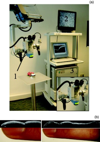
An operating microscope (Hi-R 1000; Möller Wedel GmbH, Wedel, Germany) was combined with a spectral domain optical coherence tomography system with a central wavelength of 840 nm, which allowed optical coherence tomography scans of any object in the centre of the microscopic field of vision (Figure 1a). Details of the integration of the microscope and the optical coherence tomography technology have been published elsewhere,Reference Lankenau, Klinger, Winter, Malik, Müller, Oelckers, Buzug, Holz, Weber, Bongartz, Kohl-Bareis and Hartmann13 and are only briefly reviewed here.
(a) Operating microscope with integrated optical coherence tomography system. The scan head of the optical coherence tomography system (1; also shown in inset) is connected to the camera port; further optomechanical parts (including the reference arm and the spectrometer) are housed in a separate unit (3). Optical coherence tomography images are shown together with a video image on a special screen (2). (b) Optical coherence tomography images of a human finger, using different operating microscope magnifications.

The scan head of the optical coherence tomography device was coupled to the camera port of the microscope. Optical coherence tomography scanning was performed during normal visualisation of the tissue surface. Our device was based on a commercial optical coherence tomography system (Thorlabs, Newton, New Jersey, USA) but had increased output power and delivered a light intensity of 3 mW to the tissue. The lateral and longitudinal resolution were approximately 24 and 12 µm, respectively. The optical coherence tomography system was able to adjust automatically to imaging distances of between 230 and 290 mm, via computer-controlled adjustment of the reference arm length. Approximately 1 second was required to obtain an optical coherence tomography B-scan, consisting of 1000 single A-scans. A depth range of up to 3.5 mm could be visualised. Due to the light-scattering of most biological tissues, real measurement depths varied with tissue type, and reached 1–2 mm in the larynx. A green pilot beam indicated the optical coherence tomography scanning field. The system was CE (CE marking means french: Communauté Européenne = (german) ,,Europäische Gemeinschaft“ or Conformité Européenne, ,,Conformity with the European Union guidelines”) certified for intra-operative documentation of tissue structures.
Intra-operative application
The larynx was visualised using a laryngoscope. Photomicrographs were obtained whenever appropriate. Using the operating microscopy, a nominal working distance of between 224 and 280 mm was used for optical coherence tomography measurements (using a magnification of between ×4.0 and ×7.0). In the intra-operative setting, perpendicular optical coherence tomography sections through the true vocal folds were performed, and the images were stored digitally. Optical coherence tomography measurements were performed by the surgeon alone, without assistance. The process could be started manually, and images were displayed on a dedicated screen. A green pilot beam indicated the scanning field and enabled the surgeon to define the location of the biopsy and resection plane. Performing intra-operative optical coherence tomography scanning lengthened the normal operating time by 5 to 10 minutes.
Sterile covering of the microscope enabled the surgeon to also use the optical coherence tomography system during external laryngeal surgery when appropriate.
Results
Our optical coherence tomography system employed the zoom optics of the operating microscope, such that the field of view for optical coherence tomography imaging could be changed synchronously with the conventional image. This allowed imaging of quite different tissue fields, as shown in Figure 1(b). Focusing for optical coherence tomography was also done automatically according to the focus of the image the surgeon saw in the ocular. Despite the large imaging distance, the quality of the optical coherence tomography image was similar to that achieved when using a contact probe connected to the same optical coherence tomography device.
Our optical coherence tomography system imposed no restrictions on use of the operating microscope. Optical coherence tomography images were taken during normal procedures, after selecting magnification and focus, and were displayed on a separate screen. The duration of operating time was approximately 5 minutes as a result.
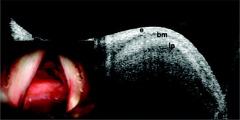
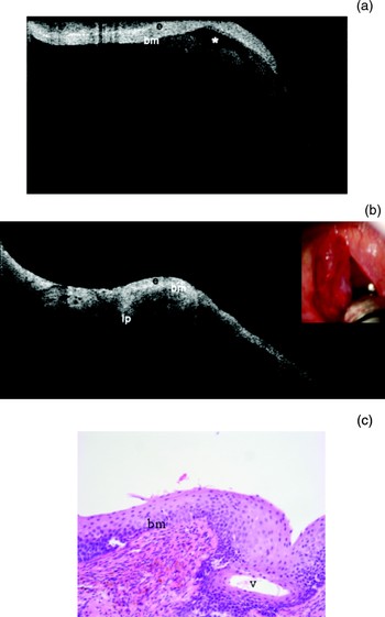
During microlaryngoscopy, optical coherence tomography technology the basement membrane to be identified in all healthy subjects (Figure 2), and also in those with benign lesions of the larynx. A representative image of a case of Reinke's oedema is shown in Figure 3(a). In this patient, optical coherence tomography scanning made it possible to identify the typical low signal intensities of the gelatinous fluid (Figure 3a). Such scanning also enabled the surgeon to identify intra-operatively regions with remaining fluid chambers. In Figure 3(a), the well demarked area low signal intensity beneath the basal membrane within the lamina propria is consistent with oedema. After incision and suction of the oedema fluid, the remaining fluid chamber disappeared (Figure 3b). The corresponding histology, obtained by resection of the excessive epithelium, showed inflammation (Figure 3c).
Optical coherence tomography scan through a vertical cross-section of a healthy true vocal fold. Inset shows the clinical appearance of healthy true vocal folds. e = epithelium; bm = basal membrane; lp = lamina propria

(a) Optical coherence tomography scan of a case of Reinke's oedema. The well demarcated area low signal intensity beneath the basal membrane, within the lamina propria (*), is consistent with oedema. (b) Corresponding optical coherence tomography scan after incision and suction of the oedema fluid; inset shows the intraoperative situs after incision of the left vocal cord. (c) Corresponding photomicrograph reveals inflammation within the epithelium (H&E; ×40). e = epithelium; bm = basal membrane; lp = lamina propria; v = vesicle

Discussion
To date, most optical coherence tomography studies of the larynx have been conducted using a rigid or flexible probe.Reference Wong, Jackson, Guo, Ridgway, Mahmood and Su1–Reference Kraft, Luerssen, Lubatschowski, Glanz and Arens3, Reference Sommer, Lankenau, Thorns, Fedder, Hüttman, Wollenberg and Papaspyrou14 This represents a substantial limitation when high precision is required in locating a biopsy site within the laryngeal epithelium. In addition, this approach is not always acceptable because of the difficulty in handling the optical coherence tomography probe while using surgical instruments. The importance of a non-contact application of optical coherence tomography technology was recognised in 2006.Reference Armstrong, Ridgway, Vokes, Guo, Perez and Jackson2 One of the big advantages of optical coherence tomography over ultrasound is the avoidance of tissue contact during scanning.
The advent of fast spectral domain optical coherence tomography technology has renewed interest in coupling optical coherence tomography devices to the operating microscope. Here, we report our approach to optical coherence tomography scanning during microlaryngoscopy. When optical coherence tomography technology is integrated into an operating microscope,Reference Geerling, Muller, Winter, Hoerauf, Oelckers and Laqua10 tissue structures can be visualised and their extensions measured during surgery.Reference Heermann, Hauger, Issing and Lenarz11 A new generation of optical coherence tomography devices based on the spectrally resolved detection of interference signalsReference Häusler and Lindner15 has dramatically increased imaging speed.Reference Leitgeb, Hitzenberger and Fercher5 These spectral domain optical coherence tomography devices have the potential to enable real-time, B-scan imaging and fast acquisition of three-dimensional volumes. Our newly developed optical coherence tomography operating microscope system addresses all the shortcomings of similar systems reported thus far,Reference Heermann, Hauger, Issing and Lenarz11, Reference Vokes, Jackson, Guo, Perez, Su and Ridgway12 namely: inferior image quality, complicated handling, restricted use of the surgical microscope, additional bulk, and unclear definition of the location of the B-scan.
It has been shown that optical coherence tomography scans can describe the microanatomy of the healthy larynx and of benign lesions.Reference Wong, Jackson, Guo, Ridgway, Mahmood and Su1 Laryngeal cancer can be identified from the loss of basement membrane integrity.Reference Armstrong, Ridgway, Vokes, Guo, Perez and Jackson2 This latter aspect of optical coherence tomography seems to have had the highest impact in the field of laryngology, as it enables early detection of microinvasive carcinoma. With identification and localisation of the focal basement membrane damage seen in microinvasive carcinoma, it should be possible to improve the accuracy of biopsies during diagnostic microlaryngoscopy. We expect that our system will allow us to benefit from this advantage.
The interpretation of optical coherence tomography images obtained intra-operatively is always dependent on the viewer's experience, and scans should be compared with conventional histology in cooperation with a pathologist. The accuracy of interpretation of optical coherence tomography images is subject to a learning curve similar to that for interpretation of ultrasound, computed tomography and magnetic resonance imaging scans.
Improvement in optical coherence tomography technology is an ongoing process. Our future research will focus on the provision of optical coherence tomography images to surgeons as they look through the microscope, and on the development of three-dimensional, real-time optical coherence tomography scanning. Real-time optical coherence tomography should provide instantaneous scanning information on the larynx. This will enable the surgeon to precisely identify the border of the lesion and also to obtain information during surgery. Optical coherence tomography guided resection may become feasible.
The method presented in this paper will be explored in a further study in patients with dysplastic, precancerous and malignant lesions of the larynx. The aim of this future, prospective study will be to assess the diagnostic sensitivity, specificity and accuracy of optical coherence tomography equipment coupled with an operating microscope, compared with data obtained by optical coherence tomography devices using special probes.
Conclusions
Optical coherence tomography scanning using equipment coupled with an operating microscope is a non-contact procedure which has important advantages over optical coherence tomography scanning conducted via special probes, in that the optical zoom of the operating microscope can be used in its complete range during surgery. In our experience of our optical coherence tomography system, the technical problems hindering clinical use were largely resolved. Our optical coherence tomography system was easy to handle intra-operatively, and no assistance was required for optical coherence tomography scanning.