Introduction
Beyond the retina, attempts to link a visual phenomenon to the activity of neurons that belong to a specific class, or else possess particular functional characteristics, have been met with only partial success. Linking hypotheses made in the late 19th century to particular neuronal classes in the retina, such as that drawn between the spectral sensitivity of vision at low (scotopic) light levels to the absorption spectrum of rhodopsin as reflected by the neural activity of rod photoreceptors have withstood repeated examination with progressively more sophisticated techniques. However, for perceptual phenomena thought to be mediated at levels in the visual pathway beyond the retina, the linking hypothesis must be refined continually as knowledge of the complexity of the organization of the visual pathways expands. Following hard on the heels of the original documentation of the selective visual response characteristics of single cells in the cat and monkey striate cortex (Hubel & Wiesel, Reference Hubel and Wiesel1962, Reference Hubel and Wiesel1968), there were numerous attempts to link a variety of perceptual phenomena to the activity of simple and complex cortical cells. Forty years on, these attempts seem naïve for a number of reasons including their shared assumption that the many perceptual phenomena they addressed were mediated solely by events in areas 17 and 18. Many of these attempts relied on correlations drawn between the response properties of cortical cells in animals and parallel specificity observed in human perceptual data, but another source of data was provided by observations made on selectively visually deprived (or exposed) animals or humans.
In parallel with the vanguard electrophysiological studies of the functional properties of cells in early visual cortical areas (areas 17 and 18) were some noteworthy reports of long-term changes in these properties consequent to early selected visual exposure. Two forms of early selective exposure were instructive, notably exclusive monocular exposure which results in near-complete shifts of ocular dominance in the cortex to favor the open eye and stroboscopic illumination that substantially reduces directional selectivity among cortical cells (Cynader & Chernenko, Reference Cynader and Chernenko1976; Pasternak et al., Reference Pasternak, Schumer, Gizzi and Movshon1985). Investigations of the perceptual consequences of different forms of selected early visual deprivation provide additional evidence to link the properties of cells in the visual cortex to certain perceptual phenomena (Mitchell, Reference Mitchell, Chalupa and Werner2004). A virtually unstated possibility at the time these developmental studies were undertaken was that the selected visual deprivation may have different consequences for visual cortical regions other than the well-documented effects observed in area 17. If this were the case, then the perceptual consequences of deprivation may reflect in part the characteristics of neurons in other, presumably higher cortical areas than areas 17 and 18. This possibility has received specific attention in recent years with respect to the neural basis of the amblyopia following early selected rearing with strabismus or anisometropia (Kiorpes & McKee, Reference Kiorpes and McKee1999) as well as to the neural basis of the deficits of motion perception following stroboscopic rearing (Pasternak et al., Reference Pasternak, Schumer, Gizzi and Movshon1985; Pasternak, Reference Pasternak1986).
In this report, we provide experimental evidence based on experiments conducted over 30 years ago, for substantial effects of monocular deprivation (MD) beyond cortical areas 17 and 18 in cats, particularly those visual areas that mediate spatial vision (Kaye et al., Reference Kaye, Mitchell and Cynader1981; Mitchell, Reference Mitchell, Payne and Peters2002) following lesions of striate and extrastriate cortex. This observation as well as more recent observations of physiological changes induced by early selected visual deprivation in higher cortical areas (Sireteanu & Best, Reference Sireteanu and Best1992; Schröder et al., Reference Schröder, Fries, Roelfsema, Singer and Engel2002; El-Shamayleh et al., Reference El-Shamayleh, Kiorpes, Kohn and Movshon2010) adds to the growing realization of the complexity of the task of identifying the site of the neural activity underlying particular perceptual phenomena (Hedges et al., Reference Hedges, Gartshteyn, Kohn, Rust, Shadlen, Newsome and Movshon2011).
Materials and methods
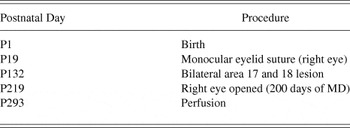
The subject of this study was a cat (C87) born and raised in a closed animal colony at Dalhousie University and was a subject in a large and as yet mainly unpublished study (Kaye et al., Reference Kaye, Mitchell and Cynader1981; Mitchell, Reference Mitchell, Payne and Peters2002) of the effects of ablation of cortical areas 17 and 18 at different ages on visual acuity, contrast sensitivity, and depth perception. Unlike the other cats, this cat was monocularly deprived, at postnatal day (P) 19, long before bilateral ablation of visual cortical areas 17 and 18 (Tretter et al., Reference Tretter, Cynader and Singer1975) at P132 days (Table 1). Vision was restored to the deprived eye about 3 months later at P219 (Table 1) to allow binocular visual input. This rearing history enhanced the opportunity to observe in the same animal effects of MD on those visual areas that mediate spatial vision after lesions of areas 17 and 18. This study was conducted a decade or more before the first published investigations of the effects of selected visual deprivation (strabismus) on cortical areas beyond the striate cortex (Sireteanu & Best, Reference Sireteanu and Best1992; Schröder et al., Reference Schröder, Fries, Roelfsema, Singer and Engel2002).
Experimental timetable

To facilitate comparison of the postlesion acuity of C87 with other animals that received the same lesion at comparable ages and tested in a similar manner, data are provided for five other cats, two (K192 and K215, cortical ablation of cortical areas 17 and 18 at 15 months) from a previous published study (Kaye et al., Reference Kaye, Mitchell and Cynader1981) and three (C41, C45, and C100, cortical lesions at respectively, 12.5, 11, and 5 months) from the larger study that have as yet been reported in only preliminary form in a book chapter (Mitchell, Reference Mitchell, Payne and Peters2002; Fig. 16-2).
All surgical and behavioral testing procedures were conducted in compliance with the National Research Council’s Guidelines for the Care and Use of Mammals in Neuroscience and Behavioral Research (2003), the Canadian Council on Animal Care’s Guide to the Care and Use of Experimental Animals (Olfert et al., Reference Olfert, Cross and McWilliam1993) and were approved by the Dalhouise University Committee on Animal Care.
Monocular deprivation
The right eyelid was sutured closed at P19 under gaseous halothane anesthesia supplemented by injections of a long-lasting local anesthetic (Marcaine 0.25%) around the palpebral aperture. The palpebral conjunctiva was first dissected free from both lid margins to expose about 3 mm of the eyelid tissue. The palpebral conjunctivae freed from the upper and lower lids were sutured together with 5-0 chromic gut. Following application of a broad-spectrum ophthalmic antibiotic (Chloromycetin l%), the exposed tissue on the underside of the eyelids was opposed and sutured together with 4-0 silk. Although the eyelid margins themselves were not trimmed, the exposed palpebral eyelid tissue near the margins healed together, as did the sutured palpebral conjunctivae, to form two occlusion layers. For detailed procedures, consult Murphy and Mitchell (Reference Murphy and Mitchell1987).
Removal of areas 17 and 18
Areas 17 and 18 were bilaterally removed at day P132 (Table 1). General anesthesia was induced with sodium pentobarbital (40 mg/kg, i.p.). The cephalic vein was cannulated for the infusion of mannitol (1 gm/kg, i.m.) to facilitate the removal of cortical tissue, and body temperature was maintained in the normal range with a heating pad. During the procedure, the cannula also permitted the administration of additional pentobarbital, when necessary, and the infusion of fluids (2.5% dextrose in half-strength lactated Ringer’s solution). After the head was stabilized, a midline incision was made in the scalp, the temporalis muscles were detached and reflected laterally, and the marginal (Doty, Reference Doty1967), posterolateral, and the junction of the middle and posterior suprasylvian gyri were exposed by craniotomy. The dura was incised and reflected laterally, and areas 17 and 18 were removed by subpial aspiration. The lateral sulcus, the medial third of the suprasylvian gyrus at the junction of its middle and posterior portions, and the fundi of the posterolateral and splenial sulci mark the boundaries of areas 17 and 18 (Tusa et al., Reference Tusa, Palmer and Rosenquist1978, Reference Tusa, Rosenquist and Palmer1979) and served as boundaries for the ablation. When the ablation was completed and hemostasis was achieved, saline-soaked sterile gelatin sponges were placed in the cortical defect, the dura and stored bone pieces were replaced, the temporalis muscles were sutured together, and the skin incision was repaired with 3-0 silk suture material.
Behavioral assessments
The behavioral measurements of visual acuity were made by the use of a jumping stand and procedures that were well established prior to the experiment (Mitchell et al., Reference Mitchell, Giffin and Timney1977). A detection task was employed whereby the animal was trained to jump to a grating in preference to a uniform gray stimulus of the same average luminance (50 cd/m2). The stimuli were large (19 cm2) square-wave gratings having a Michelson contrast close to 1.0. A descending method of limits was used beginning with suprathreshold gratings having a low spatial frequency that was increased progressively in small steps that were equated on a logarithmic scale (as many as 12 to an octave) until the cat’s performance fell to chance. Usually, the animal was flawless until close to threshold when it became agitated and could no longer achieve criterion performance, defined as a minimum of 7 correct out of a maximum of 10 trials presented at a particular spatial frequency. The acuity values specified represent the highest spatial frequency for which this criterion or better could be achieved. Training of C87 began later than was usual for kittens in this laboratory (usually at 5 weeks) when it was 3-months old. Retraining and testing of acuity began 2 months after the cortex was lesioned, and stable measurements were made 3 weeks later. The comparison animals were also trained late, and in only one case (C45) were acuity measurements made prior to the cortical lesion. For detailed procedures, consult Murphy and Mitchell (Reference Murphy and Mitchell1987).
Reversal of MD
Upon termination of the period of MD (at P219), the closed eyelid was carefully parted and the palpebral conjunctivae cut open. Because the eyelid margins were not trimmed at the time of eyelid closure, the cat was able to maintain a palpebral aperture of normal extent throughout the period of visual recovery. This surgical procedure also produced far less corneal irritation than occurs with the conventional procedure for eyelid closure that requires that the eyelid margins be trimmed, with the consequent loss of the sebaceous glands located there.
Perfusion and tissue processing
Following completion of behavioral testing, the animal was sacrificed at 293 days of age. The cat was deeply anesthetized (sodium pentobarbital, 40 mg/kg, i.p.) and perfused through the ascending aorta with 0.9% saline followed by aldehyde fixatives. The right auricle was opened to permit the drainage of solutions from the vascular system. The net effect of these procedures was to exsanguinate the cat, a method consistent with the recommendations of the American Veterinary Medical Association Panel on Euthanasia (Beaver et al., Reference Beaver, Reed, Leary, McKiernan, Bain, Schultz, Bennett, Pascoe, Schull, Cork, Francis-Floyd, Amass, Johnson, Schmidt, Underwood, Thorton and Kohn2001). All solutions were buffered to pH 7.4 with 0.1 M Sorenson’s buffer. Immediately following the perfusion, the calvaria were removed to expose the brain, which was blocked at Horsley–Clarke coronal level A22. The brain was photographed and then cryoprotected with sucrose after which it was frozen and sections cut in the coronal plane at 50 µm using a freezing microtome and collected serially through the entire brain. One section, every 250 µm, was processed for Nissl (cresyl violet stain) to label the soma and extranuclear RNA. All tissue was mounted onto gelatin-coated slides, air dried, cleared, and coverslipped.
Reconstruction of the lesion
The initial reconstruction of the cortical ablations was based on the photographed brain and the Nissl-processed coronal sections taken through visual cortex. The surviving cortex was mapped onto standardized sections illustrated by Tusa et al. (Reference Tusa, Palmer, Rosenquist and Woolsey1981). The extent of the visual cortical ablation, in retinotopic coordinates for each area, was estimated and charted onto standard, gross outline drawings of the cat cerebral cortex (Reinoso-Suárez, Reference Reinoso-Suárez1961).
Coronal sections revealing retrograde degeneration in the dorsal lateral geniculate nucleus (dLGN) were examined and the boundaries of the A-laminae and C-complex in each case were delimited according to criteria of Guillery (Reference Guillery1970). The extent of the degeneration was plotted onto standard coronal outlines of the laminated portion of the dLGN by noting retinotopic coordinates (Sanderson, Reference Sanderson1971). From these plots, the rostrocaudal extent of the degeneration was charted onto a standardized parasagittal section through the middle of the laminated portion of the dLGN. In addition, any detectable degeneration in the pulvinar and lateral posterior nuclei was recorded. Regions of the dLGN were categorized as showing intense, mild, or no degeneration. The partially spared portions of the dLGN (mildly degenerated) were either small regions that represent the lateral visual field periphery, which are connected to the sparing of area 17 on the upper bank of the splenial sulcus, or thin transition zones between intact and degenerated areas.
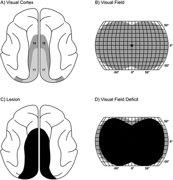
After the initial lesion assessment, the visual cortex ablation was compared with the degeneration plotted in the dLGN according to the retinotopic coordinates used as the reference for the two estimates. Commonly, the lesion as assessed from the dLGN degeneration is larger than the lesion estimated from the cortical sections, which we interpret to indicate that portions of apparently intact cortex were undercut. These undercut regions are included in the cortical lesion reconstruction (Fig. 1). For a detailed account of lesion reconstruction procedures, consult Lomber et al. (Reference Lomber, Payne, Cornwell and Pearson1993, Reference Lomber, MacNeil and Payne1995).
Lesion reconstruction of areas 17 and 18 and its impact on the visual field. (A) Dorsal view of the cat cerebrum showing the positions of areas 17 and 18. Top is anterior. (B) Combined visual field representations in areas 17 and 18 (gray). (C) Dorsal view of the cat cerebrum showing the extent of the lesion (black) in C87. Compare with (A) and note nearly complete ablation. (D) Regions of the visual field impacted by the lesions of areas 17 and 18 (black). Note that with the exception of the uppermost and far peripheral representations that the lesion affects nearly all the visual field representation in areas 17 and 18.

Results
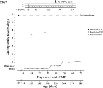
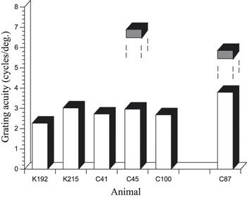
The results of the measurements of grating acuity at various times with respect to the timing of the experimental interventions are shown in Fig. 2. The data are displayed in graphical form with grating acuity plotted as a function of the time (days) since termination of the period of MD. A schematic representation of the rearing history is presented at the top of the figure. Two days before the cortical lesion, the grating acuity was 5.95 cycles/deg (solid square symbol). Following the cortical lesion, the animal eventually recovered an acuity of 3.8 cycles/deg, a value slightly higher than that observed after comparable lesions made on other animals at 5–15 months of age in this laboratory as illustrated in Fig. 3. The postlesion acuity deficit expressed with respect to that obtained prior to the lesion was comparable to that reported by Lehmkuhle et al. (Reference Lehmkuhle, Kratz and Sherman1982) but larger than that reported (0.2–0.5 octaves) by Berkley and Sprague (Reference Berkley and Sprague1979), following a lesion restricted to area 17.
The grating acuity of the nondeprived eye measured either before (filled square) or after (open squares) bilateral ablation of cortical areas 17 and 18 of a monocularly deprived kitten at 132 days of age. The data are plotted with respect to the time elapsed since opening of the deprived eye at P219 days. Data are also shown for the acuity of the deprived eye (open circles) following termination of MD. The visual history of the animal (C87) is illustrated in a schematic form at the top of the figure.

Histograms that display the grating acuity of C87 following ablation of cortical areas 17 and 18 as compared to data from five other animals tested in a similar manner after the same extensive cortical lesions. Measurements of the grating acuity prior to the lesion are shown for only two animals for which such data were obtained (C45 and C87). The acuity prior to and following the lesion are depicted respectively, as shaded and open bars. Data for K192 and K215 are reproduced from Kaye et al. (Reference Kaye, Mitchell and Cynader1981), whereas the results from the other three animals have been published as yet in only a preliminary form (Mitchell, Reference Mitchell, Payne and Peters2002).

The long (200 days) period of MD was initiated prior to the peak of the critical period of ocular dominance plasticity in area 17 (Olson & Freeman, Reference Olson and Freeman1980) and terminated nearly 3 months (87 days) following the cortical lesion. If imposed on an intact animal, such a long period of MD would produce a severe and permanent effect on the vision of the deprived eye (Mitchell, Reference Mitchell1988). Remarkably, the effects observed on C87 were just as severe; the animal appeared blind on the jumping stand for a month when using its deprived eye. During this time, it could not discriminate an open from an adjacent closed door on the jumping stand by the use of visual cues alone. For another 3 weeks, it could only pass this task, which represents at best the ability to make a crude luminance discrimination, before it acquired the ability to discriminate a grating from a uniform field of the same luminance. Measurements of grating acuity made thereafter revealed only crude form vision (an acuity of 0.15 cycles/deg) in the deprived eye just prior to termination of the experiment.
Cortical lesion reconstruction
Examination of the fixed brain showed that all or most of the marginal (Doty, Reference Doty1967) and posterolateral gyri were removed bilaterally and that the lateral bank of the lateral sulcus was additionally included in the left hemisphere ablation (Fig. 1). However, microscopical analyses revealed variable degrees of minor tissue sparing. For ablations of areas 17 and 18, when tissue is spared, it was most frequently found on (1) the most anterior portion of the marginal gyrus, (2) on the upper bank of the splenial or posterior bank of the postsplenial sulci, or (3) the inferior and lateral part of the posterolateral gyrus. The extent of the ablation of areas 17 and 18 was verified by examination of the patterns and density of retrograde degeneration in layers A and Al of the dLGN. These layers contain neurons that project solely to areas 17 and 18 (Rosenquist, Reference Rosenquist, Peters and Jones1985; Sherman, Reference Sherman, Sprague and Epstein1985). Similar analyses to determine the amount of area 19 included in the ablation could not be carried out because there is no part of dLGN that projects solely to area 19. Area 19 is an extrastriate area that shares, with the numerous other extrastriate areas, inputs from the parvocellular layers of the C-complex and medial interlaminar nucleus (Rosenquist, Reference Rosenquist, Peters and Jones1985; Sherman, Reference Sherman, Sprague and Epstein1985). However, based on direct examination of cortex, it should be noted that the extent of area 19 removed varied between the two hemispheres.
In both hemispheres, the caudal pole of the posterolateral gyrus remained intact, as did part of the upper bank of the splenial sulcus. Thus, most of areas 17 and 18 were removed, although the uppermost visual field representation was spared, as was the lateralmost visual field periphery representation. The involvement of the lateral bank of the lateral sulcus in the left hemisphere included the lower field representation of area 19 and portions of area 7. Cingulate cortex and the lateral suprasylvian visual areas remained intact in both hemispheres (Palmer et al., Reference Palmer, Rosenquist and Tusa1978).
When the visual coordinates of the defect and degeneration of areas 17 and 18 in layers A and Al were compared, via the standard retinotopic maps (Sanderson, Reference Sanderson1971; Tusa et al., Reference Tusa, Palmer, Rosenquist and Woolsey1981), the extent of the degeneration in dLGN was slightly larger than the assessed extent for the cortical defect. We interpret this difference to imply that regions of cortex that appeared intact on gross and microscopical inspection may have been undercut. The undercut cortex is considered part of the defect illustrated in the gross brains shown in Fig. 1.
The ablation included greater than 90% of areas 17 and 18, and in the left hemisphere, it extended beyond the lateral border of area 18 to include part of area 19 and beyond the medial border of area 17 to include portions of splenial visual cortex (Kalia & Whitteridge, Reference Kalia and Whitteridge1973). These minor incursions into area 19 and splenial cortex ensured removal of as much of areas 17 and 18 as possible. Lastly, there was no evidence of degeneration or shrinkage of the extrageniculate nuclei, indicating the integrity of the visual areas lateral to area 19. In total, the lesion removed areas 17 and 18 bilaterally. Functionally, the affected region included nearly the entire visual field with the exception of the uppermost representation and the extreme periphery on the left and right sides (Fig. 1).
Discussion
For a motivated cat, the discrimination of gratings of high spatial frequencies requires that they access the responses of neurons with the highest spatial resolution that in areas 17 and 18 are located in the representation of central retina (the area centralis). As the peripheral retina cannot sustain high spatial resolution, any cortical tissue that was spared by the lesion, if not undercut, could mediate only poor acuity. On the basis of the substantial visual acuity retained after the cortical lesion, together with the observation that the animal’s head posture during postlesion testing on the jumping stand did not appear to change, it can be assumed that the animal accessed the representation of the central retina in whatever visual area(s) that sustained vision after ablation of areas 17 and 18.
The severity of the immediate effects of the period of MD on the vision of the deprived eye which was reduced to blindness stands in striking contrast to the relatively minor effects of ablation of both areas 17 and 18, which reduces visual acuity by about an octave (Mitchell, Reference Mitchell, Payne and Peters2002). Indeed both the magnitude of the immediate effect of MD on the vision of the deprived eye and the very slow and limited subsequent recovery were comparable to those observed after MD of a similar onset and duration in intact animals (Mitchell, Reference Mitchell1988). It would appear that the effects of MD in those areas that mediate vision after the lesion may be just as profound as those that occur in areas 17 and 18.
Because the period of MD both preceded and continued after ablation of areas 17 and 18, its effects on the vision of the deprived eye could reflect events in the visual areas that sustained vision after the lesion at one or both of these times. Potentially, prior to the lesion, MD could exert effects on first, areas 17 and 18, second, on cortical areas to which they project, and third, on the many extrastriate cortical areas that receive visual input via pathways in parallel to the geniculostriate projections, such as via the C laminae of the dLGN, the lateral posterior nucleus or the pulvinar (Lomber et al., Reference Lomber, Payne, Cornwell and Pearson1993, Reference Lomber, MacNeil and Payne1995). Following the lesion, the deprivation could not affect the first two but could still exert changes on the extrastriate visual cortical area(s). The fact that the effects of MD on vision were so profound suggests that the functional changes induced by the deprivation in the extrastriate cortical areas may be at least as substantial as those in area 17.
In the year this study was begun (1981), there were only limited data to suggest that the effects of early MD in kittens on vision were far greater than the consequences for vision of ablation of cortical areas 17 and 18. The results from C87 make this point very clear by showing in the same animal that MD leads to a visual loss that is both profound and long-lasting, whereas cortical ablation reduces acuity by only about a factor of two. The conclusion that MD must produce severe effects in cortical areas other than 17 and 18 and particularly those neural structures that mediate vision after their ablation is now well supported by many studies. Investigations of cats reared with strabismus (Sireteanu & Best, 1992) and who demonstrate strabismic amblyopia (Schröder et al., Reference Schröder, Fries, Roelfsema, Singer and Engel2002) revealed substantial shifts of ocular dominance in the posteromedial lateral suprasylvian area (PMLS) toward the nonamblyopic eye that seemed even greater in cortical area 21a. Electrophysiological investigations of the striate cortex of amblyopic monkeys that had been reared with anisometropia provided strong evidence that the effects of early visual deprivation must extend beyond the striate cortex (Kiorpes et al., Reference Kiorpes, Kiper, O’Keefe, Cavanaugh and Movshon1998). Later work confirmed this suggestion by demonstrating substantial changes of motion processing by neurons in area MT of monkeys reared with either strabismic or anisometropic amblyopia (El-Shamayleh et al., Reference El-Shamayleh, Kiorpes, Kohn and Movshon2010). A number of imaging studies of humans with amblyopia also reveal abnormalities in various extrastriate cortical regions beyond V1 (e.g., Barnes et al., Reference Barnes, Hess, Dumoulin, Achtman and Pike2001; Lerner et al., Reference Lerner, Pianka, Azmon, Leiba, Stolovich, Loewenstein, Harel, Hendler and Malach2003; Li et al., Reference Li, Dumoulin, Mansouri and Hess2007).
There is now very good evidence that amblyopia results in deficits in many visual cortical areas in higher mammals including humans. Still, the results described here for C87 combined with the well-documented (Mitchell, Reference Mitchell, Payne and Peters2002) visual capacities of cats following bilateral lesions of 17 and 18 point to the fact that neural activity in multiple sites can support various perceptual abilities. The extensive effects of selected early visual input as well as the potential for perception to be mediated at different neural sites following deprivation points to a need for caution in the use of data from early experiential manipulations for formulation of linking hypotheses. In a larger context, elegant recent work questions the very notion of a single neural site for some perceptual phenomena; certain aspects of motion perception may depend not just on neural activity in a single cortical areas but rather interactions between the neural activity in multiple motion selective systems (Hedges et al., Reference Hedges, Gartshteyn, Kohn, Rust, Shadlen, Newsome and Movshon2011).
Acknowledgment
This work was supported by Discovery grants (7660 and 327442) to DEM and SGL from the Natural Sciences and Engineering Research Council of Canada.