The coarctation dilatation is usually achieved using fluoroscopy guidance in the catheterisation platform. In fact, two-dimensional transthoracic echocardiograph is a recognised tool during the structural heart interventions such as patent ductus arteriosus, interatrial defect, or ventricular septal defect. Reference Johnson, Sathanandam and Naik1 Nowadays, and to the best of our knowledge, this is the first experience of neonatal coarctation dilatation using TTE as the only guidance at the bed in neonatal intensive care and without fluoroscopy.
Clinical case
We describe a case of 10-day old full-term female. The patient was urgently referred from the regional hospital to our neonatal ICU since she presented a severe cardiovascular collapse. At arrival, the patient presented with bradycardia, marked hypotonia, and polypnea. The lower leg saturation was 75 % in room air with very weak pulses. Arterial blood gas analysis showed severe metabolic acidosis. Then, inotrope drugs and respiratory support were immediately started. The Doppler echocardiography was achieved by the paediatric cardiologist and established the etiological diagnosis of the described shock. It consisted of coarctation of the aorta without any additional associated malformations. Two hours after partial improvement of the situation, we decided to perform dilatation without delay.
Technical setup
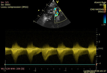
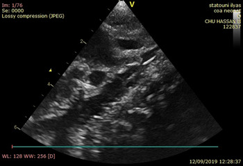
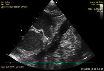
The written informed consent of the patient parents was obtained. The echocardiography examination was then performed by an experienced paediatric cardiologist using the Philips Epic machine. The coarctation was well described with good image quality ranging from supra-sternal plane to left lateral view. Accurate measurement of different segments of the aorta at the maximum systolic time was acquired and is used to select the appropriate balloon size. The suitable dimensions were as follows: horizontal aorta 7 mm, descendant aorta 9 mm, coarctation segment 2–3 mm (Figs 1 and 2). Besides, restrictive PDA right–left shunting was found with severe left ventricle dysfunction and supra-systemic pulmonary hypertension.
Echocardiography before dilatation.

Echocardiography before dilatation.

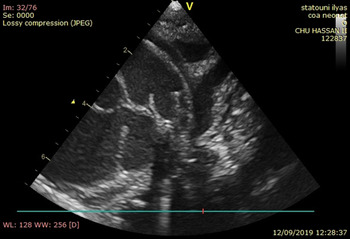
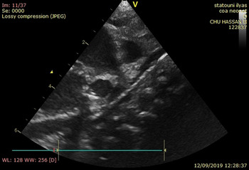
Hence, TayShak non-compliant balloon of 6 and 8 mm diameters with 3 cm length were selected based on the transverse arch and the descending aorta at the level of diaphragm measurement. The procedure was performed under general anaesthesia. The patient was intubated, femoral access was obtained easily by four French sheaths; heparin at dose of 50 UI/kg was given just after artery access. The passage of four French multipurpose catheters was controlled using the echocardiography, and was advanced over 0.018 French-guided exchange wire. The catheter crossed easily the coarctation and was pushed to the left carotid artery. The control with the echocardiography was permitted to confirm the appropriate positioning of the balloon over the coarctation (Figs 3 and 4).
Guide and balloon placement.

Guide and balloon placement.

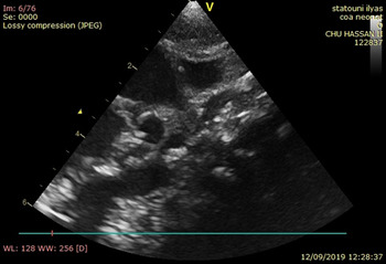
After checking the best position; the first 6 mm balloon was inflated up to its maximal diameter. The second dilatation was achieved with 8 mm TSNCB (Fig 5). In addition, we completed our procedure by PDA dilatation, which was achieved successfully using 4 mm TSNCB. We didn’t require any ventricle pacing because we acquired low contractility of the left ventricle.
Balloon dilatation and echocardiography after dilatation.

After deflation, the balloon was withdrawn carefully. The gradient was measured across the coarctation by TTE. An “aortogram” was measured easily by TTE and showed a complete relief of the coarctation (Fig 6). Indeed, good and appropriate PDA shunting was realised exclusively in the direction left to right.
Balloon dilatation and echocardiography after dilatation.

The sheath was removed and haemostasis was done with hand compression.
The patient saturation has improved after the procedure and the drugs were weaned off after 48 hours following the intervention. The patient was extubated and discharged 5 days later. Three months follow-up showed very good results without requiring any surgical intervention or restenosis. The different achieved steps of the technique are shown in (Figs 1–6).
Discussion
TTE has emerged as one of the best tools in transcatheter interventions in paediatric cardiology. It has been used as an alone guide during various cardiac interventional catheterisation. It becomes a routine and accepted technique in cardiac malformation treatment in the world with other imaging techniques and modalities. Coarctation of the aorta is one of the most common congenital cardiac heart diseases in neonate. Surgical treatment is actually the first choice in this age stage. Reference Rao2 Our initial experience demonstrated that TTE could be the only guidance in coarctation dilatation in neonatal stage.
Percutaneous dilatation of the coarctation is usually performed under fluoroscopy in catheter laboratory. This technique could be a safe and effective way to perform dilatation with or without a stent. However, high radiation exposure and contrast agent administration in neonate with cardiac failure and renal deficiency constitute an important limitation of use in such profile of cases. Reference Reddington, Booth, Shore and Rigby3,Reference Syamasundar Rao4
In our opinion, the newborn is a good candidate since demonstrating a high echocardiography echogenicity. Reference Balcı5 This allows a good evaluation of the stenosis before and after the procedure. TTE provides useful information through a repetitive and unlimited sequence of slices. Balloon dilatation could be facilitated by echocardiography without displacement of the neonate from his bed to the cath lab. In addition, TTE protocol allows evaluating cardiovascular function besides any associated complications.
This case demonstrates that the TTE guidance allows a good morphological description of the aorta and PDA compared to fluoroscopy. Another major advantage of this procedure consists of rapid recovery from metabolic and cardiac failure within a very short time. Reference Syamasundar Rao4 Finally, easy access and diminished cost of the intervention are also a major benefit.
Conclusion
TTE is a safe and secure technique allowing to assist and guide coarctation dilatation. TTE provides a good and detailed structural and haemodynamics information before, during, and after the coarctation dilatation procedure.
Percutaneous coarctation dilatation is good and safe choice with outcomes similar to achieved results using fluoroscopy. Further experiences with large number of patients should improve the confidence and the validity of the reported approach.
Acknowledgements
Authors are grateful to Mrs. Khadija Qadida for here availability to support the team during intervention and post-intervention stages.
Financial support
Nothing to declare.
Conflict of interest
The authors declare that they have no conflicts of interest.
Ethical standards
The clinical case reported inhere was carried out with best care of the ‘good clinical practice’ and according to the national ethical standards complying with the international regulation especially Helsinki declarations and National Institute of Health (USA) standards.