Introduction
Dementia is frequently accompanied by motor dysfunctions, especially in its advanced stages (Camicioli et al., Reference Camicioli, Howieson, Oken, Sexton and Kaye1998; Kurlan et al., Reference Kurlan, Richard, Papka and Marshall2000; Prehogan and Cohen, Reference Prehogan and Cohen2004; Allan et al., Reference Allan, Ballard, Burn and Kenny2005; Scherder et al., Reference Scherder2007). Paratonia, a form of hypertonia, is a motor dysfunction that is notably present in 90%–100% of people in the advanced stages of dementia (Souren et al., Reference Souren, Franssen and Reisberg1997). In 2006, a new international consensus definition was established that defined paratonia as a form of hypertonia with an involuntary variable resistance during passive movement. In contrast to Parkinson's disease, in paratonia the degree of resistance varies with the speed of movement (i.e low resistance to slow movement and high resistance to fast movement). The degree of paratonia is proportional to the amount of force applied. The severity of paratonia increases with the progression of dementia. In contrast to spasticity after stroke, the resistance of paratonia to passive movement can be in any direction, with no clear pattern in flexion or extension, and there is no clasp-knife phenomenon (Hobbelen et al., Reference Hobbelen, Koopmans, Verhey, van Peppen and de Bie2006).
Finally, paratonia results in a characteristic bed posture of flexed arms and legs and an uplifted head floating above the pillow; it is accompanied by pain, and it affects mobility and quality of life (Middelveld-Jacobs and van den Boogerd, Reference Middelveld-Jacobs and van den Boogerd1986; Souren et al., Reference Souren, Franssen and Reisberg1997).
Consequently, caregiver burden increases exponentially with increasing muscle tone and decreasing abilities of the patient (Souren et al., Reference Souren, Franssen and Reisberg1997). Passive Movement Therapy (PMT) aims to decrease high muscle tone and to sustain the range of motion in the affected joints, and it is the main therapy applied by physiotherapists in nursing homes(Pomeroy, Reference Pomeroy1994; Leemrijse et al., Reference Leemrijse, Boer and Ribbe2005; Luijckx-Comeau and Brooijmans-Reuser, Reference Luijckx-Comeau and Brooijmans-Reuser2008). Whereas some physiotherapists claim that it reduces the burden on caregivers and nurses in daily care, others are more skeptical about its beneficial effects. There is some supporting evidence for a positive effect of PMT shortly after treatment (van Wingerden, Reference van Wingerden1997; Gajdosik, Reference Gajdosik2001; Hobbelen et al., Reference Hobbelen, de Bie and van Rossum2003; Bautmans et al., Reference Bautmans, Demarteau, Cruts, Lemper and Mets2008). However, the most common frequency of PMT is two to three times a week, implying a more extended effect of the treatment (Leemrijse et al., Reference Leemrijse, Boer and Ribbe2005; Luijckx-Comeau and Brooijmans-Reuser, Reference Luijckx-Comeau and Brooijmans-Reuser2008). A pilot study (n = 15) with PMT at a frequency of three times a week with a follow-up after three weeks showed an unexpected trend in which muscle tone increased in the PMT group compared to the controls (Hobbelen et al., Reference Hobbelen, de Bie and van Rossum2003). Although the participants of this pilot study were probably not a homogeneous sample, the results emphasize the need to study the effects of PMT in this frail population.
The recent international consensus definition of paratonia and the development of the Paratonia Assessment Instrument (PAI) as a valid and reliable assessment instrument for the diagnosis of paratonia enable further research in a more homogeneous population (Hobbelen et al., Reference Hobbelen, Koopmans, Verhey, van Peppen and de Bie2006; Reference Hobbelen, Koopmans, Verhey, Habraken and de Bie2008). Our null hypothesis was that PMT administered at a frequency of three times a week has no effect on the severity of paratonia caregiver's burden, nor on the pain experienced by patients during morning care after 2 and 4 weeks.
Methods
Design
The study was a single-blinded multicenter randomized clinical trial with a four-week follow-up period (Hobbelen et al., Reference Hobbelen, Verhey, Bor, de Bie and Koopmans2007). Participants were randomly assigned to either the PMT group or the control group. Interventions were administered three times a week for four weeks in a row. Assessments with the primary and secondary outcome measures took place at baseline, after two weeks (6 treatments) and after four weeks (12 treatments). The local ethical committee, CMO Arnhem-Nijmegen, the Netherlands, approved the study and filed it under nr. 2006/1567, ABR file nr. NL13777.091.06. The trial is registered at Current Controlled Trials ISRCTN43069940.
Participants
For this study, 19 physical therapy departments in nursing homes in the Netherlands were approached. Only after consent for participation by the board of directors from each institution did recruitment of eligible patients start. Recruitment took place in the participating nursing homes by physical therapists trained to diagnose paratonia with the PAI and to assess the severity of paratonia with the Modified Ashworth Scale (MAS; see below for further explanation) (Waardenburg et al., Reference Waardenburg, Elvers, van Vechgel and Oostendorp1999). The PAI is a dichotomous assessment instrument with which an examiner can establish the presence (or absence) of paratonia by successively moving all four limbs passively in flexion and extension while the participant is in a sitting position or supine in bed (Hobbelen et al., Reference Hobbelen, Koopmans, Verhey, Habraken and de Bie2008). The PAI has an interrater reliability of Cohen's κ ranging from 0.625 to 1.
Nursing home residents were included if (1) they met the DSM-IV-TR Criteria for dementia, (2) they were diagnosed to have paratonia (see appendix), and (3) they had moderate to severe paratonia defined as a score on the MAS of at least 2 ( = more marked resistance to passive movement) in at least one limb. Patients were excluded if (1) they were prescribed antipsychotic medication, (2) they received PMT less than 4 weeks prior to the start of the trial, (3) they had an unstable health problem or disease prior to admission or during the trial, or (4) they showed signs of challenging behavior toward the therapist and/or the intervention.
Participants were only included after written proxy consent.
Randomization
After computerized and concealed block randomization (block size of 4), patients were assigned to one of two groups. For every participating institution, a new randomization list was made at the Department of Epidemiology at Maastricht University. At every institution, patients were permanently numbered in order of received proxy consent. The randomization code (per patient) was only available to the assigned therapists and was kept secret from all other personnel involved, including the primary investigator.
Intervention
Patients were assigned to either the group that received PMT or the control group, which received no PMT. PMT was provided in a standardized way by trained local physical therapists. During the first part of passive movement, the therapists were instructed to move the affected limb slowly, with an emphasis on lowering muscle resistance. Next, the therapists had to reach the end range of motion and possibly stretch the structures three to five times very lightly without causing pain. In the training sessions the therapists were instructed to lessen the tension immediately if the patients showed any signs of pain. Pain was indicated by an increase of the rhythm of breathing, a more tense expression, an increase of the muscle tone or a verbal hint. The patients were positioned comfortably supine in bed while the therapists started PMT with the left arm, moving the shoulder and, subsequently, the elbow in flexion and extension (up and down). Next, the same movements were made in the right arm. Subsequently, the left leg and right leg were moved, the hip and knee in flexion, extension, and abduction/external rotation (only the hip being rotated, with the hip flexed at 45o), though with no spinal movements. The duration of PMT was approximately 20 minutes per patient per session (Hobbelen et al., Reference Hobbelen, Verhey, Bor, de Bie and Koopmans2007). In each participating nursing home, the therapists were trained to perform PMT in two training sessions of 2 hours each with patients eligible for this trial. Note that these patients were only included in the trial at least four weeks after the training session. The treatment group and the control group were visited by the therapists between 8 a.m. and 10 a.m. before being washed and dressed by the nursing staff, three times a week for four weeks. The intervention was spread out over five days (Monday – Friday) allowing for a maximum of two consecutive treatment days. Nursing staff and all assessors were blinded for treatment allocation.
To guarantee the blinding of the nursing staff, the therapists were instructed to close the door and place a “do not disturb” sign on it along with a note of their presence. The control treatment consisted of positioning the participant comfortably supine in bed and sitting silently alongside the bed for an equal duration of a PMT treatment.
Although it was impossible to blind the participants, all participants were in the advanced stages of dementia with limited ability to communicate, therefore minimizing the chances of revealing their treatment assignment to nursing staff and assessors.
Assessments
Primary outcome measure
The Modified Ashworth Scale (MAS) was used to assess the severity of paratonia. The MAS has been tested and shows acceptable reliability for assessing the severity of paratonia (intra-rater reliability: Kendall's Tb 0.62–0.80 and inter-rater reliability: Kendall's Tb 0.72–0.77) (Waardenburg et al., Reference Waardenburg, Elvers, van Vechgel and Oostendorp1999). It is based on a 5-point ordinal scale ranging from 0 to 4, in which 0 = no resistance to passive movement, 1 = slight resistance during passive movement, 2 = more marked resistance to passive movement, 3 = considerable resistance to passive movement, and 4 = severe resistance, such that passive movement is impossible.
The assessors were local physical therapists trained on the job in two training sessions of 1 hour each. They administered the MAS between 8 a.m. and 10 a.m. before the patients were washed and dressed by nursing staff at baseline (T0; one day prior to treatment start); after 2 weeks (T1; 1 day after treatment 6); and after 4 weeks (T2; 1 day after treatment 12). Flexion/extension and abduction/adduction of the shoulders were assessed, as was the flexion/extension of the elbows and the flexion/extension and abduction/adduction of the hips with the knees in a fixed position (depending on the patients’ abilities in extension and flexion). With the participant comfortably supine in bed, the assessments started in the left arm, and then went to the right arm, the left leg, and finally the right leg.
Secondary outcome measures
Pain during morning care was a secondary outcome measure and assessed with the Pain Assessment Checklist for Seniors with Limited Ability to Communicate (PACSLAC-D). The PACSLAC-D is an observational assessment instrument that lists 24 items clustered into three categories: nonverbal facial signs (10 items), total resistance (6 items), and emotional state (8 items) (Fuchs-Lacelle and Hadjistavropoulos, Reference Fuchs-Lacelle and Hadjistavropoulos2004; Zwakhalen et al., Reference Zwakhalen, Hamers, Abu-Saad and Berger2006; Reference Zwakhalen, Hamers and Berger2007). This version, a reduction of the original 60-item PACSLAC scale, had high levels of internal consistency for the complete scale (Cronbach's α range 0.82–0.86) and for all subscales (α range 0.72–0.82) (Zwakhalen et al., Reference Zwakhalen, Hamers and Berger2007).
Within an hour after the first, sixth, and twelfth treatments, an observer, typically a physical therapist (PT), occupational therapist (OT), or assistant PT or OT, assessed the PACSLAC-D during the first 10 minutes of morning care for all participants. A cut-off score of 4 or higher was judged as an indication of pain (Zwakhalen et al., Reference Zwakhalen, Hamers and Berger2007).
To gain insight into the effects of PMT on the caregivers’ burden, we used two assessment instruments. First, the observed and experienced clinical changes were assessed by nursing staff using the Clinical Global Impressions scale (CGI) (Schneider et al., Reference Schneider1997). This scale is a widely used, simple, reliable, and valid tool to measure treatment response in dementia (Reisberg, Reference Reisberg2007). At baseline, the nurses were asked to rate the severity of the participants’ stiffness in comparison to all other patients with paratonia on the ward on a 7-point scale, ranging from “normal, no stiffness at all” to “most severe stiffness.” After 2 and 4 weeks (1 day after treatments 6 and 12), they were asked to rate the participants again on the same scale with an additional question, the CGI-change score, to rate the global improvement on a 7-point scale, ranging from “very much improved” to “very much worse,” in which we translated the qualification of “improved/stable/worse” into “easier/same/heavier” in care.
Additionally, we used a modification of the Dutch Patient Specific Complaint (PSC) list, in which the nurses were asked to address the three most difficult items in daily care and rate these items on a visual analogue scale (VAS) of 100 mm, ranging from “no trouble at all” to “impossible.” This was completed and rated one day prior to the start of the treatment, and with their original score visible, the same three items were rated again one day after treatments 6 and 12 (Beurskens et al., Reference Beurskens, de Vet and Koke1996). The original PSC assessment scale is used for measuring the functional status of a patient and has been validated for use in a wide variety of diseases (Beurskens et al., Reference Beurskens, de Vet and Koke1996). The PSC has a high inter-rater reliability (ICC = 0.92–0.91); it is responsive to change; and it correlates significantly the VAS for pain (r = 0.70–0.80) (Beurskens et al., Reference Beurskens1999).
At baseline, age, sex, use of medication, type of dementia, and severity of the dementia were registered. Severity of dementia was assessed with the Global Deterioration Scale, which consists of seven stages of cognitive decline: stage 1 = no cognitive decline, stage 2 = very mild cognitive decline, stage 3 = mild cognitive decline, stage 4 = moderate cognitive decline, stage 5 = moderately severe, stage 6 = severe, and stage 7 = very severe cognitive decline (Reisberg et al., Reference Reisberg, Ferris, de Leon and Crook1982).
Analysis
All data were analyzed with SPSS 16.0 for Macintosh. For the analysis, we summed the Ashworth score for all movement directions of both arms with a maximum score of 48 points, and we subsequently summed the Ashworth score for all movements of both legs with a maximum score of 32 points. We considered these to be continuous data. Various levels can be distinguished in this research: time, patient, and institution. Due to the progressive nature of dementia a correction should be made on time. Furthermore, the treatments of patients in a particular institution are not independent of each other, so for this, a correction is also necessary. The mixed effects regression method using the direct likelihood method is particularly suitable to handle such data (Tan, Reference Tan and Osborne2008). So to account for dependency between the variables and between the different levels in this trial, and the independent variables the Ashworth score, PACSLAC-D, and the PSC were analyzed with mixed effects linear models on three levels: time level nested within patient level nested within institution (Tan, Reference Tan and Osborne2008). To account for the dependency of the three subsequent questions regarding caregiver's strain as measured by the PSC, the data were fully cross-classified with time at first level.
In all analyses, we accounted for differences in the various types and stages of dementia, the baseline assessments, and a natural time effect. To test if PMT had a different effect in the different nursing homes or on the different types of dementia or stages of the disease, we entered these factors as interaction terms in the models.
Careful analysis (not presented here) suggested that the missing-at-random assumption was plausible. No data were imputed.
Finally, the CGI was analyzed with cross-tabulation χ2. For all analyses, we considered p-values < 0.05 to be statistically significant. The analysis was carried out according to the intention-to-treat principle.
Power analysis was carried out and resulted in an α of 0.05 and power of 80%, in a sample size of 69 patients per group (taking into account a drop-out percentage of 10%) (Hobbelen et al., Reference Hobbelen, de Bie and van Rossum2003).
Results
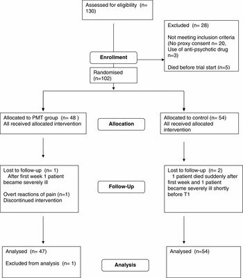
Twelve nursing homes in both rural and urban regions of the Netherlands participated in the trial. A total of 130 patients were considered eligible, of whom 110 (85%) agreed by proxy consent to participate. Ultimately, 102 of them participated. Due to overt reactions of pain, the intervention in one participant was discontinued. Data from 101 participants were analyzed. Data collection took place between April 2007 and April 2009.
The number of participants varied between the nursing homes. In one institution, 26 participants were randomized, and in another, 20 participants were randomized; in the remaining ten institutions, the number of participants varied between 1 and 10. A total of 24 therapists performed PMT, 19 assessed the MAS, 21 assessed pain, and 35 nurses assessed caregiver burden.
Figure 1 shows the study flow chart according to the CONSORT statement (Altman et al., Reference Altman2001).
Study flow chart.

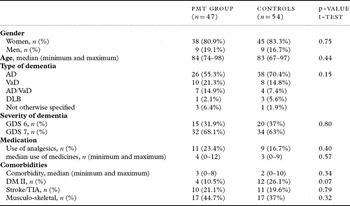
Of the participants, 82.2% (n = 83) were female. The mean age was 84 years (range 67–98 years), and most of the participants (65.3%, n = 66) were in the most severe stage of dementia (GDS 7), with 34.7% (n = 35) in GDS 6 stage. Sixty-three percent (n = 64) had Alzheimer's dementia (AD), 18% (n = 18) had vascular dementia (VaD), 11% (n = 11) had a combination of AD and VaD, 4% (n = 4) had a diagnosis of dementia with Lewy bodies, and in 4% (n = 4) of patients, dementia was not otherwise specified.
Table 1 contains the patient characteristics and distributions between the control and PMT groups.
Baseline patient characteristics

AD = Alzheimer's disease; VaD = vascular dementia; DLB = dementia with Lewy bodies; GDS = Global Deterioration Scale; DM II = Diabetes Mellitus type II.
The effect of PMT on severity of paratonia
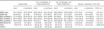
After 2 and 4 weeks, the mean change showed an increase of paratonia in both arms and legs (Table 2).
Baseline and changes in clinical outcome

Note: Data are mean (SD) or difference (SD) or median (minimum and maximum). MAS = Modified Ashworth Scale; PSC = modified Patient specific complaints; PACSLAC-D = Pain assessment checklist for seniors with limited ability to communicate-Dutch version; CGI = Clinical Global Impression; CGI-C- Clinical Global Impression-Change.
MAS arms max. score 48; mean change positive value indicate increase of paratonia.
MAS legs max. score 32; mean change positive value indicate increase of paratonia.
PSC max. score 100; mean change negative value indicate decrease of carer's burden.
PACSLAC-D max. score 24; mean change negative value indicate decrease of pain.
CGI 7-point ordinal scale; mean change positive value indicate worsening of paratonia.
CGI-C 7-point ordinal scale; score of 4 indicating no change.
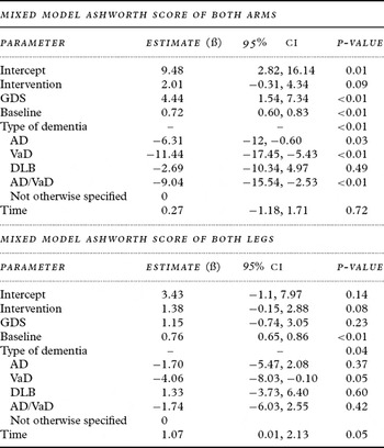
However, the mixed model showed that the increase of paratonia (indicated by a positive ß) in the PMT group was not statistically significant after 2 or 4 weeks, either for both arms (ß = 2.01, 95% confidence interval (CI) −0.31, 4.34, p = 0.09) or for both legs (ß = 1.37, 95% CI −0.15, 2.88, p = 0.08) (Table 3).
Mixed model estimated Ashworth scores of arms and legs. 3-level mixed-effects linear regression model with a random intercept at both the institution and patient levels

CI = confidence interval; GDS = Global Deterioration Scale; AD = Alzheimer's disease; VaD = vascular dementia; DLB = dementia with Lewy bodies.
Note: A positive ß indicates an increase on the Ashworthscore, a negative ß indicates a decrease on the Ashworthsscore.
The effect of PMT on caregiver strain
Clinical global impression scores
The median score for both groups was 5 (marked stiffness) at baseline and remained stable during the trial period with a slight decrease of stiffness in the PMT group and increase of stiffness in the controls (PMT: –0.3, SD 1.3; controls: + 0.2, SD 1.2) (Table 2).
After 2 and 4 weeks, the median score of improvement/worsening on the CGI change score was similar for both groups; Pearson χ2 analysis showed no statistically significant difference between the PMT and control groups (p = 0.60 after 2 weeks; p = 0.14 after 4 weeks).
Patient specific complaint scores
Specifically focusing on the caregiver's strain at the individual level of the participant, in a mixed-effects linear regression model, all participants improved and a significant interaction was found between PMT and GDS (p < 0.02). The GDS 7 patients remained stable during the trial, whereas GDS 6 patients appeared to improve over the course of 4 weeks. This improvement was significantly less in the PMT group than in the control group.
The effect of PMT on pain experienced during care giving
At baseline, 49.5% (n = 49) of participants had a PACSLAC-D score greater than or equal to 4, indicating possible pain during the observation. The mean pain score was 7.10 (SD = 3.7; minimum and maximum values 4–18). The participants with pain were equally distributed over both groups (PMT group: 47%, n = 21; control group: 53%, n = 28). The pain experienced during caregiving within an hour after the intervention decreased in the PMT group but decreased even more so in the control group. The mixed model analyses showed that only the first measurement was significantly related with the differences in pain score during the trial (ß = 0.67, standard error = 0.05, p < 0.001).
Discussion
In both groups, PMT and controls, we observed an increase of paratonia, but this was not statistically significant. Although PMT is currently used to reduce the severity of paratonia, caregiver burden, and pain experienced during morning care, the data from this study suggest that there are no beneficial effects of PMT for patients with moderate or severe paratonia.
We cautiously point out that the higher Ashworth score in the PMT group could have been caused by as-of-yet undetected microtraumata induced during the stretching of tissues of the frail elderly. This hypothesis is supported by animal studies indicating that older muscle tissue, if stretched in an activated condition, is very susceptible to injury at the sarcomere level (Brooks and Faulkner, Reference Brooks and Faulkner1996).
There appears to be a conflict between the results of the MAS, in which increased muscle tone was found in the PMT group, and the results of the CGI, in which the nursing staff noted a decrease in stiffness. This discrepancy can be explained by the different approaches of the two assessments. As indicated in the Introduction, PMT appears to have positive short-term effects shortly after treatment. These effects have been explained by the viscoelasticity of the tissues and are present 20 to 30 minutes after PMT (Van Wingerden, Reference van Wingerden1997; Gajdosik, Reference Gajdosik2001). Nurses were instructed to wash and dress the patients shortly after PMT. The MAS assessments took place on the mornings in which no PMT was given. It is possible that the nurses actually felt the short-term positive effects during their activities and reported them in the CGI score.
This study is the first multicenter randomized controlled trial to investigate a physiotherapeutic intervention for dementia patients with paratonia. A major strength of this research is that it was a pragmatic multicenter trial performed in a real setting with physical therapists and nurses dealing with the problems of paratonia on a daily basis. It is well known that these types of trials struggle to achieve both internal and external validity (Godwin et al., Reference Godwin2003). Limiting the inclusion criteria and optimizing randomization were used to realize both. In contrast with most pragmatic trials, we also successfully blinded all of the assessors. The pragmatic trial construct enabled all therapists to make pragmatic decisions with, for example, a sudden change of the treatment days to ensure three treatments a week and the assessments on the right day. Extrapolation of the results is, therefore, possible.
The treatment protocol training and the good organization to ensure blinding give strength to the results obtained.
The mixed linear effects analysis used is generally seen as best practice for longitudinal data and accounts for the correlated structure of the data (Tan., Reference Tan and Osborne2008).
Furthermore, by treating the different nursing homes as a level in these analyses, we were able to account for possible different interpretations of the ordinal levels of the MAS, our primary outcome.
Limitations
Our power analysis indicated that we needed 138 participants for this trial, and so, with only 110, this study was underpowered. Nevertheless, given the fact that there was a greater worsening evident in those participants who received PMT, it is even more debatable whether it would have been ethical to proceed and reach the target numbers. An additional 28 participants could have modified this negative trend, but they are unlikely to have turned it into a positive trend. Given the fact that the main goal of PMT is to subdue the high muscle tone and improve daily care, no effect is still negative from this perspective.
To recruit a sufficient number of participants, we included more institutions than we originally planned (Hobbelen et al., Reference Hobbelen, Koopmans, Verhey, Habraken and de Bie2008). The fact that more physiotherapists were involved in giving PMT and administering the assessments lowers the reliability of this study but expands the generalizability of our findings.
The separate randomization lists per institution caused a small deviation in the sample size of the two groups, which was slightly aggravated by the discontinuation of PMT in one participant. We believe, however, that this had no negative effect on the outcome of this trial.
To obtain insight into the effect of PMT on caregiver burden, we incorporated the CGI and PSC to be filled in by nursing staff. The CGI was used to capture a global picture of the effect of PMT on caregiver burden, translating their qualification of “decreased/stable/improved” into “easier/same/heavier.” A limitation in this study may be that the inter-rater agreement of the CGI is not known sufficiently, and that the assessments were not videotaped, which could have increased the reliability. The CGI was nevertheless chosen because it is quick and easy to use and the ecological validity is more clinically relevant, which was important for this research (Berk et al., Reference Berk2008). The questions from the PSC were directly focused on the strain of daily care, allowing us to obtain a better picture of the effect of PMT. However, the results of the PSC indicating that all participants improved, particularly the control group, remain unexplained. In retrospect, we doubt the validity of this instrument in this setting because we transferred the original PSC into a proxy assessment scale. Therefore, we think that the results might be biased, and proper validation of this instrument is required in future studies.
Conclusion
PMT has no beneficial effect on the severity of paratonia after 2 weeks or after 4 weeks, nor does it have a beneficial effect on pain experienced during care giving shortly after the treatment. There is no indication that caregiver strain decreases as a consequence of PMT. PMT is, therefore, not recommended as an intervention in severe paratonia. Future studies should focus on the effect of alternative interventions in severe paratonia and especially on the development of preventive intervention in earlier stages.
Conflict of interest declaration
This study was supported by a grant from the Vitalis WoonZorg Groep, Eindhoven, the Netherlands (a non-profit foundation).
The first author, J. Hobbelen, was a part-time employee at the Vitalis WoonZorg Groep in Eindhoven.
Description of authors' roles
J. Hobbelen designed the study, coordinated the study, analyzed and interpreted the data, and prepared the paper. F. Tan performed the statistical analyses and helped write the paper. F. Verhey, R. Koopmans, and R. de Bie supervised the design of the study and the data collection, interpreted the results of the analysis, and helped write the paper.
Acknowledgments
This study was supported by a grant from the Vitalis WoonZorg groep Eindhoven, the Netherlands (a non-profit foundation). We thank all participants and their relatives for their cooperation in this trial. We thank all participating elderly healthcare institutions: “de Zorgboog” Helmond, “AxionContinu” Utrecht, “Zorgroep Elde” Boxtel, “Stichting Nieuwebrug” Ammerzoden, “stichting Zorgcirkel” Purmerend, “stichting Quarijn” Doorn, “stichting zorggroep Noord West Veluwe” Harderwijk, “stichting Meavita” Den Haag, “Het Parkhuis” Dordrecht, “Rivas Zorggroep” Leerdam, “stichting Circonflex” Zevenbergen and “Vitalis WoonZorg Groep” Eindhoven. Furthermore, we thank all physiotherapists and other personnel who actively contributed to this trial. We thank José van Lier for her enthusiastic contribution to this trial by adopting a part of this trial for her own academic education, and finally Bob Wilkinson from the Maastricht University Translation and Editing Department.