Pulmonary artery sling, known as anomalous left pulmonary artery from the right artery, is a vascular anomaly in which the left pulmonary artery arises aberrantly from the proximal part of the right pulmonary artery and courses posterior to the trachea to reach the left hilum.Reference Kirklin and Barrett-Boyes1 We report an intriguing case with a “subtype” of pulmonary arterial sling diagnosed by three-dimensional computed tomographic angiography.
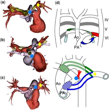
A female infant was born at 40 weeks’ gestation weighing 2734 grams. Echocardiographic examination revealed a ventricular septum defect, as well as high “take-off” of the left pulmonary artery. We performed three-dimensional computed tomographic angiography to clarify potential bronchial stenosis instead of no breathing problem, which revealed the patient's proximal pulmonary arteries (Fig 1a) to exist with a relatively large distance from the pulmonary trunk (figs, circle) to the left distal pulmonary artery (figs, diamond), as well showing original pulmonary artery sling (Fig 1b), as compared with normal arteries (Fig 1c). We performed a catheterisation examination and found no pressure gradient between the proximal left pulmonary artery and main trunk. Normally, the distal pulmonary arteries arise from their respective lung buds and join the proximal pulmonary arteries, which have formed from the left sixth aortic arch. We hypothesised that a pulmonary artery sling may occur when the proximal part of the left sixth aortic arch fails to develop its normal connections to the left lung bud (yellow part) and a “collateral” compensatory vessel from either the right (Fig 1d, green arrow) or the main pulmonary trunk thereby develops (Fig 1d, blue arrow).
Three-dimensional computed tomographic angiography ((a) this case; (b) original pulmonary arterial sling; (c) normal position), (d): diagram showing normal aortic arch development at 5 weeks (upper) and maturity (lower). Roman numerals indicate the embryonic aortic arches. The grey zone is normally involuted, the red zone shows the patent ductus arteriosus. Ao, aorta; PA, pulmonary artery.