Introduction
The maxillary sinus is one of the most commonly affected sinuses in patients with chronic rhinosinusitis.Reference Georgy and Peters1,Reference Banglawala, Schlosser, Wenztel, Walsh, Overton and Soler2 Patients with maxillary sinusitis suffer from clinical symptoms, such as nasal discharge, posterior nasal discharge, nasal obstruction, offensive odour, headache, and either buccal pain, tooth pain or both. Because the floor of the maxillary sinus is anatomically located adjacent to the roots of the maxillary molars, odontogenic inflammation around dental roots is likely to spread into the maxillary sinus. Thus, maxillary sinusitis can be not only rhinogenic but primarily odontogenic in origin.Reference Lopatin, Sysolyatin, Sysolyatin and Melnikov3,Reference Mehra and Murad4 The prevalence of odontogenic maxillary sinusitis is reported to be approximately 10–40 per cent of all maxillary sinusitis cases,Reference Lopatin, Sysolyatin, Sysolyatin and Melnikov3,Reference Brook5,Reference Melén, Lindahl, Andréasson and Rundcrantz6 and its incidence has been increasing.Reference Hoskison, Daniel, Rowson and Jones7,Reference Longhini and Ferguson8
The aetiology of odontogenic maxillary sinusitis can be untreated dental infection, radicular cysts, dental caries, odontogenic foreign bodies, inappropriate root canal treatments and unsuitable implant treatments.Reference Lee and Lee9 In Japan, odontogenic maxillary sinusitis is comprehensively diagnosed based on clinical symptoms, presence of causative teeth and findings on radiological imaging such as dental panoramic radiography and computed tomography (CT)Reference Bomeli, Branstetter and Ferguson10–Reference Patel and Ferguson12 by dentists or otolaryngologists. However, the differential and definite diagnosis of maxillary sinusitis of odontogenic origin currently depends on close collaboration between departments of dentistry and otolaryngology.
Diagnostic criteria and appropriate terms for odontogenic maxillary sinusitis have not been well established, and these terms can be used in both a broad sense to include non-infection and a narrow sense indicating infection alone. Furthermore, there is no consensus for proper management after diagnosis.Reference Aukštakalnis, Simonavičiūtė and Simuntis13 Treatments for odontogenic maxillary sinusitis caused by untreated dental caries and causative teeth of inappropriate root canal treatment have not yet been established.
Otolaryngologists and dentists both use conservative therapy as current first-line treatment. However, because the prognosis of teeth with periapical lesions on previous root-filling treatment is dismal,Reference Sjögren, Hägglund, Sundqvist and Wing14 most general practitioners in the dental clinic have performed tooth extraction. Functional endoscopic sinus surgery (FESS) is also indicated for severe maxillary sinusitis.Reference Lopatin, Sysolyatin, Sysolyatin and Melnikov3,Reference Crovetto-Martínez, Martin-Arregui, Zabala-López-de-Maturana, Tudela-Cabello and Crovetto-de la Torre15
Based on this background, the purpose of this study is: (1) to investigate the clinical course, diagnosis and treatment of maxillary sinusitis patients undergoing extraction of causative maxillary teeth proximate to the maxillary sinus floor; (2) to determine critical indicators for the requirement of FESS; and (3) to propose clinically beneficial management for odontogenic maxillary sinusitis. The benefits of clinical management that have been learnt from our experience are also fully discussed.
Materials and methods
Patients
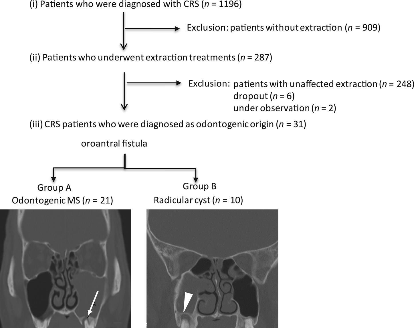
Between March 2013 and June 2018, adult patients who completely fulfilled three conditions were strictly selected and retrospectively analysed in this study (Figure 1). These three conditions were: (1) patients who were diagnosed with chronic rhinosinusitis by otorhinolaryngologists at the Department of Otorhinolaryngology Head and Neck Surgery, Hyogo College of Medicine, Japan; (2) patients who underwent maxillary tooth extraction by dentists at the Department of Dentistry and Oral Surgery, Hyogo College of Medicine; and (3) patients with maxillary sinusitis considered to be odontogenic in origin because extracted maxillary molars were proximate to the lesioned maxillary sinus root on sinonasal CT.
Diagram of the inclusion strategy of patients analysed in this study. The arrow in the group A image points to presence of a bony defect (with oroantral fistula). The arrowhead in the group B image points to absence of a bony defect (without oroantral fistula). CRS = chronic rhinosinusitis; MS = maxillary sinusitis

Data were collected from patients’ medical records by a third party at the Information Centre at Hyogo College of Medicine. Consequently, 31 adult patients comprising 20 men and 11 women with a mean age (± standard deviation (SD)) of 58.0 ± 11.9 years at the time of the diagnosis of maxillary sinusitis (range, 30–80 years) were analysed.
Patients with tumour-associated disease, trauma, history of any previous sinonasal surgery and without history of maxillary tooth extraction were excluded from this study.
Patients were further divided into two groups according to diagnosis: patients with odontogenic maxillary sinusitis (group A) and patients with radicular cysts (group B), based on the presence or absence of oroantral fistula on CT, respectively.
This study utilised a case series design and conformed to the regulations of the ethics committee of Hyogo College of Medicine (approval number: 1512). This study was performed in accordance with the principles of the Declaration of Helsinki.
Diagnosis
Chronic rhinosinusitis was diagnosed when nasal respiratory symptoms, including nasal obstruction, rhinorrhoea, post-nasal drip, pain and cough were observed for more than three months based on the guidance provided by the Japan Rhinologic Society, a previous European position paperReference Fokkens, Lund, Mullol, Bachert, Alobid and Baroody16 and current guidelines in the USA.Reference Rosenfeld, Andes, Bhattacharyya, Cheung, Eisenberg and Ganiats17 Endoscopic findings of mucopurulent nasal discharge and inflamed mucosae in the middle nasal meatus before treatment at the first visit were also adequately referenced. Computed tomography, the ‘gold standard’ in the diagnosis of maxillary sinus disease because of its high resolution and ability to distinguish between bone and soft tissue, was used for the evaluation of radiological findings.Reference Simuntis, Kubilius and Vaitkus18,Reference Little, Long, Loehrl and Poetker19
Maxillary sinusitis was defined as CT scans demonstrating partial or total opacification of the maxillary sinus. Furthermore, when a maxillary bone defect between the lesioned maxillary sinus floor and periapical root was observed on CT, maxillary sinus lesions were carefully diagnosed as odontogenic in origin by both experienced dentists and rhinologists as well as radiologists.
In this study, we defined and classified the patients into the following two groups: patients with odontogenic maxillary sinusitis with oroantral fistula (group A) and patients with radicular cysts without any oroantral fistula (group B; Figure 1).
Once maxillary teeth were recognised as the causative teeth by intraoral and CT findings, tooth extraction was performed under local anaesthesia by oral surgeons. To assess whether sinusitis improved after tooth extraction, a second sinonasal CT scan was performed at least one month later (mean ± SD was 2.2 ± 1.6 months after extraction) when local post-extraction wounds had stabilised. Functional endoscopic sinus surgery was indicated and subsequently performed under general anaesthesia by rhinologists for patients whose symptoms and maxillary sinus opacification did not improve on second CT examination. Patients who underwent FESS were classified as the FESS group in this study.
Endoscopic appearance scores
To statistically evaluate the severity and change in the maxillary sinus mucosal condition in the FESS group, the endoscopic appearance during and after FESS was scored, according to previous reports.Reference Tsuzuki, Hinohira, Takebayashi, Kojima, Yukitatsu and Daimon20,Reference Tsuzuki, Hashimoto, Okazaki and Sakagami21 During and after FESS, the score was determined using the following scale: normal (0 points); oedema (1 point); and polyps (2 points) in the maxillary sinus, using a 4-mm diameter rigid endoscope (0° and 70°; Karl Storz Endoscopy, Tokyo, Japan) by three expert rhinologists.
Computed tomography score
To assess disease severity for chronic rhinosinusitis, sinonasal CT findings were scored following the international scoring system proposed by Lund and Mackay.Reference Lund and Mackay22 The maxillary, frontal, and anterior and posterior ethmoid and sphenoid sinuses were scored as: 0 points (no opacification), 1 point (partial opacification) or 2 points (complete opacification). The ostiomeatal complex was scored as: 0 points (not opaque) or 2 points (with opacification). The CT score was the summation of the score at each site. The maximum possible total CT score was 12 points per side. Focusing on the inflamed maxillary sinus, the CT score of the maxillary sinus on the lesion side (0, 1 and 2 points) was particularly analysed in each group.
Statistical analysis
Clinical characteristics between groups A and B were retrospectively analysed. For continuous parameters, the Mann–Whitney U test was employed to assess between-group differences (group A vs. B). For categorical parameters, Fisher's exact test was employed to assess between-group differences (group A vs. B). Comparisons of changes in scores over time were analysed using the Wilcoxon signed rank sum test. Data are presented as the mean ± SD unless otherwise indicated. All p-values are two-sided and values of p < 0.05 were considered to indicate statistical significance. All statistical analyses were performed using Stat Flex statistical software (version 6; Osaka, Japan).
Results
Patient background
Group A consisted of 21 patients (14 men and 7 women; mean age: 60.1 ± 10.7 years). Group B consisted of 10 patients (6 men and 4 women; mean age: 53.6 ± 13.6 years). No significant differences were observed in age and sex between the two groups.
For clinical symptoms at the initial visit (patient numbers in groups A and B), mucopurulent rhinorrhoea (n = 8 and 4), pain including facial and buccal pain and headache (n = 6 and 4) and nasal obstruction (n = 6 and 1) were observed. Three asymptomatic patients diagnosed by chance were included based on CT findings only (n = 2 and 1).
Functional endoscopic sinus surgery was performed in 48 per cent (15 of 31) of patients after tooth extraction. Functional endoscopic sinus surgery was significantly more frequently required for patients in group B (100 per cent, 10 of 10 patients) than those in group A (24 per cent, 5 of 21 patients) (p < 0.0001).
Extracted causative teeth
Molars were predominantly extracted because they were the teeth causing chronic rhinosinusitis in both groups. In group A, the first and second molars were equally and predominantly observed as causative teeth (52 per cent, 11 of 21 each), followed by premolars (14 per cent, 3 of 21). In group B, the first, second and third molars were equally observed as causative teeth (40 per cent, 4 of 10). Data in both groups include duplication.
Severity of chronic rhinosinusitis
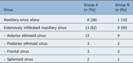
The severity of chronic rhinosinusitis on CT before and after tooth extraction was analysed. Before extraction treatment, CT in groups A and B showed association with widespread inflammation from the maxillary sinus to other sinuses in 62 per cent (13 of 21 patients) and 90 per cent (9 of 10 patients) of cases, respectively (Table 1). The most involved sinus, as a result of maxillary sinusitis, was the anterior ethmoid sinus in both groups. In both groups, CT scores significantly improved after extraction (p = 0.0002 in group A and p = 0.0037 in group B) (Table 2). CT scores in group A were significantly lower than those in group B after extraction (p = 0.0037). Focusing on the maxillary sinus opacification score, there was no significant difference between groups A and B before tooth extraction (p = 0.5937) (Table 3). Subsequently, the score in group A significantly improved after extraction (p < 0.0001) but did not improve in group B (p = 0.3559).
Severity of chronic rhinosinusitis with odontogenic origin

Computed tomography scores before and after tooth extraction

Data of the computed tomography (CT) scores are represented with mean ± standard deviation. Patients without CT before or after extraction were excluded. *n = 21; †n = 10; ‡significant difference between groups A and B; **significant differences between pre-and post-extraction
Maxillary sinus Computed tomography scores before and after tooth extraction

Data of the computed tomography (CT) scores are represented with mean ± standard deviation. Patients without CT before or after extraction were excluded. *n = 21; †n = 10; ‡significant difference between groups A and B; **significant differences between pre-and post-extraction
Maxillary sinus conditions in the FESS group
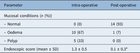
In order to evaluate maxillary sinus mucosal conditions, endoscopic appearances during and after FESS were scored and compared in the FESS group (Table 4). Regarding the change in maxillary sinus mucosal conditions, oedema (n = 10) and polyps (n = 5) that were observed during FESS were mainly improved to normal appearance (n = 14) in the post-operative stage on endoscopy. Consequently, the endoscopic score during FESS (1.3 ± 0.5) significantly decreased after FESS (0.1 ± 0.3) (p < 0.001) at 11.5 ± 2.3 months post-operatively (range, 3–31 months) indicating the effectiveness of FESS with a surgical improvement ratio of 93 per cent (14 of 15 patients).
Endoscopic findings in maxillary sinus mucosae

*significant difference in endoscopic score between intra- and post-operative score. SD = standard deviation
Discussion
This study demonstrated the clinical characteristics of patients with chronic rhinosinusitis who underwent causative maxillary tooth extraction proximate to the maxillary sinus floor. In order to aetiologically identify patients with chronic rhinosinusitis with odontogenic origin, we focused on the existence of oroantral fistula. We therefore classified patients as having odontogenic maxillary sinusitis with oroantral fistula (group A) or radicular cysts without oroantral fistula (group B).
In Japan, therapeutic strategies for odontogenic maxillary lesions are varied because odontogenic maxillary sinusitis is separately treated by Departments of Dentistry and Otolaryngology. In addition, most of the previous studies on treatment for odontogenic maxillary sinusitis were limited reports from small populations.Reference Aukštakalnis, Simonavičiūtė and Simuntis13,Reference Ungar, Yafit, Kleinman, Raiser and Safadi23 Thus, in this study, we sought to propose a management policy from our clinical experience.
When compared to previous epidemiological analyses, our data were similar concerning patient age (primarily patients were aged between 40 and 60 years old) and molars were the causative teeth.Reference Simuntis, Kubilius and Vaitkus18,Reference Arias-Irimia, Barona-Dorado, Santos-Marino, Martínez-Rodriguez and Martínez-González24,Reference Maillet, Bowles, McClanahan, John and Ahmad25 As for sex, the male predominance observed in our study compared with previous studies may be explained by the fact that we focused on patients who underwent tooth extraction.
Recently, in the clinical management of chronic rhinosinusitis, an increasing number of patients with eosinophilic chronic rhinosinusitis, which is characterised by bilateral nasal polyposis with abundant eosinophil infiltration, ethmoid sinus predominance, blood eosinophilia and high risk of recurrence, have become important.Reference Tokunaga, Sakashita, Haruna, Asaka, Takeno and Ikeda26,Reference Fujieda, Imoto, Kato, Ninomiya, Tokunaga and Tsutsumiuchi27 Differentiating between eosinophilic and non-eosinophilic patients is critical for the analysis of chronic rhinosinusitis pathogenesis.Reference Tsuzuki, Hashimoto, Okazaki and Sakagami21 Most eosinophilic patients who are bilaterally ethmoid sinus dominant show initial inflammatory involvement in the posterior sinonasal area, such as the posterior ethmoid sinus and olfactory clefts,Reference Tokunaga, Sakashita, Haruna, Asaka, Takeno and Ikeda26,Reference Fujieda, Imoto, Kato, Ninomiya, Tokunaga and Tsutsumiuchi27 whereas most non-eosinophilic patients who are unilaterally maxillary sinus dominant show initial inflammatory changes and impairment of ventilation in the anterior sinonasal area, such as the anterior ethmoid sinus and frontal sinus.Reference Zinreich, Kennedy, Rosenbaum, Gayler, Kumar and Stammberger28,Reference Bolger, Butzin and Parsons29 As most cases of odontogenic maxillary sinusitis are characterised by unilateral maxillary sinus,Reference Albu and Baciut30 all patients in this study had unilateral lesions that were diagnosed as non-eosinophilic.
Anatomical proximity facilitates the spread of inflammation from the maxillary sinus ostium to the anterior ethmoid sinus.Reference Crovetto-Martínez, Martin-Arregui, Zabala-López-de-Maturana, Tudela-Cabello and Crovetto-de la Torre15,Reference Andric, Saranovic, Drazic, Brkovic and Todorovic31,Reference Costa, Emanuelli, Robiony, Zerman, Polini and Politi32 This study showed that the anterior sinus was the most involved area in maxillary sinusitis, followed by the frontal sinus.Reference Ungar, Yafit, Kleinman, Raiser and Safadi23 For the severity of chronic rhinosinusitis on CT, patients with oroantral fistula showed significantly improved CT findings after tooth extraction. One possible reason could be additional irrigation through the Schneiderian membrane during extraction. Although the severity of sinusitis in patients without oroantral fistula also improved after tooth extraction, maxillary sinusitis significantly remained even after extraction compared to patients with oroantral fistula. It is also possible that radicular cysts and maxillary sinusitis coexisted by chance rather than being caused by the spread of inflammation.
Although maxillary sinusitis might also be relieved by concurrent medical treatments for chronic rhinosinusitis, remnant maxillary sinusitis required additional surgical therapy of FESS. Consequently, not only severe chronic rhinosinusitis but also absence of oroantral fistula on imaging can be critical factors for the requirement of FESS in the management of odontogenic chronic rhinosinusitis. In the FESS group, the endoscopic appearance of maxillary sinus mucosae dramatically improved after FESS with a ratio of 93 per cent, which is similar to previously reported ratios of 93–100 per cent.Reference Aukštakalnis, Simonavičiūtė and Simuntis13,Reference Crovetto-Martínez, Martin-Arregui, Zabala-López-de-Maturana, Tudela-Cabello and Crovetto-de la Torre15 The usefulness of FESS for maxillary sinusitis is considered.
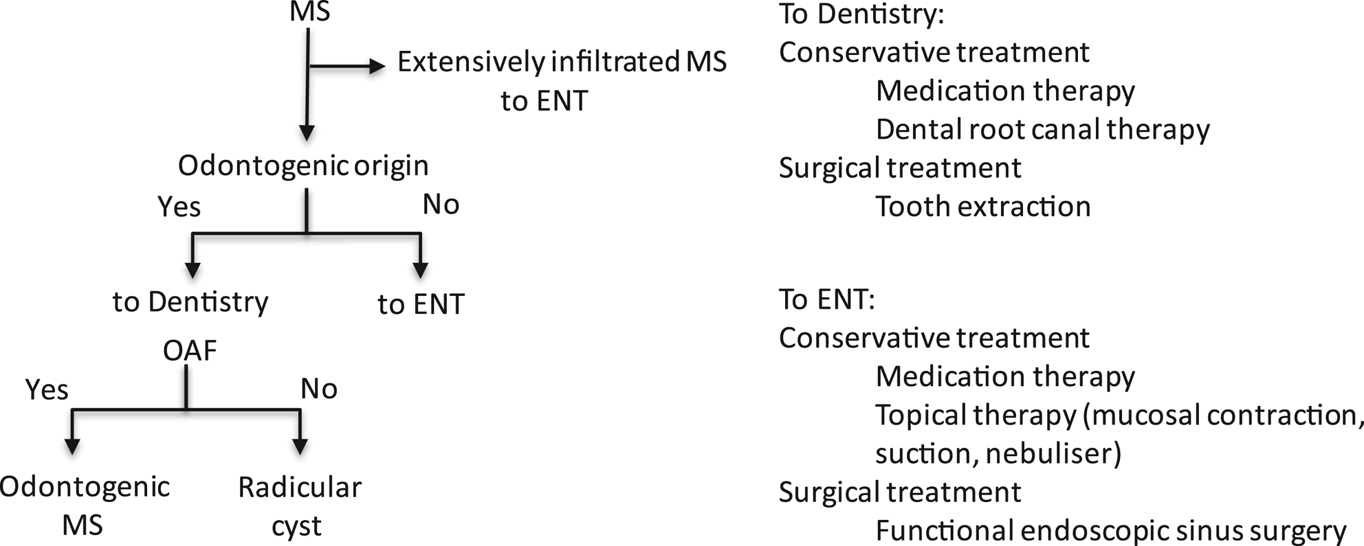
We propose an algorithm for the management of sinusitis with odontogenic origin, as shown in Figure 2 from the current case series. Patients with extensively infiltrated maxillary sinusitis, such as pansinusitis, are likely to be initially consulted at the Department of Otorhinolaryngology, whereas patients who have maxillary sinusitis with suspected odontogenic origin are likely to be consulted at the Department of Dentistry. Presence or absence of oroantral fistula can greatly influence the clinical course. Tooth extraction is recommended for treatment of odontogenic maxillary sinusitis because inappropriate root canal treatment can cause secondary periodontal cysts. A lower success rate (62 per cent) was reported for roots with periapical lesions, particularly maxillary molars with anatomically complex root canals, that were previously filled and treated.Reference Sjögren, Hägglund, Sundqvist and Wing14 Thus, most oral surgeons select tooth extraction versus endodontic treatment. Although maxillary sinusitis can be sufficiently resolved by dental infection removal alone, some cases require concomitant FESS.Reference Aukštakalnis, Simonavičiūtė and Simuntis13 Functional endoscopic sinus surgery also has operative indication when maxillary sinusitis-related symptoms do not improve even after tooth extraction. Patients with extensively infiltrated maxillary sinusitis are more likely to be indicated for FESS than those with maxillary sinus alone. As shown in this study, patients without oroantral fistula were more likely to undergo FESS than those with oroantral fistula even after tooth extraction. Whereas some studies have emphasised dental surgery as the primary treatment modality for odontogenic sinusitis, other studies suggested that FESS alone may be an effective treatment approach.Reference Wang, Nichols, Poetker and Loehrl33,Reference Jiam, Goldberg, Murr and Pletcher34 Recently, expanded endoscopic approaches, such as antrostomy in inferior nasal meatus and modified medial maxillectomy have been performed instead of the Caldwell–Luc procedure for full removal of the lesions.Reference Costa, Emanuelli, Robiony, Zerman, Polini and Politi32,Reference Suzuki, Nakamura, Nakayama, Inagaki, Murakami and Takemura35,Reference Nakayama, Otori, Asaka, Okushi and Haruna36 Although extended intervention can lead to complete resolution, clinicians must seek treatments that can obtain maximum effects with minimal invasiveness to avoid unnecessary expansion therapy. Taken together, multidisciplinary therapeutic strategies comprising medical treatment, dental surgery and FESS are suggested for patients with odontogenic originated sinusitis.Reference Aukštakalnis, Simonavičiūtė and Simuntis13,Reference Little, Long, Loehrl and Poetker19
Algorithm diagram for the management of maxillary sinusitis. MS = maxillary sinusitis; OAF = oroantral fistula

Because diagnostic criteria and appropriate treatment for odontogenic maxillary sinusitis remain unclear, this study aimed to accurately determine the clinical course in odontogenic chronic rhinosinusitis. However, it was difficult to reliably extract data for chronic rhinosinusitis patients with conservative dental treatments. Therefore, we strictly focused on patients with chronic rhinosinusitis with odontogenic origin who received extraction of causative teeth in the study period as shown in Figure 1 (3 per cent, 31 of 1196 patients).
Although the number of analysed patients was reduced because of the strict conditions set, we believe that this study provides significant results on a reliable population of patients with odontogenic sinusitis with dental surgery as second-line therapy. This study was retrospective in nature and focused on patients who underwent tooth extraction at one tertiary hospital; therefore, large-scale, multicentre prospective studies are required to confirm our findings. To further develop an appropriate therapeutic strategy, full investigation of other important aetiologies, including inappropriate root canal treatment, presence of odontogenic foreign bodies or implant treatment is next required in the future.
• Odontogenic maxillary sinusitis is caused by untreated dental caries and causative teeth of inappropriate root canal treatment
• Diagnostic criteria and appropriate treatment for odontogenic maxillary sinusitis have not been well established
• Whereas tooth extraction can improve odontogenic sinusitis, functional endoscopic sinus surgery (FESS) is also required when sinusitis remains after extraction
• Absence of oroantral fistula and severe sinus inflammation on imaging can be critical factors for the requirement of FESS in the management of odontogenic chronic rhinosinusitis
• FESS has beneficial effects for patients with maxillary sinusitis-related symptoms even after tooth extraction
• Multidisciplinary approaches comprising medical treatment, dental surgery and FESS are suggested to be beneficial for management of sinusitis with odontogenic origin
Acknowledgements
We gratefully acknowledge the technical assistance from Ms Yumi Kida and Mrs Midori Tanide. This work was supported by Grants-in-Aid for Scientific Research (KAKENHI grant numbers: JP25462671 and JP16K11220) from the Japan Society for the Promotion of Science; Grant-in-Aid for Researchers, Hyogo College of Medicine, 2018 (K Hashimoto) and the Practical Research Project for Rare/Intractable Diseases from the Japan Agency for Medical Research and Development (grant number: JP 16ek0109062).
Competing interests
None declared