INTRODUCTION
Salivarian trypanosomes are capable of adapting and developing mechanisms to avoid immune destruction (Jokiranta et al. Reference Jokiranta, Jokipii and Meri1995). These trypanosomes evade the host immune response by sequentially changing its variant surface glycoprotein (VSG), a strategy-denominated antigenic variation (Zambrano-Villa et al. Reference Zambrano-Villa, Rosales-Borjas, Carrero and Ortiz-Ortiz2002). Each trypanosome carries a large repertoire of VSG genes coding for approximately 1000 VSG variants (Donelson, Reference Donelson2003), but at any one time expresses only a single VSG gene. These parasites keep one step ahead of the host immune system by periodically switching from the expression of one VSG on their surface to the expression of another immunologically distinct VSG. This repeated antigenic change on the trypanosomes' outer coat allows them to evade the host humoral response, resulting in successive surges of parasitaemia, a condition similar to being infected successively by related, but not identical, pathogens.
Due to their intravascular and extracellular location, the prevailing immune reaction of the vertebrate host towards infection by these trypanosomes is mediated by the production of specific antibodies, the activation of the classical complement system, and by phagocytic responses (Taylor, Reference Taylor1998). The complement system consists of a set of serum proteins in charge of many of the cytolytic and inflammatory effects of humoral immunity, and as such acts as a first-line defence mechanism against trypanosomes. The activation of the terminal components of the complement system culminates in the formation of pores in the parasite plasma membrane by the membrane attack complex (Benz et al. Reference Benz, Schmid, Wiedmer and Sims1986), which allows Ca2+ to enter and initiate changes in its cytosolic concentration (Campbell and Luzio, Reference Campbell and Luzio1981; Campbell et al. Reference Campbell, Daw, Hallett and Luzio1981; Morgan and Campbell, Reference Morgan and Campbell1985; Laffafian et al. Reference Laffafian, Davies, Campbell and Hallett1995; Newsholme et al. Reference Newsholme, Adogu, Soos and Hales1993). This abrupt increase of Ca2+ concentration is extremely toxic for the parasite. Additionally, this phenomenon produces an influx of water into the parasites, which leads to death via osmotic lysis.
Increasing antibody titres eventually lead to complement lysis; however, the remaining parasites survive and establish the infection thanks to their antigenic variation strategy. To date, no evidence has been reported to demonstrate that the host immune system directly induces switching of the trypanosome outer layer of VSG, but it is known that it contributes to an environment in which new and temporarily unrecognized serotypes can prosper, giving rise to new waves of parasitaemia (Donelson, Reference Donelson1995). Additionally, antigenic variation in trypanosomatids has been related to Ca2+ signalling processes, since the release of the surface coat protein is dependent on the presence of extracellular Ca2+ (Voorheis and Martin, Reference Voorheis and Martin1981; Voorheis et al. Reference Voorheis, Bowles and Smith1982; Bowles and Voorheis, Reference Bowles and Voorheis1982).
Trypanosoma evansi and Trypanosoma vivax are the major aetiological agents of animal trypanosomosis in Venezuela, and generate a significant economic impact in livestock. T. vivax affects predominantly bovines while T. evansi causes the disease known as derrengadera or surra in equines. Recently, Uzcanga et al. (Reference Uzcanga, Mendoza, Aso and Bubis2002, Reference Uzcanga, Perrone, Noda, Pérez-Pazos, Medina, Hoebeke and Bubis2004) have demonstrated that sera from T. evansi-infected horses and T. vivax-infected bovines immunorecognized a purified VSG variant from the TEVA1 Venezuelan isolate of T. evansi and suggested the potential use of this VSG variant as a diagnostic reagent. Interestingly, anti-VSG polyclonal antibodies generated against this variant immunolabelled the surface of both T. evansi and T. vivax (Uzcanga et al. Reference Uzcanga, Perrone, Noda, Pérez-Pazos, Medina, Hoebeke and Bubis2004). Here, fluorometric ratio imaging from single isolated T. evansi parasites (Mendoza et al. Reference Mendoza, Mijares, Rojas, Ramos and Dipolo2001) were used to explore the effects of sera from trypanosome-infected animals and anti-VSG antibodies on the parasite Ca2+ homeostasis. Our results indicated that the interaction of anti-VSG antibodies with the parasite surface promotes a Ca2+ signal, which probably is involved in the T. evansi response against the host immune system, and most likely regulates the interactions between the parasite and its host. A preliminary report of this study has already been published (Mendoza et al. Reference Mendoza, Uzcanga, Pacheco, Bubis and Mijares2004a).
MATERIALS AND METHODS
Bovine sera
Two bovines were infected by intravenous injection of a suspension containing approximately 106T. vivax parasites from a cryopreserved Venezuelan isolate. Blood samples were taken every day, for a 2-month period, in order to determine parasitaemia by the microhaematocrit method (Woo, Reference Woo1970). Blood samples were also taken in the absence of anti-coagulant to obtain sera containing anti-T. vivax antibodies and the complement system proteins. The progress of the infection was followed by indirect enzyme-linked immunoabsorbent assays using a T. evansi clarified fraction as antigen, which was prepared as described below (Aray et al. Reference Aray, Uzcanga, Soto and Mendoza1998; Camargo et al. Reference Camargo, Uzcanga and Bubis2004). Animal sera were decomplemented by heating to 58°C for 30 min or by the addition of 180 μm rosmarinic acid.
Parasite preparation and purification
Cryopreserved samples of T. evansi from the Venezuelan TEVA1 isolate (Desquesnes and Tresse, Reference Desquesnes and Tresse1996) were expanded in adult albino rats (Sprague-Dawley). When the number of parasites reached 106–108 trypanosomes/ml, the blood was extracted from the rats by cardiac puncture using EDTA as anti-coagulant. Trypanosomes were purified by anion-exchange chromatography using a fibrous DEAE-cellulose column (Lanham and Godfrey, Reference Lanham and Godfrey1970) equilibrated with PBS-G buffer (57 mm Na2HPO4, 3 mm NaH2PO4, 43·8 mm NaCl, 1% glucose, pH 8). Parasites eluting from the column were collected by centrifugation at 1475 g, for 20 min, and washed 3 times with PBS-G buffer.
Purification of a VSG from Trypanosoma evansi
T. evansi parasites (2·7×1011) were extracted on ice, by sonication (2 cycles, 30 sec each, with a 30 sec resting period in between) using 2 ml of a 5 mm Tris-HCl buffer (pH 7·2) containing 1 mm benzamidine, 1 mm PMSF, 5 mm EDTA, and 1 mm iodoacetamide. The resulting homogenate was centrifuged at 15 000 g for 30 min, at 4°C, to obtain a supernatant containing the clarified antigenic fraction and the corresponding pellet. The clarified antigenic fraction was loaded onto a 50 ml Q-Sepharose column connected in tandem with a 10 ml S-Sepharose column, which previously had been equilibrated with 50 mm Tris-HCl buffer (pH 7·2). The T. evansi VSG eluted in the flow-through fraction as previously described by Uzcanga et al. (Reference Uzcanga, Mendoza, Aso and Bubis2002, Reference Uzcanga, Perrone, Noda, Pérez-Pazos, Medina, Hoebeke and Bubis2004).
Protein sequencing
Purified VSG was separated by sodium dodecyl sulfate-polyacrylamide gel electrophoresis (SDS-PAGE) on 15% polyacrylamide gels (Laemmli, Reference Laemmli1970) and electroblotted to polyvinylidene difluoride membranes (Immobilon-P, Millipore) in 10 mm 3-(cyclohexylamino)-1-propanesulfonic acid (CAPS, pH 11), 10% methanol (HPLC grade), and 0·1 mm sodium thioglycolate, for 1 h at 250 mV. Polypeptide bands were subjected to sequencing according to Matsudaira (Reference Matsudaira1987), using an automated protein microsequencer Prosize 493 cLC (PE-Applied Biosystems).
Preparation of polyclonal antibodies against purified Trypanosoma evansi VSG
The purified VSG was used to raise polyclonal antibodies in ascitic fluids of 7 female Balb/c mice, according to the procedure described by Bubis et al. (Reference Bubis, Millan and Martinez1993). Briefly, mice were pre-immunized twice (at 7 and 9 weeks of age) using lipid A and muramyl dipeptide as adjuvants, and under conditions that did not involve ascites fluid formation. Then, when the mice were 10 weeks old, we followed the standard protocol described by Tung (Reference Tung1983).
Purification of anti-VSG antibodies from ascitic fluids
Purified VSG (0·56 mg) was adsorbed on a nitrocellulose paper (11 cm×7 cm), which was blocked using TBST buffer (10 mm Tris-HCl, pH 8, 150 mm NaCl, 0·05% Tween 20) with 5% skimmed milk, and was subsequently incubated for 2 h with mouse ascitic fluids containing anti-VSG polyclonal antibodies (20 ml of a dilution of 1:50). Anti-VSG specific antibodies were eluted employing 20 ml of 100 mm glycine-HCl (pH 2·8) by gentle shaking for 20 min. The eluted antibodies were neutralized by adding 670 mg of solid Tris and were then dialysed overnight against 20 mm phosphate buffer (pH 8). We obtained 3·3 mg of purified anti-VSG-specific antibodies.
Western blot analysis
Proteins separated by SDS-PAGE (Laemmli, Reference Laemmli1970) were electrotransferred from the gels to nitrocellulose sheets (0·45 μm pore size) according to the procedure described by Towbin et al. (Reference Towbin, Staehelin and Gordon1979). The nitrocellulose filters were incubated with either mouse ascitic fluid or affinity-purified anti-VSG antibodies, and developed using alkaline phosphatase-conjugated secondary antibodies against mouse IgG. The protein bands were visualized by the addition of NBT and BCIP. The specificity of the ascitic fluids was evaluated by competiton with increasing concentrations of purified VSG (0·1–30 μg) on blots containing either parasite homogenates or purified VSG.
Fluorescence measurements
Isolated parasites (106 cell/ml) were incubated with 8 μM of 1-[2-(5-carboxyoxazol-2-yl)-6aminobenzofuran-5oxyl]-2-(2′amino-5′-methylphenoxy)-ethane-N,N,N′,N′-tetraacetic acid/acetoxy-methyl ester (Fura-2/AM) in PBS-G buffer for 30–45 min, at room temperature (22°C), with gentle and continuous agitation. Loaded parasites were washed 3 times with PBS-G to remove excess Fura-2/AM. Then, the suspension of Fura-2/AM-loaded parasites was added to an open experimental chamber mounted in an inverted microscope connected to a fluorescence imaging apparatus. The cover-slips of the chamber bottom were treated with poly-L-lysine solution (0·01% v/v) to allow partial adhesion of the parasites. Excitation wavelengths at 340 and 380 nm were used and the emitted light was measured at 510 nm. Fluorescence ratios and images were collected every 500 ms through a steeper-switch light source via a charge-coupled device video-camera coupled to an image intensifier. Single parasites could be discriminated through video-acquisition software. Calibration of the Fura-2 fluorescence ratio in terms of [Ca2+]i was carried out as described previously by Grynkiewicz et al. (Reference Grynkiewicz, Poenie and Tsien1985). Test solutions were delivered to the parasite via pressure ejection from a carbonated pipette using a perfusion system, which is connected to a computer and a temperature control system (Mendoza et al. Reference Mendoza, Mijares, Rojas, Ramos and Dipolo2001, Reference Mendoza, Mijares, Rojas, Rodríguez, Urbina and DiPolo2002). Since the solutions can be rapidly changed, the effect of various conditions can be analysed on the same single parasite. The composition of the various solutions employed and the number of parasites in which [Ca2+]i was measured are indicated in the figure legends. In all cases the temperature was maintained at 30°C and all solutions contained 300 mm NaCl to prevent non-specific interactions. In order to determine the difference in calcium concentration, the final calcium level was measured when the steady state was reached. The results of fluorescence experiments were expressed as mean values ± standard error of the mean.
Variation of intracellular calcium concentrations induced by affinity-purified anti-VSG antibodies in parasite populations
To evaluate the effect of purified anti-VSG antibodies (66 μg/ml) on cell populations, measurements of intracellular Ca2+ concentration were performed in the presence of 1·8 mm extracellular Ca2+. Briefly, a suspension of 2×10⁶ cells/ml previously loaded with Fura-2/AM (8 mm) was monitored in a stirred cuvette, at 30°C, using a ratiometric fluorescence spectrophotometer Perkin-Elmer LS-55 (Colina et al. Reference Colina, Flores, Rojas, Acosta, Garrido, Israel, Castillo, DiPolo and Benaim2005), and [Ca2+]i was estimated as described by Grynkiewicz et al. (Reference Grynkiewicz, Poenie and Tsien1985).
In vivo direct immunofluorescence
Purified parasites were incubated at 37°C, for 30 min, with decomplemented anti-T. evansi VSG antibodies, and then with fluorescein-conjugated anti-mouse IgG secondary antibodies. The suspension of parasites was added to an experimental chamber containing a cover-slip that was treated with a solution of poly-L-lysine (0·01% v/v) to allow partial adhesion of the parasites. The parasites were viewed using a fluorescence microscope directly connected with a photographic camera.
Effect of Ca2+ on the release of Trypanosoma evansi VSG
Parasites (1×10⁶ cells) were incubated with Ca2+ (1·8 mm) in the presence or absence of the A-23187 ionophore (2·5 μg/ml). The time-course of VSG release was evaluated by removing an aliquot (150 μl) every 5 min. Following centrifugation at 8000 g for 1 min, the supernatants were analysed by Western blot using anti-VSG antibodies. Live parasites were quantified on a Neubauer counting chamber.
RESULTS
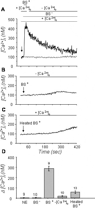
Since T. evansi and T. vivax have exhibited a very high immunological cross-reactivity (Aray et al. Reference Aray, Uzcanga, Soto and Mendoza1998; Uzcanga et al. Reference Uzcanga, Mendoza, Aso and Bubis2002, Reference Uzcanga, Perrone, Noda, Pérez-Pazos, Medina, Hoebeke and Bubis2004; Mendoza et al. Reference Mendoza, Uzcanga, Pacheco, Bubis and Mijares2004a), fluorometric ratio imaging from single T. evansi parasites (Mendoza et al. Reference Mendoza, Mijares, Rojas, Ramos and Dipolo2001) was used here to evaluate changes in the parasite [Ca2+]i following perfusion with sera from T. vivax-infected bovines. In the presence of extracellular Ca2+, a short exposure of T. evansi parasites to sera from bovines infected with T. vivax yielded a fast and substantial raise on their [Ca2+]i. Fig. 1A shows a representative experiment in which the parasite [Ca2+]i was increased from 100 to 400 nm following exposure, for 5 sec, to bovine sera withdrawn 25 days after infection. This effect terminated and was reversed when the extracellular Ca2+ was eliminated (Fig. 1A). Additionally, cell lysis was generated after an extended exposure (>10 sec) of the parasites to sera from infected bovines in the presence of Ca2+ (data not shown). Parasites were also exposed to sera from T. vivax-infected bovines in a calcium-free medium that contained 100 μm EGTA. As shown in Fig. 1B, only a slight increase in the parasite [Ca2+]i (~25 nm) was obtained under these conditions. These results indicated that the main source for the Ca2+ that is mobilized during the parasite-serum interaction is extracellular. On the contrary, neither sera from healthy animals (Fig. 1A) nor sera from bovines living in France, which is a trypanosomosis non-endemic area (data not shown), affected the T. evansi [Ca2+]i, which was maintained approximately at its basal value of 100 nm. Similar results were obtained with control parasites without any addition (data not included). When T. evansi parasites were exposed to decomplemented sera from infected animals, only a small increase of ~50 nm in the parasite [Ca2+]i was observed (Fig. 1C). Thus, the activation of the classical complement system pathway is probably responsible for the generation of most of the rise in the [Ca2+]i reported here. Fig. 1D summarizes the mean values of [Ca2+]i obtained under the various conditions described. These values were the following: 288±23 nm when individual parasites were exposed to sera from infected bovines in the presence of external Ca2+, 19±6 nm for the exposure of parasites to sera from infected bovines in the absence of Ca2+, and 57±13 nm for the exposure of parasites to decomplemented infected sera in the presence of external Ca2+.
Effect of sera from infected and non-infected animals on the [Ca2+]i in single isolated Trypanosoma evansi cells loaded with Fura-2/AM. Arrows show the time of addition of the sera. (A) Changes in the parasite [Ca2+]i following addition of sera from a bovine infected with T. vivax (BS+) or from a healthy bovine (BS−). Sera (1:100) were initially added in the presence of 1·8 mm external Ca2+ (+[Ca2+]o) and then this ion was removed using a solution containing no calcium and 100 μm EGTA (−[Ca2+]o). (B) Changes in parasite [Ca2+]i after addition of BS+ in a solution containing no calcium and 100 μm EGTA (−[Ca2+]o). (C) Changes in the parasite [Ca2+]i after addition of BS+ previously heated at 58°C, to inactivate the complement, in the presence of 1·8 mm external Ca2+ (+[Ca2+]o). (D) Histograms corresponding to the intracellular Ca2+ increase (expressed as Δ[Ca2+]i) observed following sera perfusion under the various conditions. Also shown is the effect of sera from bovines living in a non-endemic area (NE). On top of each bar is indicated the number of parasites in which [Ca2+]i was measured.

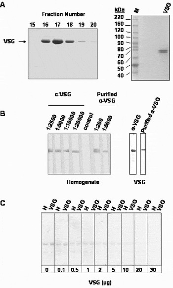
Since sera from infected bovines recognized VSG molecules from T. evansi (Uzcanga et al. Reference Uzcanga, Perrone, Noda, Pérez-Pazos, Medina, Hoebeke and Bubis2004) and the dense surface coat formed by the closely packed monolayer of VSG has been shown to be essential for the parasite's life in the host's bloodstream, we purified the soluble form of a particular T. evansi VSG variant to prepare specific polyclonal antibodies and to evaluate their effect on the parasite [Ca2+]i. Fig. 2A illustrates a Coomassie blue-stained gel containing the resulting chromatography fractions (left). As seen in this figure, we obtained a highly pure VSG that migrated as a single polypeptide band. However, in some preparations, 2 polypeptide bands were obtained (right). To demonstrate that these two polypeptides represented isoforms of the same VSG, we determined their partial primary structure by Edman degradation and found that the first 10 amino acid residues were identical (data not shown). This amino acid sequence corresponded to that previously reported by Uzcanga et al. (Reference Uzcanga, Perrone, Noda, Pérez-Pazos, Medina, Hoebeke and Bubis2004), which displayed a significant homology with a putative T. brucei VSG gene. The purified VSG shown in Fig. 2A was then employed to raise ascites polyclonal antibodies in mice. Western blot analyses show that these antibodies, at various dilutions, specifically recognized VSG from parasite whole-cell homogenates (Fig. 2B, left). Purified VSG was also recognized by these ascitic fluids (Fig. 2B, right). Additionally, no recognition of purified VSG or VSG from parasite extracts was obtained by anti-VSG antibodies (dilution 1:5000) that were pre-incubated with 5 μg or more of purified VSG (Fig. 2C). These results confirmed the specificity of the ascites anti-VSG polyclonal antibodies.
Purification of VSG and evaluation of the specificity of the anti-VSG polyclonal antibodies. (A) Trypanosoma evansi VSG eluted in the flow-through fraction as described by Uzcanga et al. (Reference Uzcanga, Mendoza, Aso and Bubis2002, Reference Uzcanga, Perrone, Noda, Pérez-Pazos, Medina, Hoebeke and Bubis2004). Left: SDS-PAGE separation of the fractions containing the chromatographically purified VSG (fractions 15–20). Right: In some cases, the purified VSG appeared as 2 polypeptide bands on gels. (B) Specific recognition of VSG by ascitic fluids (α-VSG) and affinity-purified anti-VSG antibodies (purified α-VSG) on both, parasite homogenates (Left) and purified VSG (Right). (C) Blots containing parasite homogenates (H, 25 μg of total protein) and purified VSG (VSG, 5 μg) were developed using ascitic fluids pre-incubated with increasing amounts of purified VSG (0–30 μg).

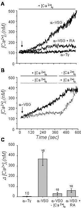
We also explored the effect of specific anti-VSG polyclonal antibodies on the parasite [Ca2+]i employing fluorometric ratio imaging. Fig. 3A illustrates that a exposure (600 sec) of a single T. evansi parasite to mouse ascitic fluids (dilution 1:100) containing anti-VSG antibodies generated an increase from 100 to 500 nm in its [Ca2+]i. Cell lysis was also produced following an extended exposure of parasites to these antibodies (data not included). In contrast, control parasites without any addition maintained their [Ca2+]i at about 100 nm (data not shown). Fig. 3B clearly shows that this effect is dependent on the presence of extracellular Ca2+. In this particular experiment, exposure of a single parasite to anti-VSG antibodies produced an increase of about 400 nm in its [Ca2+]i in 420 sec, when extracellular calcium was present in the medium. This effect was substantially reduced to 30 nm when Ca2+ was eliminated from the extracellular medium by adding EGTA, and was reversed when Ca2+ was reloaded in the extracellular medium. Interestingly, when calcium was recharged in the extracellular medium, the increase on the parasite [Ca2+]i was much faster, generating cell lysis after only 200 sec of exposure (Fig. 3B). Fig. 3C sums up the mean values of the increases observed on the parasite [Ca2+]i attained under these conditions. These values were 369±65 and 30±6 nm when individual parasites were exposed to anti-VSG antibodies in the presence or absence of external Ca2+, respectively.
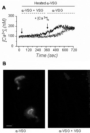
Effect of anti-VSG polyclonal antibodies (α-VSG) on the [Ca2+]i in single isolated parasites loaded with Fura-2/AM. Arrows show the time of addition of the antibodies. (A) Changes in the [Ca2+]i after addition of ascitic fluid containing α-VSG or antibodies against the γ-subunit of bovine transducin (α-Tγ), a non-related protein. Also shown is the effect of adding α-VSG in the presence of 180 μm rosmarinic acid (RA). In all cases, ascites were added in the presence of 1·8 mm external Ca2+ (+[Ca2+]o). (B) Changes in the [Ca2+]i after addition of α-VSG in the presence of 1·8 mm external Ca2+ (+[Ca2+]o). Following 6 min incubation, this ion was removed using a calcium-free solution (− [Ca2+]o). In addition, α-VSG were added initially in a Ca2+-free solution (− [Ca2+]o), and subsequently the [Ca2+]o was raised to 1·8 mm (+[Ca2+]o). (C) Histograms corresponding to the intracellular Ca2+ increase (expressed as Δ[Ca2+]i) observed following ascites perfusion under the various conditions. On top of each bar is indicated the number of parasites in which [Ca2+]i was measured.

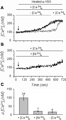
When decomplemented ascitic fluid was employed in the presence of extracellular calcium, a decrease of approximately 300% in the parasite [Ca2+]i influx was obtained (Fig. 4A). A similar effect was acquired when 180 μm rosmarinic acid was added to these antibodies (Fig. 3A), obtaining a mean value of 60±7 nm in the parasite [Ca2+]i (Fig. 3C). These results suggested that most of the increase in the T. evansi [Ca2+]i produced by the ascites containing the anti-VSG antibodies is mediated by the activation of the classical complement pathway. However, a small increase in the parasite [Ca2+]i of about 100 nm was still observed after decomplementation of the anti-VSG antibodies, which must be directly caused by the specific anti-VSG antibodies present in the ascites. Fig. 4A also compares the effect produced by the decomplemented ascitic fluid containing the anti-VSG antibodies in the presence and absence of extracellular Ca2+. Similar experiments were performed in the presence of Ca2+ and Ni2+ (Fig. 4B). The rise in parasite [Ca2+]i was substantially reduced in the absence of Ca2+ and in the presence of Ni2+. In addition, the inhibitory effect of Ni2+ was reversed when Ni2+ was eliminated from the extracellular medium and Ca2+ was re-added (Fig. 4B). Either with Ni2+ or in the absence of external calcium, an increase of about 19 nm in the parasite [Ca2+]i was obtained. Fig. 4C presents the main values of the parasite [Ca2+]i variations attained under these various conditions. These values were 98±14 nm in the presence of extracellular Ca2+, 19±3 nm when both Ca2+ and Ni2+ were present in the incubation medium, and 15±3 nm in the presence of 100 μm EGTA (Fig. 4C); and were measured after the steady state was reached. These results indicated that approximately 80% of the effect generated by the anti-VSG antibodies was produced by the influx of extracellular Ca2+, probably through some Ca2+-specific channels located at the parasite membrane, while a less significant percentage of the effect (~20%) was produced by the release of this ion from parasite intracellular storage organelles.
The effect of Ni2+ on the intracellular Ca2+ increase induced by decomplemented anti-VSG antibodies (α-VSG) in single isolated Trypanosoma evansi parasites loaded with Fura-2/AM. (A) Variations in the [Ca2+]i after addition of specific antibodies against the parasite VSG protein previously heated at 58°C, in the presence or absence of 1·8 mm external Ca2+ (+/−[Ca2+]o). (B) Changes in the parasite [Ca2+]i following addition of α-VSG, previously heated at 58°C, in the presence of 5 mm Ni2+ (+[Ni2+]o). Following 8 min incubation, Ni2+ was removed and parasites were reincubated with 1·8 mm external calcium (+[Ca2+]o). (C) Histograms corresponding to the intracellular Ca2+ variation (expressed as Δ[Ca2+]i) observed after perfusion with decomplemented α-VSG under the various conditions. On top of each bar is indicated the number of parasites in which [Ca2+]i was measured.

In contrast, decomplemented mouse ascitic fluids containing antibodies generated against the γ-subunit of bovine visual transducin, a non-related protein, did not affect the T. evansi [Ca2+]i (Fig. 3A). Values obtained of the parasite [Ca2+]i following exposure to ascitic fluids containing anti-bovine γ-subunit of transducin, did not statistically differ from basal concentration values acquired under control conditions (Fig. 3C). Therefore, the presence of specific anti-VSG antibodies is necessary to produce the effect on the parasite [Ca2+]i attained by these ascites. This was corroborated by comparing the effects on the parasite [Ca2+]i when the anti-VSG antibodies were applied in the presence or absence of the corresponding purified VSG (Fig. 5A). The purified T. evansi VSG completely inhibited the effect of the antibodies because no variation in the levels of the parasite [Ca2+]i was observed under this condition. Mean values acquired of the parasite [Ca2+]i following exposure to ascitic fluids pre-incubated with the purified VSG variant did not statistically differ from basal concentration values obtained under control conditions, demonstrating the specific responsibility of the anti-VSG antibodies on the effect initially seen. Fig. 5B shows the in vivo immunofluorescent recognition of the parasite surface by these antibodies. Trypanosomes displayed fluorescent patches on the flagellum and the anterior face of the cell. As expected, anti-VSG antibodies pre-incubated with the purified VSG did not label the parasites. Therefore, the interaction of anti-VSG antibodies with VSG molecules located on the parasite surface eventually caused an increase in its [Ca2+]i as a signal that is probably involved in the parasite response against the host immune system.
Effect of decomplemented anti-VSG antibodies (α-VSG) pre-incubated with the purified soluble VSG protein on the [Ca2+]i in single parasites loaded with Fura-2/AM. (A) No change in the parasite [Ca2+]i was obtained after addition of decomplemented α-VSG pre-incubated with the purified soluble Trypanosoma evansi VSG protein. Following 6 min incubation, parasites were washed and re-incubated with decomplemented α-VSG. The increase in [Ca2+]i was measured on 14 individual parasites. All experiments were performed in the presence of 1·8 mm external Ca2+ (+[Ca2+]o). (B) In vivo immunolabelling showing anti-VSG antibodies-induced raft patching of VSG on the parasite surface (left). Raft patching of VSG was hindered when anti-VSG antibodies were pre-incubated with the soluble VSG protein (right). Scale bar=6 μm.

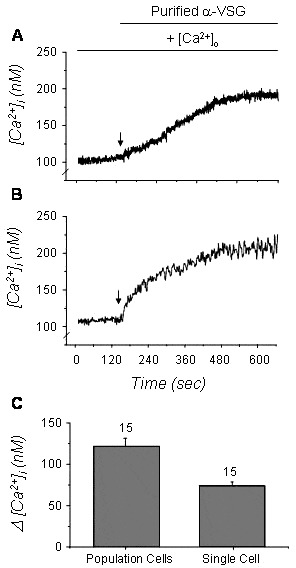
Anti-VSG antibodies were affinity purified from mouse ascitic fluids using the purified VSG antigen immobilized on nitrocellulose. As illustrated in Fig. 2B, analyses by Western blot showed that affinity-purified anti-VSG antibodies, at various dilutions, specifically recognized VSG from whole-cell T. evansi lysates (left). Purified VSG was also recognized by these antibodies (right). Affinity-purified anti-VSG antibodies were then used to perform direct measurements of their effect on the parasite [Ca2+]i. Fig. 6A is a representative experiment showing that exposure of a single T. evansi parasite to a concentration of 66 μg/ml of affinity-purified anti-VSG antibodies generated an increase from 100 to ~190 nm in its [Ca2+]i. The mean value of the parasite [Ca2+]i variations attained under this condition was 74±9 nm in the presence of extracellular Ca2+, after the steady state was reached (Fig. 6C). No response was obtained using 6·6 or 33 μg/ml of the purified antibodies (data not included). Since in the single cell approach only a single parasite partially immobilized with poly-L-lysine can be assayed, we repeated these measurements using the more conventional bulk cuvette setup. This fluorescence spectrophotometric arrangement allows us to determine the effect of the affinity-purified anti-VSG antibodies on a parasite cell population, where the signals from a suspension containing 2×10⁶ cells/ml are averaged. Fig. 6B shows a representative experiment in which the [Ca2+]i of a population of parasites (0·8×106 cells) was increased from 105 to ~200 nm following exposure to 33 μg of affinity-purified anti-VSG antibodies, in the presence of 1·8 mm extracellular Ca2+. We obtained a mean value for the [Ca2+]i variation of 122±15 nm, when populations of T. evansi parasites were exposed to purified anti-VSG antibodies in the presence of external Ca2+ (Fig. 6C). In conclusion, affinity-purified antibodies against VSG were able to elicit comparable calcium fluxes in T. evansi using both the single cell and the bulk cuvette set ups.
Effect of affinity-purified anti-VSG antibodies (purified α-VSG) on the parasite [Ca2+]i. Purified α-VSG antibodies were added to single cells (A) or cell populations (B), which were loaded with Fura-2/AM (8 mm) in the presence of extracellular Ca2+ (1·8 mm). Arrows indicate the time of α-VSG antibody addition. Shown is a representative trace of 5 independent experiments that were conducted on 3 separate preparations. (C) Histograms corresponding to the intracellular Ca2+ variation (expressed as Δ[Ca2+]i) observed after the addition of the purified α-VSG antibodies when the [Ca2+]i reached a stable state. All the experiments were carried out at 30°C. On top of each bar is indicated the number of parasites in which [Ca2+]i was measured.

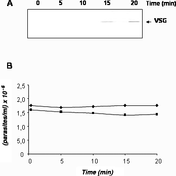
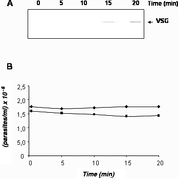
VSG has been shown to be involved in antigenic variation and shedding, which cause release of this protein from the parasite surface coat. Since the Ca2+ signal reported here is generated by the interaction between VSG molecules and specific antibodies, we evaluated the direct effect on the VSG release of the increase of [Ca2+]i caused by the Ca2+ ionophore A-23187. Western blot analyses using anti-VSG antibodies were conducted to detect the time-course of the release of VSG from bloodstream forms of T. evansi, in the presence of Ca2+ (1·8 mm) and A-23187 (2·5 μg/ml). As shown in Fig. 7A, VSG was released at 15 and 20 min. However, no free VSG was observed in the absence of A-23187 (data not shown). No changes in the number of motile parasites (Fig. 7B) and on the parasite morphology (data not included) occurred during the time-period of the experiment.
Induction of VSG release from Trypanosoma evansi by Ca2+ influx. (A) Release of VSG was detected by Western blot at various times (0–20 min), in the presence of the A-23187 ionophore (2·5 μg/ml). (B) During the time-course, live parasites were quantified in the presence (■) or absence (◆) of A-23187 (2·5 μg/ml). All these experiments included 1·8 mm extracellular Ca2+.
DISCUSSION
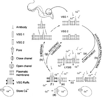
Here, we showed that anti-T. vivax antibodies and anti-VSG ascitic fluid reacted with T. evansi ex vivo and initiated an increase in its [Ca2+]i. This effect was abrogated by the addition of EGTA, showing that it is dependent on the presence of extracellular calcium. Accordingly, sera from healthy animals were not capable of increasing the parasite intracellular Ca2+ levels. Parasite lysis produced by infected sera and anti-VSG antibodies was sensitive to both, heat and rosmarinic acid, indicating that this effect was mediated by the complement system (Fig. 8a). Physiological evidence has demonstrated that the action of the complement system is dependent on the presence of extracellular Ca2+ since the activation of this system culminates in the formation of pores in the plasma membrane, which permits the entry of Ca2+ (Campbell et al. Reference Campbell, Daw and Luzio1979; Newsholme et al. Reference Newsholme, Adogu, Soos and Hales1993; Laffafian et al. Reference Laffafian, Davies, Campbell and Hallett1995). Destruction of T. vivax parasites by the complement system has been previously reported in bovines (Murray and Urquhart, Reference Murray and Urquhart1977; De Gee et al. Reference De Gee, Shah and Doley1982; Mahan et al. Reference Mahan, Hendersoshot and Black1986). However, it has been demonstrated that salivarian trypanosomes have mechanisms for complement evasion. During infection, T. congolense releases factors that directly activate the bovine complement cascade at the C1 level resulting in depletion of complement components (Nielsen et al. 1978). It has also been proposed that the direct activation of the classical complement pathway observed in humans by a T. brucei VSG could lead to considerable depletion of complement components (Musoke and Barbet, Reference Musoke and Barbet1977). Additionally, reductions in sera of the levels of total haemolytic complement from the alternative pathway and C3 from the classical route have been also reported in T. congolense- and T. vivax-infected cattle (Nielsen, Reference Nielsen and Tizard1985). Thus, under hypocomplementaemia conditions, a small proportion of trypanosomes expressing the predominant VSG variant might survive once the complement system has decreased (Fig. 8b).
Scheme of possible Ca2+ signal transduction routes induced by anti-VSG antibodies. (a) Parasite lysis by the complement classical pathway. (b) Under hypocomplementaemia conditions, α-VSG did not produce parasite lysis but (c) were capable of inducing a calcium signal. Both, a release of this ion from parasite intracellular stores (d) and an influx of Ca2+ through ion channels located on its plasmatic membrane (e) were observed. These Ca2+ responses might be a consequence of lipid raft-associated VSG clusters induced by specific α-VSG (f). The antigenic variation strategy has been correlated with a Ca2+ signal in trypanosomatids (g). Additionally, VSGs are naturally released by a process known as VSG shedding (h), which could be associated with calcium signalling.

When the complement system was completely inactivated by either heat treatment or addition of rosmarinic acid, in order to simulate hypocomplementaemia conditions, an increase in the [Ca2+]i was still obtained after exposure of parasites to either animal infected sera or anti-VSG antibodies (Fig. 8c). This effect was dependent on specific anti-VSG antibodies since pre-incubation of these antibodies with the purified VSG completely abolished the increase in the parasite [Ca2+]i. Moreover, similar effects were generated by affinity-purified anti-VSG antibodies using either single cell or parasite population measurements. Since addition of Ni2+, a common inactivator of membrane calcium channels, only partially inhibited the increase of [Ca2+]i, two sources must be responsible for the Ca2+ signal reported here: (1) a release of Ca2+ from parasite intracellular stores, such as the endoplasmic reticulum, the mitochondria or the acidocalcisomes (Fig. 8d) (Moreno and Docampo, Reference Moreno and Docampo2003; Mendoza et al. Reference Mendoza, Mijares, Rojas, Rodríguez, Urbina and DiPolo2002, Reference Mendoza, Mijares, Rojas, Colina, Cervino, DiPolo and Benaim2004b), and (2) an influx of this ion through channels located on its plasma membrane (Fig. 8e). In 2005, the genome sequence of T. brucei was published (Berriman et al. Reference Berriman, Ghedin, Hertz-Fowler, Blandin, Renauld, Bartholomeu, Lennard, Caler, Hamlin, Haas, Bohme, Hannick, Aslett, Shallom, Marcello, Hou, Wickstead, Alsmark, Arrowsmith, Atkin, Barron, Bringaud, Brooks, Carrington, Cherevach, Chillingworth, Churcher, Clark, Corton, Cronin, Davies, Doggett, Djikeng, Feldblyum, Field, Fraser, Goodhead, Hance, Harper, Harris, Hauser, Hostetler, Ivens, Jagels, Johnson, Johnson, Jones, Kerhornou, Koo, Larke, Landfear, Larkin, Leech, Line, Lord, Macleod, Mooney, Moule, Martin, Morgan, Mungall, Norbertczak, Ormond, Pai, Peacock, Peterson, Quail, Rabbinowitsch, Rajandream, Reitter, Salzberg, Sanders, Schobel, Sharp, Simmonds, Simpson, Tallon, Turner, Tait, Tivey, Van Aken, Walker, Wanless, Wang, White, White, Whitehead, Woodward, Wortman, Adams, Embley, Gull, Ullu, Barry, Fairlamb, Opperdoes, Barrell, Donelson, Hall, Fraser, Melville and El-Sayed2005) and a putative sequence for a calcium channel protein has been reported [GeneDB website (http://www.genedb.org), sequence number Tb10.70.4750]. Accordingly, calcium entry into T. brucei bloodstream trypomastigotes has been reported to be apparently regulated via a novel signalling pathway involving phospholipase A2-mediated generation of arachidonic acid and stimulation of a plasma membrane-located calcium channel (Eintracht et al. Reference Eintracht, Maathai, Mellors and Ruben1998; Catisti et al. Reference Catisti, Uyemura, Docampo and Vercesi2000).
In higher eukaryotes and kinetoplastid parasitic protozoa, several plasma membrane proteins, particularly those anchored by a glycosyl-phosphatidylinositol (GPI) moiety, are associated with sphingolipid/esterol-rich microdomains termed lipid rafts (Brown and London, Reference Brown and London1998; Simons and Ikonen, Reference Simons and Ikonen1997; Nolan et al. Reference Nolan, Jackson, Biggs, Brabazon, Payes, Van Laethem, Paturiaux-Hanocq, Elliot, Voorheis and Pays2000; Denny et al. Reference Denny, Field and Smith2001). VSG like other GPI-anchored proteins have been reported to be located in these rafts (Denny et al. Reference Denny, Field and Smith2001; Denny and Smith, Reference Denny and Smith2004). The presence of membrane protein clusters has been extensively used for lipid raft characterization (Harder et al. Reference Harder, Scheiffele, Verkade and Simons1998). CD59, a GPI-anchored protein, was still able to mediate Ca2+ intracellular signalling events following antibody-induced ligation, and subsequently was clustered in these microdomains (Hiscox et al. Reference Hiscox, Hallett, Morgan and van den Berg2002). Pizzo et al. (Reference Pizzo, Giurisato, Tassi, Benedetti, Pozzan and Viola2002) also demonstrated in Jurkat cells that anti-CD59 antibodies induced Ca2+ signalling and that the CD59-induced Ca2+ response was sensitive to treatment with methyl-β-cyclodextrin. These results suggested that the signalling pathway initiated by cross-linking of this raft-associated protein was altered specifically by disruption of the integrity of these rafts. Interestingly, on application of homologous antiserum to a given T. brucei VSG, the VSG antigen moves to the flagellar pocket region that overlays the Golgi apparatus (Barry, Reference Barry1979; Webster, Reference Webster, Russo and Black1990). This redistribution, known as capping, might be related to the presence of lipid rafts in these parasites. Interactions between lipid raft resident molecules, such as VSG, can also be studied by patching, a process in which these molecules are cross-linked by specific antibodies, facilitating association of rafts into a complex that is visible by microscopy (Denny and Smith, Reference Denny and Smith2004). This phenomenon has been observed in various protozoa (Doyle et al. Reference Doyle, Behin, Mauel and Rowe1974; Silva et al. 1975; Dwyer, Reference Dwyer1976). Here, we demonstrated by in vivo immunofluorescence labelling that anti-VSG antibodies induced the formation of patches of VSG on the parasite surface. Similar to the results reported by Hiscox et al. (Reference Hiscox, Hallett, Morgan and van den Berg2002), the Ca2+ signal we observed might be a consequence of lipid raft-associated VSG clusters induced by specific anti-VSG antibodies (Fig. 8f).
Which processes may be regulated by these Ca2+ pathways?. To date, the only stage that has been described for T. evansi is the trypomastigote form, and consequently this parasite does not undergo differentiation. Therefore, the Ca2+ signal reported here could be associated with (i) VSG endocytosis and recycling, (ii) antigenic variation, or (iii) shedding. Removal of anti-VSG antibodies from the parasite cell surface might be mediated by endocytosis, as a mechanism of evasion of the host immune response. O'Beirne et al. (Reference O'Beirne, Lowry and Voorheis1998) demonstrated that bloodstream forms of T. brucei, suffered a cycle of aggregation-disaggregation in the presence of anti-VSG antibodies. The disaggregation required energy and was dependent upon normal endosomal activity. Also, endocytosis and recycling of the GPI-anchored VSG in T. brucei has been described via large clathrin-coated vesicles (Engstler et al. Reference Engstler, Thilo, Weise, Grünfelder, Schwarz, Boshart and Overath2004). Since the mechanism of disaggregation described by O'Beirne et al. (Reference O'Beirne, Lowry and Voorheis1998) was found to be independent of Ca2+, the endosomal pathway is probably not correlated with the calcium signal we reported here. On the other hand, the antigenic variation strategy has been correlated with a Ca2+ signal in trypanosomatids, because the release of their surface coat protein is dependent on the presence of extracellular Ca2+ (Fig. 8g). Voorheis et al. (Reference Voorheis, Bowles and Smith1982) detected the release of [3H]acetimido-labelled VSG from bloodstream forms of T. brucei in the presence of calcium ions together with the calcium ionophore A-23187. We also showed that the [Ca2+]i increase by ionophores led to VSG release. All these results suggested that VSG release requires an increase in the parasite [Ca2+]i, presumably by the opening of controlled Ca2+-channels located on the parasite plasma membrane. Additionally, it has been reported that VSG molecules are naturally released during culture of T. brucei bloodstream forms (Bulow et al. Reference Bulow, Nonnengasser and Overath1989). This process is known as VSG shedding (Seyfang et al. Reference Seyfang, Mecke and Duszenko1990). In our model, released VSG may compete with live trypanosomes for antibody binding and consequently avoid parasite lysis (Fig. 8h).
In conclusion, a new methodological approach has been employed to determine parasite signalling routes induced by the host immunological response. This study is quite significant since it is the first to identify a ligand coupled to a calcium signal, which leads to a cellular response in salivarian trypanosomes. More experiments need to be performed in order to understand the physiological and immunological intracellular mechanisms, which are responsible for the activation, modulation and control of various aspects of host innate and acquired immunity during infection.
This work was supported by grants from FONACIT, No. G-2000001152, No. G-2001000637 and No. LAB-2000001639, and from Decanato de Investigación y Desarrollo, Universidad Simón Bolívar, No. S1-IC-CB-017-06. We thank Dr Johan Hoebecke (Université Louis Pasteur Strasbourg, Strasbourg, France) for performing the protein sequencing and Ronald Ramírez for helping with the illustrations.