Introduction
Mammalian ovarian follicles are highly specialized structures that support the growth and development of oocytes. Follicles leave the resting primordial follicle pool and continue to grow, although only very small numbers of follicles ever ovulate. In fact, more than 99% of all ovarian follicles in mammals will never ovulate; instead, they will undergo atresia at various stages of follicular development (Mao et al., Reference Mao, Wu, Smith, Mccauley, Cantley, Prather, Didion and Day2002).
Progression through successive stages of folliculogenesis is dependent on effective two-way communications between oocyte and granulosa cells and between granulosa and theca cells. It also requires appropriately timed endocrine signals, notably pituitary gonadotrophins (FSH/LH) and metabolic hormones, which act on receptors on the two somatic cell types and interact with local autocrine/paracrine signalling pathways (Knigh & Glister, 2003). While much is known about antral follicle development, relatively little is known about the development of preantral follicles, i.e., primordial, primary and secondary follicles. The development of preantral follicles has been considered independent of gonadotrophins because these follicles develop in FSH β subunit (Kumar et al., Reference Kumar, Wang, Lu and Matzuk1997) and FSH receptor knockout mice (Dierich et al., Reference Dierich, Sairam, Monaco, Fimia, Gansmuller, LeMeur and Sassone-Corsi1998). However, preantral follicles are sensitive to FSH because their receptors are expressed in bovine granulosa cells (Xu et al., Reference Xu, Garverick, Smith, Smith, Hamilton and Youngquist1995). In addition, mRNA for FSH receptors is expressed by the granulosa cells of secondary follicles in many species (Dufour et al., Reference Dufour, Cahill and Mauleon1979; Webb et al., Reference Webb, Campbell, Garverick, Gong, Gutierrez and Armstrong1999; McNatty et al., Reference McNatty, Fidler, Juengel, Quirke, Smith, Heath, Lundy, O'Connell and Tisdall2000). Moreover, full-length transcripts of FSH receptors were detected in the ovaries of neonatal mice from day 3 to day 15 (O'Shaughnessy et al., Reference O'Shaughnessy, McLelland and McBride1997), but in caprine, expression of FSH receptor (FSHR) in ovarian follicles has not yet been described.
Some studies have demonstrated that FSH stimulates the growth of oocytes enclosed in preantral follicles cultured in vitro in various species (murine: Cortvrindt et al., Reference Cortvrindt, Hu, Liu and Smitz1998a; bovine: Itoh et al., Reference Itoh, Kacchi, Abe, Sendai and Hoshi2002; caprine: Matos et al., Reference Matos, Lima-Verde, Luque, Maia, Silva, Celestino, Martins, Báo, Lucci and Figueiredo2007), indicating the importance of FSH during early follicular development. In mice, both gonadotropins (FSH and LH) promote growth and differentiation of granulosa cells in the culture of preantral follicles, as well as oocyte growth and maturation (Cortvrindt et al., Reference Cortvrindt, Hu, Liu and Smitz1998a, Reference Cortvrindt, Hu and Smitzb). Matos et al. (Reference Matos, Lima-Verde, Luque, Maia, Silva, Celestino, Martins, Báo, Lucci and Figueiredo2007) showed that 50 ng/ml FSH is sufficient to promote the activation of primordial follicles and further follicular growth during the culture of goat ovarian cortical tissue. In addition, FSH (50 ng/ml), associated or not with LH (1 ng/ml), maintained the ultrastructural integrity of goat primordial follicles after culturing cortical tissue (Saraiva et al., Reference Saraiva, Celestino, Chaves, Martins, Bruno, Lima-Verde, Matos, Silva, Porfirio and Báo2008). Conversely, effects of FSH on early folliculogenesis in vivo have been reported in rodents (Roy & Greenwald, Reference Roy and Greenwald1996; McGee et al., Reference McGee, Perlas, LaPolt, Tsafriri and Hsueh1997). Recently, Campbell et al. (2004) provided the first in vivo evidence that gonadotropins can affect the development of preantral follicles in ovine, a large monovulatory species that has a prolonged period of preantral follicle development. Furthermore, FSH has been identified as a regulator of granulosa cell apoptosis (Chun et al., Reference Chun, Eisenhauer, Minami, Billig, Perlas and Hsueh1996).
Although the importance of FSH for follicular development is well established for antral follicles, the effects of this hormone on the in vitro development of large preantral follicles were not yet evaluated. To better understand the role of FSH in the development of caprine preantral follicles, this work was performed: (1) to evaluate the expression of FSHR in the different stages of goat follicle development immediately after follicular isolation and after in vitro culture; and (2) to investigate whether variations in FSH concentrations during the in vitro culture period have a beneficial role on the viability, growth and antral cavity formation of goat preantral follicles.
Material and methods
Source of ovaries
Ovaries were collected at a local slaughterhouse from 25 (1- to 3-year-old) mixed-breed goats and used to perform Experiments 1 (five pairs), 2 (16 pairs) and 3 (four pairs). Immediately postmortem, the ovaries were washed in 70% alcohol followed by two rinses in Minimum Essential Medium (MEM) supplemented with 100 μg/ml penicillin and 100 μg/ml streptomycin. The ovaries were then transported within 1 h to the laboratory in MEM at 4°C (Chaves et al., Reference Chaves, Martins, Saraiva, Celestino, Lopes, Correia, Verde, Matos, Báo and Name2008). Unless indicated otherwise, the culture media and other chemicals used in the present study were purchased from Sigma Chemical Co.
Experiment 1: Expression of mRNA for FSH receptor in goat follicles
Primary and secondary follicles, were isolated from 10 ovaries using the mechanical procedure described previously (Lucci et al., Reference Lucci, Amorim, Báo, Figueiredo, Rodrigues, Silva and Gonçalves1999). The ovaries were quickly cut individually into small fragments using a tissue chopper (Mickle Laboratory Engineering) adjusted to 75 μm. The fragments were then placed in phosphate-buffered saline (PBS) containing 5% bovine serum albumin, aspirated 40 times using a large Pasteur pipette (diameter ~1600 μm) and aspirated another 40 times with a smaller pipette (diameter ~600 μm). The suspension was then filtered successively through 500- and 100-μm nylon mesh filters. After repeated washing to completely remove the stromal cells, 30 primary or secondary follicles were carefully selected based on the morphological shape and number of granulosa cell layers around the oocyte and placed in separate Eppendorf tubes. All samples were stored at −80°C until the RNA was extracted.
For the second group, cumulus–oocyte complexes (COCs) aspirated from small (1–3 mm) antral follicles were recovered from 16 ovaries. Compact COCs were selected from the follicle content as described by van Tol & Bevers (Reference van Tol and Bevers1998). Thereafter, groups of 10 COCs were stored at −80°C until RNA extraction. To collect mural granulosa and theca cell complex, small follicles (n = 30) were isolated from ovaries (n = 5) and dissected free from stromal tissue with forceps as previously described (van Tol & Bevers, Reference van Tol and Bevers1998). The follicles were then bisected and mural granulosa/theca were collected and stored at −80°C. The ovarian follicles were classified as: (1) primary (a single layer of cuboidal granulosa cells); (2) secondary (two or more layers of cuboidal granulosa cells); or (3) small antral follicles (<3 mm in diameter; with multiple granulosa cells enclosing an antrum). The diameter of follicles was calculated according to the method described by van den Hurk et al. (Reference van den Hurk, Dijkstra, Hulshof and Vos1994).
The isolation of total RNA was performed using the Trizol plus purification kit (Invitrogen). According to the manufacturer's instructions, 1 ml of Trizol solution was added to each frozen sample and the lysate was aspirated through a 20-gauge needle before centrifugation at 10 000 g for 3 min at room temperature. Thereafter, all lysates were diluted 1:1 with 70% ethanol and subjected to a mini-column. After the RNA bound to the column, DNA digestion was performed using RNase-free DNase (340 Kunitz units/ml) for 15 min at room temperature. After washing the column three times, the RNA was eluted with 30 μl RNase-free water.
Prior to reverse transcription, the eluted RNA samples were incubated for 5 min at 70°C and chilled on ice. Reverse transcription was then performed in a total volume of 20 μl, which was comprised of 10 μl of sample RNA, 4 μl 5× reverse transcriptase buffer (Invitrogen), 8 units RNase out, 150 units Superscript III reverse transcriptase, 0.036 U random primers (Invitrogen), 10 mM DTT and 0.5 mM of each dNTP. The mixture was incubated for 1 h at 42°C, for 5 min at 80°C and then stored at −20°C. Negative controls were prepared under the same conditions but without the inclusion of the reverse transcriptase.
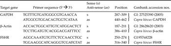
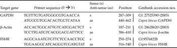
Quantification of the mRNA for FSHR was performed using SYBR Green. PCR reactions were composed of 1 μl cDNA as a template in 7.5 μl of SYBR Green Master Mix (PE Applied Biosystems), 5.5 μl of ultra-pure water and 0.5 μM of each primer. The primers were designed to perform amplification of mRNA for FSHR. GAPDH and β-actin (Table 3) were used as endogenous controls for normalization of gene expression. The thermal cycling profile for the first round of PCR was: initial denaturation and activation of the polymerase for 15 min at 94°C, followed by 40 cycles of 15 s at 94°C, 30 s at 60°C and 45 s at 72°C. The final extension was for 10 min at 72°C. All reactions were performed in a real-time PCR Mastercycler (Eppendorf). The delta-delta-CT method was used to transform CT values into normalized relative expression levels.
Experiment 2: In vitro culture of isolated preantral follicles
In the laboratory, surrounding fat tissue and ligaments were stripped off from the ovaries (n = 32). Ovarian cortical slices (1 to 2 mm in diameter) were cut from the ovarian surface using a surgical blade under sterile conditions. The ovarian cortex was subsequently placed in a fragmentation medium, consisting of MEM plus HEPES. Preantral follicles ≥150 μm and ≤ 180 μm in diameter were visualized under a stereomicroscope (SMZ 645, Nikon) and manually dissected from strips of ovarian cortex using 26 gauge (26G) needles. After isolation, follicles were transferred to 100-μl drops containing fresh medium under mineral oil to further evaluate the follicular quality. Follicles with a visible oocyte, surrounded by granulosa cells, an intact basement membrane and no antral cavity were selected for culture.
After selection, follicles were individually cultured in 25-μl drops of culture medium in petri dishes (60 × 15 mm, Corning). The culture medium was called α-MEM+ and consisted of α-MEM (pH 7.2–7.4) supplemented with 1.25 mg/ml bovine serum albumin (BSA), ITS (insulin 10 μg/ml, transferrin 5.5 μg/ml and selenium 5 ng/ml), 2 mM glutamine, 2 mM hypoxanthine and 50 μg/ml of ascorbic acid under mineral oil. Incubation was conducted at 39°C, 5% CO2 in air, for 18 days. Fresh media were prepared immediately before use and incubated for 1 h prior to use. Preantral follicles obtained from each animal were randomly distributed in the following treatments: α-MEM+ alone (cultured control) or associated with fixed concentrations of recombinant FSH (rFSH: 100 or 1000 ng/ml) or in α-MEM+ that was supplemented with increased concentrations of FSH throughout culture periods, corresponding to sequential media. These media were denominated: (a) sequential medium 1: FSH 100 ng/ml (from day 0 to day 6), FSH 500 ng/ml (from day 6 to day 12) and FSH 1000 ng/ml (from day 12 to day 18); and (b) sequential medium 2: FSH 500 ng/ml (from day 0 to day 9) and 1000 ng/ml (from day 9 to day 18). Every other day, 5 μl of the culture media were added to the drops. The culture was replicated four times, and approximately 30 follicles were used per treatment involving a total of 159 follicles.
Follicles were classified according to morphological characteristics, and those showing morphological signs of degeneration, such as darkness of oocytes and surrounding cumulus cells or those with misshapen oocytes were classified as degenerated. Oocyte and follicular diameter were measured only in healthy follicles at x and y dimensions (90°) by using an ocular micrometer (×100 magnification) inserted into a stereomicroscope (SMZ 645 Nikon) before and after 18 days of culture. Antral cavity formation was defined as a visible translucent cavity within the granulosa cell layers. Regarding follicular growth, the daily increase in follicular diameter was calculated as follows: the diameter of viable follicles at day 18 minus the diameter of viable follicles at day 0 divided by the number of days of in vitro culture (18 days).
In vitro maturation of oocytes from cultured preantral follicles
At the end of the 18-day culture period, all healthy follicles were carefully and mechanically opened with 26G needles under stereomicroscope for oocyte recovery. Only oocytes ≥110 μm, with homogeneous cytoplasm and surrounded by at least one compact layer of cumulus cells were selected for in vitro maturation (IVM). The recovery rate oocyte (RRO) was calculated by dividing the number of oocytes ≥110 μm by the total number of follicles at day 18 of culture and multiplying the result by 100. The selected COCs were washed three times in a maturation medium composed by TCM199 supplemented with 10% fetal calf serum, 100 μg/ml of LH, 5 μg/ml of rFSH, 1 μg/ml of 17β-estradiol and 1 ng/ml of epidermal growth factor (EGF). In our laboratory using this medium, the mean maturation rate of cumulus oocyte complexes recovered from antral follicles is 80%. After washing, the oocytes were transferred to 50 μl drops of maturation medium, under mineral oil and then incubated for 26 h at 39°C with 5% CO2 in air. At the end of the maturation period, oocytes were analysed under fluorescence microscopy for assessment of viability and chromatin configuration.
After the 26 h maturation period, live/dead fluorescence staining was performed on oocytes. The oocytes were quickly incubated in 100 μl droplets of MEM containing 4 μM calcein-AM, 2 μM ethidium homodimer-1 (Molecular Probes, Invitrogen) and 100 μM Hoechst 33342 (bisbenzimide trihydrochloride) at 37°C for 15 min. Afterwards, the oocytes were washed three times in MEM and examined under a DMLB fluorescence microscope (Leica). The emitted fluorescence signals of calcein-AM, ethidium homodimer-1 and Hoechst were collected at 488 and 568 nm, respectively. Oocytes were considered live if the cytoplasm was stained positively with calcein-AM (green) and if chromatin was not labelled with ethidium homodimer-1 (red).
Experiment 3: Expression of mRNA for FSH receptor in cultured follicles
Based on the best results obtained after the in vitro culture, a new experiment was performed. On this end, late preantral follicles were isolated from eight ovaries and cultured in sequential medium 1, as described in item Experiment 2: In vitro culture of isolated prenatal follicles. In this experiment, after 6 and 12 days of culture, two groups of viable follicles were frozen at −80°C to further evaluate the mRNA expression for FSHR, using the same methodology described earlier in item Experiment 1: Expression of mRNA for FSH receptor in goat follicles.
Statistical analysis
The data of mRNA expression for FSHR in goat follicles were analysed by paired Student's t-test (p < 0.05). Data from follicle survival, antrum formation and meiotic resumption after in vitro culture were expressed as percentages and compared by the chi-squared test. Follicular growth rates following the culture were compared using Student's t-test (SAS, 1999). Results were expressed as the mean ± SEM and differences were considered to be significant when p < 0.05.
Results
Messenger RNA expression of FSH-R in goat ovarian follicles
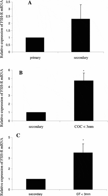
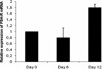
Prior to the culture, real-time PCR demonstrated an increase in the steady-state level of mRNA for FSH-R with the growth from primary to secondary follicle stages, but the differences were not statistically significant (p > 0.05; Fig. 1A). Messenger RNA levels for FSH-R in CCOs (Fig. 1B) or in granulosa/theca cells from small antral follicles (<3 mm) were significantly greater than those observed in secondary follicles (Fig. 1B,C). For in vitro cultured follicles, no significant difference in the levels of expression of FSH-R mRNA was observed after 6 or 12 days of culture when compared with day 0 (Fig. 2).
Expression of FSHR mRNA in goat ovarian follicles (means + SEM). (A) primary and secondary follicles. (B) secondary and COCs from small antral follicles (<3 mm). (C) secondary and granulosa/theca cells from small antral follicles. *p < 0.05.

Expression of FSHR mRNA in goat cultured follicles for 0, 6 and 12 days (means + SEM) in the sequential medium 1.

Follicle development after in vitro culture
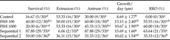
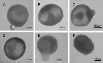
Table 1 shows the rate of follicular survival, oocyte extrusion, antral cavity formation, daily follicular growth and the percentage of oocytes destined to IVM after 18 days of culture of late caprine preantral follicles in fixed concentrations of FSH or sequential media. After 18 days of culture, sequential medium 1 significantly increased the percentage of follicular survival and reduced (p < 0.05) the precocious extrusion of oocytes compared with the other treatments (Table 1; Fig. 3D). From day 6 of culture onward, antral cavity formation was observed (Fig. 3A–D) in all treatments, including the control group. However, at the end of the culture, sequential medium 1 significantly increased the rate of antrum formation, compared with the other treatments (p < 0.05).
Percentage of survival, oocyte extrusion, antrum formation, rate of daily follicular growth and percentage of oocytes destined to IVM after 18 days of culture of caprine preantral follicles in different concentrations of FSH or sequential media

a–cDiffer among treatments. FSH, follicle stimulating hormone; RRO, recovery rate oocyte.
Stereomicrography caprine preantral follicles cultured in vitro (A,B). Normal follicles from FSH 100 ng/ml group after 6 and 10 days of culture, respectively, showing the antral cavity formation. (C) Normal follicle with well-defined antral cavity from the sequential medium 1 treatment after 8 days of culture. (D) Antral follicle after 14 days of culture in sequential medium 1. (E) Follicle from control group showing an extruded oocyte after 6 days of culture. (F) Degenerated follicles after 12 days of culture in medium containing FSH 1000 ng/ml.

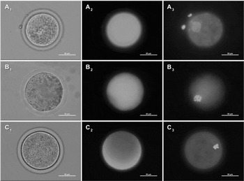
All tested treatments induced an increase of follicular growth throughout the culture; however, only sequential medium 1 significantly increased the rate of daily follicular growth, compared with the control. It is important to emphasize that when follicles were cultured in control medium (MEM+), no oocyte reached diameter ≥100 μm, which makes IVM not feasible. Figure 4A,B show the normal morphology of oocytes destined to IVM after in the vitro culture of preantral follicles in sequential medium 1. All treatments containing FSH significantly increased (p < 0.05) the percentage of oocytes larger than 100 μm, compared with the control. However, FSH (1000 ng/ml) and sequential medium 1 increased the rate of oocytes destined to IVM in relation to sequential medium 2. Despite that, sequential media 1 and 2 promoted the higher percentage of meiotic resumption, with a greater rate of oocytes showing germinal vesicle breakdown (p < 0.05) (Table 2 and Fig. 4C). Nevertheless, the formation of a metaphase plate, which corresponds to an oocyte in metaphase I, was observed only after culturing in sequential medium 2 (Table 2).
Morphology of oocytes destined to IVM, after in vitro culture of caprine preantral follicles in sequential medium 1. Oocyte visualization in a bright field (A1, B1, C1) and characterization of a viable oocyte after staining with calcein-AM (A2, B2, C2) and germinal vesicle (A3), germinal vesicle breakdown (B3) and metaphase I (C3) after Hoechst 33342 staining under fluorescence.

Percentage of oocytes showing germinal vesicle (GV), germinal vesicle breakdown (GVBD) and in metaphase I (MI) after IVM

a–cDiffer among treatments.
Discussion
This study reported for the first time the effect of FSH addition to the culture medium in fixed concentrations, or in increasing concentrations in a sequential way, on the in vitro development of isolated caprine preantral follicles. FSH is the main reproductive hormone in mammals, and is necessary for both the gonadal development and maturation, and the production of gametes during the fertile phase of life (Chappel & Howles, Reference Chappel and Howles1991; Simoni & Nieschlag, Reference Simoni and Nieschlag1995). In the ovary, FSH acts through binding to a G protein-coupled receptor superfamily, localized exclusively in granulosa cells (Gudermann et al., Reference Gudermann, Nürnberg and Schultz1995), beginning this expression in primary follicles (Méduri et al., Reference Méduri, Charnaux, Driancourt, Combettes, Granet Vannier, Loosfelt and Migrom2002).
In this study, changes in the pathway of FSHR mRNA expression in caprine follicles were demonstrated during real-time RT-PCR analysis, indicating that the levels of expression of these receptors increase with follicular development in vivo. These results suggest that there is a physiological need for increasing concentrations of FSH during in vitro follicle growth. Therefore, we decided to make and test the sequential media of the current study.
After 18 days of culture in sequential medium 1, an increase in the rates of follicular survival was observed. These results confirmed the previous findings of several authors (mouse: Cortivrindt et al., Reference Cortvrindt, Hu, Liu and Smitz1998; caprine: Matos et al., Reference Matos, Lima-Verde, Luque, Maia, Silva, Celestino, Martins, Báo, Lucci and Figueiredo2007; Saraiva et al., Reference Saraiva, Celestino, Chaves, Martins, Bruno, Lima-Verde, Matos, Silva, Porfirio and Báo2008; and swine: Mao et al., Reference Mao, Smith, Rucker, Wu, McCauley, Cantley, Prather, Didion and Day2004) who emphasized the importance of FSH as a survival factor during preantral follicle culture in vitro. Probably, the highlighted role of FSH is due to its ability to induce the expression of apoptosis inhibitor protein by granulosa cells (Wang et al., Reference Wang, Rippstein and Tsang2003). In addition, FSH is important for the regulation of almost all growth factors involved with follicular survival (Markström et al., Reference Markstrom, Svensson, Shao, Svanberg and Billig2002). Nevertheless, a reduction of follicle viability was observed when FSH was added in higher initial concentrations (sequential medium 2 or fixed concentration: 1000 ng/ml). It is likely that this phenomenon is associated with a decreased responsiveness to FSH and reduced mRNA receptor levels, due to a transcriptional down-regulation or decreased stability of receptor mRNA, effects of negative feedback induced by high FSH concentrations (Tilly et al., Reference Tilly, Kowalski, Schomberg and Hsueh1992; Tisdall et al., Reference Tisdall, Watanabe, Hudson, Smith and McNatty1995; Xu et al., Reference Xu, Garverick, Smith, Smith, Hamilton and Youngquist1995). Furthermore, the results obtained after culture demonstrated that the levels of FSHR expression did not increase at day 12, which suggests that there is no need for high concentrations of this hormone in the culture.
Primer pairs used as endogenous controls for normalization of gene expression in real-time PCR
Although the different treatments have stimulated the growth and antrum formation in this study, the most satisfactory results were obtained with sequential medium 1, which stimulated in a more efficient way follicular development, reducing the percentage of oocyte extrusion. This reduction probably occurs because of either maintenance or a gradual increase in the levels of FSHR expression, which keep the sensibility of the follicles to this hormone. Therefore, an addition of 100 ng/ml of FSH to the culture medium was sufficient to warrant follicular survival and antrum formation in the sixth day of culture. As the antral cavity was formed, an increase in FSH concentration to 500 ng/ml was essential for early antral follicle survival because the follicles became FSH dependent in this phase (Hirshfield, Reference Hirshfield1991). However, as observed in day 12, the levels of mRNA FSHR expression practically duplicated. Therefore, to ensure an elevated rate of follicular survival and to warrant follicular and oocyte development, a new increase in FSH concentration to 1000 ng/ml was essential. We can observe with this result that a convenient FSH concentration is a crucial point for the survival and development of follicles that are differentiating in the antral stage (Braw & Tsafriri, Reference Braw and Tsafriri1980; Markström et al., Reference Markstrom, Svensson, Shao, Svanberg and Billig2002).
The interaction of FSH with its receptor can result in direct and indirect effects. Among the direct effects is the activation of genes that coded to the stimulation of cell proliferation and steroid synthesis (Hunzicker-Dunn & Maizels, Reference Hunzicker-Dunn and Maizels2006). Indirectly, FSH contributes to the performance of other agents that promote follicular growth, such as EGF, insulin-like growth factor-1 (IGF-1) and LH, regulating their expression (van den Hurk & Zhao, Reference van den Hurk and Zhao2005). Thus, a great variety of studies have confirmed that FSH acts positively in the in vitro development of preantral follicles in different species (mouse: Cortvrindt et al., Reference Cortvrindt, Smitz and van Steirteghem1997, Reference Cortvrindt, Hu, Liu and Smitz1998; bovine: Gutierrez et al., Reference Gutierrez, Ralph, Telfer, Wilmut and Webb2000; swine: Wu et al., Reference Wu, Nayudu, Kiesel and Michelmann2000; bubaline: Gupta et al., Reference Gupta, Ramesh, Manjunatha, Nandi and Ravindra2008). However, in all of these studies, FSH was used only in fixed concentrations; i.e. opposite to our results, in which it was verified that the increase in follicular sensibility to FSH requires greater concentrations of this hormone during culture. This phenomenon may be associated with the biphasic regulation of FSH receptor expression in the ovary; i.e., low concentrations of FSH increase the number of FSH-binding sites parallel to the increased of FSH receptor mRNA levels. High doses of FSH down-regulate FSH receptor-binding sites and mRNA levels, suggesting a suppression of gene expression and protein synthesis concomitant to the increased receptor occupancy massive and internalization (Hsueh & LaPolt, Reference Hsueh and LaPolt1992; LaPolt et al., Reference LaPolt, Tilly, Aihara, Nishimori and Hsueh1992). Therefore, it is suggested that an excessive exposure to FSH may result in a suboptimal follicular response and consequent reduction of the beneficial effects of this hormone (LaPolt et al., Reference LaPolt, Tilly, Aihara, Nishimori and Hsueh1992).
In addition, the absence of the FSH effect may be due to its action on the regulation of glucose metabolism by glycolysis or Krebs’ cycle because this influence varies according to the degree of maturation and the need for follicles in different stages of development (Roy & Terada Reference Roy and Terada1999), and excessive or insufficient concentrations of FSH may negatively affect this mechanism of regulation.
The presence of FSH increases oocyte diameters in all treatments when compared with control, leading to the acquisition of structures with diameters convenient to IVM. This result confirmed the findings of Itoh et al. (Reference Itoh, Kacchi, Abe, Sendai and Hoshi2002), who worked with FSH in bovine preantral follicles, and Eppig and O'Brien (1998b) who studied FSH in mice. It is important to note that sequential medium 1 and 2 were the most effective in promoting meiosis resumption, leading to the recovery of the highest rate of oocytes in the stage of germinal vesicle breakdown. However, only sequential medium 2 allowed the progression up to metaphase I, which was an occasional finding. In the current study, all oocytes destined to IVM showed one or more layers of cumulus cells. It is well established that gonadotropins stimulated these cells to produce growth factors that are essential to the complete development and further oocyte maturation in vivo and in vitro (Moor & Dai, Reference Moor and Dai2001; Gilchrist et al., Reference Gilchrist, Ritter and Armstrong2004). FSH promotes follicular growth, prevents atresia and acts indirectly in follicular and oocyte maturation, inducing the synthesis of LH receptors (LHR). It also stimulates mRNA expression for the main components in the production of maturation inductor steroids, such as cytochrome P450 cholesterol side-chain cleavage (P450scc) mRNA in the bovine, murine, porcine and human species (Urban et al., Reference Urban, Garmey, Shupnik and Veldhuis1991; Adriaens et al., Reference Adriaens, Cortvrindt and Smitz2004, Sasson et al., Reference Sasson, Rimon, Dantes, Cohen, Shinder, Land-Bracha and Amsterdam2004; Hunzicker-Dunn et al., Reference Hunzicker-Dunn and Maizels2006; Silva et al., Reference Silva, Hamel, Sahmi and Price2006).
Some factors suggest that the action of FSH in oocyte maturation may be mediated by cumulus cells because FSHR mRNA is present in granulosa and cumulus cells but not in bovine oocytes isolated from antral follicles (diameter of 2 to 8 mm) (van Tol et al., Reference van Tol, van Eijk, Mummery, van den Hurk and Bevers1996). Others studies also reported that FSH can induce the production of a signalizing substance in somatic cells, which stimulates meiosis resumption because its addition to the medium enhances the rates of germinal vesicle breakdown in oocytes enclosed in cumulus cells, but not in denuded oocytes (Byskov et al., Reference Byskov, Andersen, Hossaini and Guoliang1997).
In conclusion, the use of defined concentrations of FSH, added in specific moments of the culture and in a progressive way, is essential to the in vitro development of caprine preantral follicles. The dynamic culture medium described in the present study can be used as basis for evaluating the interaction between FSH and several substances involved in the process of folliculogenesis. Increased understanding of the sequential regulatory events involving FSH and its interaction with other substances on follicular development may be important to optimize the gametogenic potential of females, through the improvement of immature follicle culture conditions.
Declaration of interest
The authors declare that there is no conflict of interest that would prejudice the impartiality of this scientific work.
Acknowledgements
The authors thank Cleidson Manoel Gomes da Silva for his technical support in fluorescence and all the members of the laboratory. This work was supported by CNPq, CAPES and FINEP. Márcia Viviane Alves Saraiva is a recipient of a grant from CAPES (Brazil).