Introduction
Eosinophilic angiocentric fibrosis is a rare benign disorder of the upper respiratory tract and orbit associated with submucosal thickening and subsequent fibrosis.Reference Lund, Howard and Wei1 The term was first coined in 1985 when Roberts and McCann described three cases, two in the nose and one affecting the subglottis, although the condition was actually first described two years earlier.Reference Roberts and McCann2, Reference Holmes and Panje3 It is slow growing and progressive, with characteristic histological appearances. There is a perivascular inflammatory infiltrate, rich in eosinophils, with subsequent concentric fibrosis in a so-called ‘onion-skin’ pattern.Reference Roberts and McCann2 The aetiology remains unknown, although recent evidence suggests that eosinophilic angiocentric fibrosis may represent part of the spectrum of immunoglobulin (Ig) G4-related disease.Reference Ferry and Deshpande4, Reference Deshpande, Khosroshahi, Nielsen, Hamilos and Stone5
We report a series of nine cases of sinonasal eosinophilic angiocentric fibrosis treated at our institution between 1995 and 2013. We review the literature on the possible aetiology of this rare progressive condition and its management options.
Materials and methods
A review of nine patients treated for eosinophilic angiocentric fibrosis at our institution between 1995 and 2013 is presented. The first four cases were reported previously.Reference Paun and Lund6
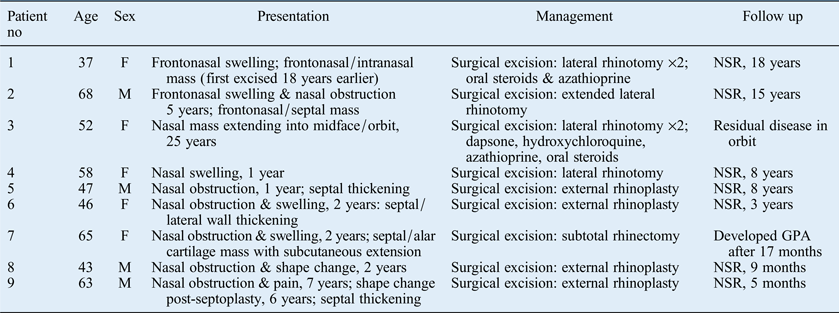
In all, there were five female and four male patients, with a mean age at presentation of 53 years (range 43–68 years; Table I). Presenting symptoms were a frontonasal or intranasal mass (three patients), septal thickening (two patients) or nasal obstruction (two patients). All were treated surgically, either via a lateral rhinotomy (four patients) or external rhinoplasty (5 patients) approach. Two patients were also treated with various anti-inflammatory and immunosuppressive agents during the course of their disease.Reference Paun and Lund6 One patient (case one) was started on oral steroids and azathioprine when eosinophilic angiocentric fibrosis recurred three years after her first lateral rhinotomy. The lesion responded very well but she was unable to tolerate the side effects of the medication, and therefore went on to undergo further surgical resection. The other patient treated medically (case three) had residual disease within the orbit, and was given dapsone, hydroxychloroquine and azathioprine over a period of some years; none had any effect on the lesion. Oral steroids were also given, with some effect, but the patient did not wish to continue the treatment.
Summary of nine cases of sinonasal eosinophilic angiocentric fibrosis

No = number; NSR = no sign of recurrence; GPA = granulomatosis with polyangiitis (Wegener's granulomatosis)
Of the nine patients, seven had no sign of recurrent disease after an average of 7.6 years (range, 5 months to 18 years). One patient (case three) was known to have residual disease within the orbit after surgery but declined further treatment or follow up. One patient was diagnosed with granulomatosis with polyangiitis (GPA, Wegener's granulomatosis) 17 months after undergoing subtotal rhinectomy. Her case is described in more detail below, as well as a less complicated case for comparison.
Results
Case seven
A 66-year-old Caucasian woman was referred from her local ENT department with an 18-month history of left nasal obstruction and swelling over the right side of her nose, associated with a fluctuating skin colour change. There was no history of pain, bleeding or epiphora and she felt well. Upon examination, there was a raised violaceous lesion over the right nasal dorsum and lateral wall, and a smooth swelling on the left of the caudal septum (Figure 1). Routine blood tests, including a full blood count, erythrocyte sedimentation rate (ESR) and angiotensin-converting enzyme level, were normal, as was her renal function. Antineutrophilic cytoplasmic antibody (ANCA) testing was negative. Computed tomography (CT) showed a mass affecting the septum and nasal tip. A recent biopsy had been interpreted elsewhere as showing non-specific inflammatory change, but review by a specialist head and neck pathologist in our unit confirmed a diagnosis of eosinophilic angiocentric fibrosis. Typical features were seen, including dense fibrosis with concentric whorls around vessels and active chronic inflammation featuring eosinophils; there was no evidence of granuloma formation or vasculitis. The same histological features were seen in a further biopsy of the thickened caudal septum. In view of the extensive involvement of the right lateral nasal wall and skin, the patient was offered subtotal rhinectomy, with either a prosthesis or nasal reconstruction; she chose the latter option.
Case seven: clinical photograph at the time of diagnosis. Published with permission.
Subtotal rhinectomy was performed in May 2011, with immediate reconstruction using a paramedian forehead flap, rib graft and a radial forearm free flap. Histopathology was consistent with eosinophilic angiocentric fibrosis, showing a diffuse fibrosing inflammatory process involving the soft tissue of the nasal septum, skin and subcutaneous tissue with extension into the surrounding fibroadipose tissue and skeletal muscle. There was perivascular fibrosis with concentric inflammatory cells and a characteristic ‘onion-skin’ appearance. The skin appearance was similar to that of granuloma faciale. The pathologist considered such extensive distribution to be unusual. There were also foci of vasculitis with scattered giant cells, necrosis and granulomatous microabscesses. A right cervical lymph node was also excised, and found to contain foci of eosinophilic angiocentric fibrosis.
The procedure was complicated by a post-operative pulmonary embolism, which was treated with warfarin for six months. A post-operative infection with development of a fistula in the right alar region was treated with long-term antibiotics and several minor debridements over the following 12 months. It appeared to have healed by May 2012. Unfortunately, the rib graft was lost as a result of the infection (Figure 2), and a second stage reconstruction with auricular cartilage was planned, to be performed locally at the patient's request. However, there was concern regarding disease progression in July 2012 (Figure 3), and further biopsies were taken. These showed ulceration with acute and chronic inflammation associated with focal perivascular fibrosis and a prominent eosinophilic infiltrate. However, there was also possible thrombosis and a single granuloma with multinucleated giant cells, suspicious for granulomatosis with polyangiitis (GPA, Wegener's). The patient was therefore referred back to our institution, and further debridement was undertaken in October 2012. Histological analysis showed a dense granulomatous inflammatory infiltrate comprising neutrophils, plasma cells and eosinophils. There was extensive fibrosis and a fibrinous exudate. Moderate-sized vessels showed necrotising vasculitis; microabscesses and multinucleated giant cells were also seen, consistent with an active vasculitic process. Repeat blood tests showed a mildly elevated ESR (43 mm/hour) and a positive ANCA test, although both proteinase 3 and myeloperoxidase assays were negative.
Case seven: Clinical photograph showing loss of reconstruction. Published with permission.
Case seven: clinical photograph showing disease progression. Published with permission.
The patient was urgently referred to the vasculitis team, which treated her with high dose methylprednisolone, rituximab and intravenous pulsed cyclophosphamide, with good effect. Her ESR returned to normal and all subsequent ANCA tests were negative. She continued taking azathioprine and co-trimoxazole, and by February 2013 the induration had resolved and her nasal mucosa was healthy. She has since opted for a prosthetic nose.
Case nine
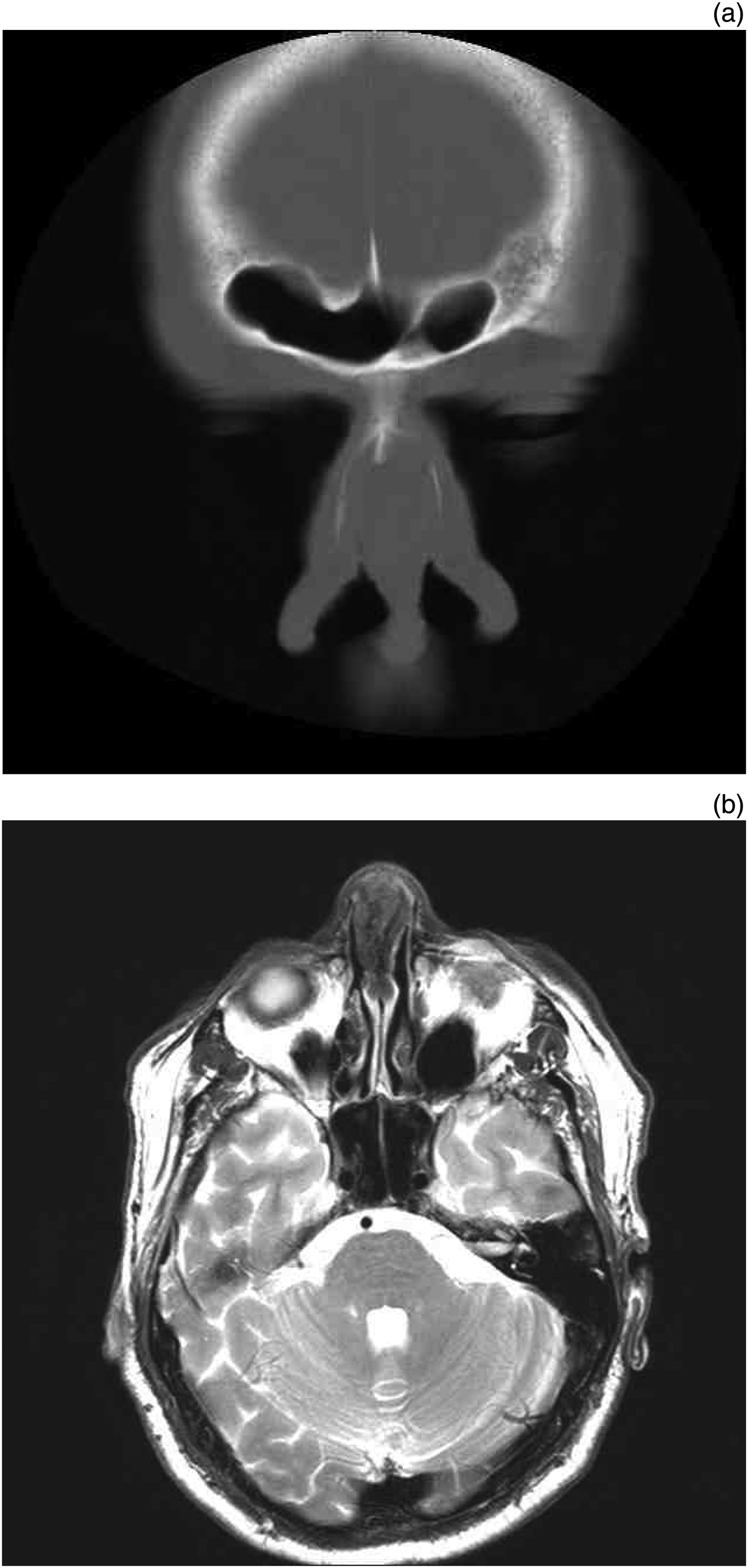
A 63-year-old man was referred with a 7-year history of nasal obstruction and pain. He had undergone septoplasty six years earlier, but the obstruction and pain had continued, and he developed a septal perforation with subsequent saddling of the nose. Examination revealed a thickened septum posterior to the perforation, and biopsy of this confirmed a diagnosis of eosinophilic angiocentric fibrosis. Computed tomography (Figure 4a) and magnetic resonance imaging (MRI; Figure 4b) showed evidence of abnormal soft tissue within the nasal septum, but this did not appear to extend into the subcutaneous tissues or the paranasal sinuses. Haematological tests were normal, including a negative ANCA test and normal IgG4 levels.
Case nine. (a) Coronal computed tomography scan showing homogenous thickening of the caudal septum. (b) T2-weighted axial magnetic resonance imaging scan showing hypointense thickening of the caudal septum.
The disease was resected via an external rhinoplasty approach. The most caudal 10 mm of his quadrilateral cartilage was of normal thickness on palpation, so a 5 mm caudal strut was left intact; the remaining cartilaginous septum was resected, along with most of the posterior bony septum. There was a plane between the disease and the nasal bones, so it was possible to ‘shell out’ the abnormal tissue from beneath the nasal bones, leaving them intact. Figure 5 shows the resected septum, with abnormal thickening. The resulting nasal deformity (Figure 6) was primarily reconstructed using an osseocartilaginous rib graft.Reference Christophel and Hilger7 Five months after surgery there was no evidence of recurrent disease.
Case nine: resection specimen. (EAF = eosinophilic angiocentric fibrosis)
Case nine. (a) Clinical photograph after resection of the involved septum. (b) Clinical photograph showing resultant cosmetic deformity.
Discussion
This is the largest single-institution series of sinonasal eosinophilic angiocentric fibrosis reported to date. Long-term follow-up data confirm that the ideal management to prevent progression of residual disease is complete surgical resection.
Eosinophilic angiocentric fibrosis is a rare benign, slow-growing condition of the upper respiratory tract. It is most commonly seen within the nasal cavity and midfacial soft tissues, although orbital and subglottic cases have been described.Reference Lund, Howard and Wei1, Reference Roberts and McCann2, Reference Leibovitch, James, Wormald and Selva8, Reference Karligkiotis, Volpi, Ferreli, Cerati, Kagkelari and Meloni9 Within the nose, it commonly affects the nasal septum and lateral nasal wall, but can involve the other cartilages of the nose and the nasal bones.Reference Karligkiotis, Volpi, Ferreli, Cerati, Kagkelari and Meloni9–Reference Narayan and Douglas-Jones11 With continued growth, it may extend into the paranasal sinuses and/or orbit.Reference Paun and Lund6, Reference Jain, Robblee, O'Sullivan-Mejia, Lea, Heller and Faquin12 Bilateral nasolacrimal duct obstruction has also been reported secondary to extensive sinus and orbital involvement.Reference Valenzuela, Whitehead, Brown and Sullivan13 It often extends into the subcutaneous tissues of the nose, and can involve the skin, which takes on a histological appearance consistent with granuloma faciale. Indeed, it has been suggested to represent a submucosal variant of granuloma faciale.Reference Burns, Roberts, De Carpentier and Zarod14–Reference Holsinger, Magro, Allen, Powell and Agrawal16
The aetiology of eosinophilic angiocentric fibrosis remains unknown, but previous trauma, including nasal surgery, has been suggested to act as a trigger; however, this has not been proven and does not apply to any of our patients.Reference Slovik, Putterman, Nash and Sion-Vardy17, Reference Nigar, Dhillon, Carr and Matin18 Allergic and parasitic aetiologies have also been proposed, but there is no evidence to support this apart from high tissue levels of eosinophils.Reference Lund, Howard and Wei1, Reference Karligkiotis, Volpi, Ferreli, Cerati, Kagkelari and Meloni9 Other authors have suggested a viral or drug-related trigger.Reference Holsinger, Magro, Allen, Powell and Agrawal16 Alternatively, an immunological cause is possible; eosinophilic angiocentric fibrosis has been linked to rheumatoid factor and inflammatory bowel disease.Reference Slovik, Putterman, Nash and Sion-Vardy17
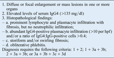
More recently, emerging evidence suggests that eosinophilic angiocentric fibrosis may belong to the IgG4-related disease spectrum, also known as IgG4-related sclerosing disease.Reference Ferry and Deshpande4, Reference Deshpande, Khosroshahi, Nielsen, Hamilos and Stone5, Reference Carruthers, Stone and Khosroshahi19 This systemic immune-mediated disease is characterised by numerous IgG4-postive plasma cells and T-cell infiltration of various organs, most frequently the pancreas and hepatobiliary tree, often (but not always) with elevated serum IgG4 levels.Reference Carruthers, Stone and Khosroshahi19, Reference Lindau, Su, Kobayashi and Smith20 Clinical diagnostic criteria for IgG4-related disease are shown in Table II. The mainstay of treatment for IgG4-related disease is systemic steroids.Reference Carruthers, Stone and Khosroshahi19
Diagnostic criteria for immunoglobulin G4-related diseaseReference Carruthers, Stone and Khosroshahi19
IgG4 = immunoglobulin G4; hpf = high-powered field
In the head and neck, IgG4-related disease includes chronic sclerosing sialadenitis, Riedel's thyroiditis and orbital pseudotumour.Reference Ferry and Deshpande4, Reference Deshpande, Khosroshahi, Nielsen, Hamilos and Stone5, Reference Lindau, Su, Kobayashi and Smith20 However, it has also been reported to occur in the nose and paranasal sinuses.Reference Deshpande, Khosroshahi, Nielsen, Hamilos and Stone5, Reference Lindau, Su, Kobayashi and Smith20 Most plasma cells seen in the early inflammatory infiltrate of eosinophilic angiocentric fibrosis have been shown to be IgG4 positive, prompting speculation that eosinophilic angiocentric fibrosis is a member of the IgG4-related disease spectrum.Reference Deshpande, Khosroshahi, Nielsen, Hamilos and Stone5, Reference Carruthers, Stone and Khosroshahi19, Reference Lindau, Su, Kobayashi and Smith20 Whilst eosinophils are usually seen in IgG4-related disease, they are not as abundant as in eosinophilic angiocentric fibrosis. Reports have described patients with classical histological features of eosinophilic angiocentric fibrosis associated with markedly elevated plasma IgG4 levels.Reference Deshpande, Khosroshahi, Nielsen, Hamilos and Stone5 Plasma IgG4 levels were measured in the last three patients in our series and were normal in each. Pathological specimens have not yet been subjected to immunohistochemical analysis.
Our series shows that presentation is most commonly with progressive nasal obstruction and/or swelling of the septum and nose, usually over the lateral nasal wall. This can progress to involve the cheeks, glabella or frontal region. In our series, the diagnosis was sometimes made after septoplasty when the abnormally thickened septum was biopsied, and surgeons should have a low threshold for performing a biopsy in such cases. Presenting symptoms and signs may also include epiphora, proptosis, diplopia and/or skin changes.Reference Leibovitch, James, Wormald and Selva8, Reference Jain, Robblee, O'Sullivan-Mejia, Lea, Heller and Faquin12 If left untreated, the process can lead to marked cosmetic deformity and functional problems. If the orbit is involved, then visual symptoms may result from the mass effect.Reference Lund, Howard and Wei1 There are no reported cases of malignant transformation.
Radiological changes are non-specific, showing a homogeneous soft tissue infiltrative process without a clear capsule. On CT, this is usually isoattenuated to grey matter (Figure 4a), as it is on T1-weighted MRI scans (Figure 4b).Reference Yang, Wang, Wang and Wang21 Appearances on T2-weighted images are reported to be more variable, being hypointense in two-thirds of cases and isointense in one-third.Reference Yang, Wang, Wang and Wang21 Classically, the septum is thickened anteriorly (Figure 4). Bony erosion is infrequent, although the nasal bones and lamina papyracea were involved in some of our patients, as well as in other reports.Reference Pereira, Millas, Reis-Filho, Maeda and Franco10, Reference Narayan and Douglas-Jones11
Histological appearances are pathognomonic. Two distinct histological stages of the disease are recognised, and may coexist within the same specimen.Reference Karligkiotis, Volpi, Ferreli, Cerati, Kagkelari and Meloni9, Reference Holsinger, Magro, Allen, Powell and Agrawal16, Reference Owa, Boyle and Gallimore22 The first involves a dense inflammatory infiltrate, with predominant eosinophils, and associated vasculitis of the submucosal small vessels.Reference Kosarac, Luna, Ro and Ayala23 Plasma cells and T lymphocytes are also seen, but true granulomas and necrosis are not a feature.Reference Jain, Robblee, O'Sullivan-Mejia, Lea, Heller and Faquin12 As the disease progresses, eosinophils become even more predominant and progressive concentric perivascular fibrosis forms characteristic ‘onion-skin’ whorls.Reference Paun and Lund6, Reference Kosarac, Luna, Ro and Ayala23
Differential diagnosis includes chronic granulomatous diseases such as granulomatosis with polyangiitis (GPA, Wegener's) and sarcoidosis, fibrosing forms of invasive fungal disease, Kimura's disease, angiolymphoid hyperplasia with eosinophilia, erythema elevatum diutinum, and neurogenic and mesenchymal tumours.Reference Lund, Howard and Wei1, Reference Pereira, Millas, Reis-Filho, Maeda and Franco10, Reference Kosarac, Luna, Ro and Ayala23 There is a single previous report of eosinophilic angiocentric fibrosis associated with granulomatosis with polyangiitis (GPA, Wegener's); however, the granulomatosis with polyangiitis (GPA) had been diagnosed and successfully treated with immunosuppressive therapy five years before the patient presented with new symptoms including progressive nasal obstruction.Reference Loane, Jaramillo, Young and Kerr24 Despite this, it may be that the original diagnosis was incorrect and the immunosuppressive treatment had temporarily suppressed the eosinophilic angiocentric fibrosis. One of our patients (case seven) had an appropriate clinical picture and classical histological appearances of eosinophilic angiocentric fibrosis in multiple biopsies, with no evidence of granuloma formation and a negative ANCA test. However, a subtotal rhinectomy specimen showed foci of vasculitis with scattered giant cells, necrosis and granulomatous microabscesses suggestive of granulomatosis with polyangiitis, and she went on to become systemically unwell with local disease progression and a positive ANCA test more than 12 months post-resection. It is therefore essential that an experienced head and neck pathologist review the original biopsy specimens, although even this is not infallible. Blood should be sent for ANCA, eosinophil and IgG4 testing in all patients. Patients should be followed carefully, although there is no consensus as to how long this should continue. The diagnosis should be reconsidered if the clinical condition changes.
Complete surgical excision is the first-line treatment but is often difficult because of the extent and location of the disease. However, any residual disease, particularly macroscopic disease, will continue to progress and cause further symptoms. The ‘recurrent’ disease reported in the literature is more likely to be progressive residual disease.Reference Thompson and Heffner25 In our series of nine patients, seven showed no sign of recurrent disease after an average of 7.6 years (range, 5 months to 18 years), following radical surgical excision via either an external rhinoplasty or lateral rhinotomy approach. It is therefore important to aim for complete resection, even if reconstruction will be more difficult. Progressive residual disease may require multiple further resections, with their attendant complications and need for reconstruction. The surgical approach used depends on the site and extent of the disease. An external rhinoplasty approach is suitable for septal and nasal cartilage involvement but lateral rhinotomy would be required for disease extending into the frontonasal region or orbit because margins can be difficult to determine. For this reason, an endoscopic approach would be suitable only for very limited well-demarcated disease, as complete resection is vital.
• Eosinophilic angiocentric fibrosis is a rare benign but progressive disorder of the upper respiratory tract associated with submucosal thickening and fibrosis
• This study is the largest single-institution series of sinonasal eosinophilic angiocentric fibrosis to date
• The aetiology of eosinophilic angiocentric fibrosis remains unknown
• Emerging evidence suggests it may be a member of the immunoglobulin G4-related disease spectrum
• Long-term follow-up data confirm that complete surgical resection is the ideal management
• Medical therapy with rituximab may prove useful for residual or unresectable disease
The use of various immunosuppressive medications has been reported as an alternative to surgery, for example when patients are unfit or refuse surgery, or when complete surgical resection is not possible. Some success has been reported for oral steroids, and these were temporarily helpful in two of our patients (cases one and three); however, both patients discontinued treatment because of side effects.Reference Paun and Lund6 Azathioprine and hydroxychloroquine were not effective in the same two patients. Dapsone has been helpful in granuloma faciale but did not lead to any improvement when used to treat one of our patients.Reference Van de Kerkhof26 More recently, use of the monoclonal antibody rituximab has been suggested because it has been successfully used to treat IgG4-related disease after failure of medical treatment with oral steroids.Reference Deshpande, Khosroshahi, Nielsen, Hamilos and Stone5, Reference Carruthers, Stone and Khosroshahi19, Reference Lindau, Su, Kobayashi and Smith20, Reference Khosroshahi, Bloch, Deshpande and Stone27 As eosinophilic angiocentric fibrosis is now thought by many to represent IgG4-related disease of the paranasal sinuses (despite the lack of eosinophils), the potential for treatment with rituximab in recalcitrant or recurrent cases, or those in whom surgery is not possible, warrants further investigation.
Conclusion
Eosinophilic angiocentric fibrosis is a rare, benign but progressive condition, and current first-line management is complete surgical excision. In the future, medical therapy with rituximab may prove useful for residual or unresectable disease. Care should be taken with the diagnosis, and chronic granulomatous conditions including granulomatosis with polyangiitis (GPA, Wegener's) should be excluded. Follow up should be continued for at least two years, ideally longer, and the diagnosis should be reconsidered if the clinical condition changes.
Acknowledgements
We would like to thank Dr Amrita Jay and Dr Andrew Gallimore, Consultant Head and Neck Histopathologists at University College Hospital London, who reviewed all specimens and diagnosed eosinophilic angiocentric fibrosis in these nine patients. We would also like to thank Dr Timothy Beale, Consultant Head and Neck Radiologist at University College London Hospital, for reviewing the imaging data.