Introduction
Seed storage physiology is typically classified as orthodox or recalcitrant based on seed responses to decreasing water content and storage temperature. Orthodox seeds survive longer with increased drying or reduced storage temperature (Roberts, Reference Roberts1973), while recalcitrant seeds generally lack a quiescent period and die if dried below about − 15 MPa (Berjak and Pammenter, Reference Berjak and Pammenter2008). Seeds in another category, called ‘intermediate’ (Ellis et al., Reference Ellis, Hong and Roberts1990, Reference Ellis, Hong and Roberts1991a), do not maintain viability with extreme drying and freezer storage and are hypothesized to exhibit desiccation tolerance intermediate to recalcitrant and orthodox types, undergo deleterious lipid phase transitions at subzero temperatures, or are prone to rapid ageing despite high tolerance to desiccation (Sacandé et al., Reference Sacandé, Hoekstra, van Pijlen and Groot1998; Crane et al., Reference Crane, Miller, Van Roekel and Walters2003, Reference Crane, Kovach and Gardner2006; Walters and Koster, Reference Walters, Koster, Jenks and Wood2007; Daws and Pritchard, Reference Daws and Pritchard2008; Kochanek et al., Reference Kochanek, Steadman, Probert and Adkins2009).
Water relations of orthodox and recalcitrant seeds are comparable during the early stages of embryogenesis (Vertucci and Farrant, Reference Vertucci, Farrant, Kigel and Galili1995; Kermode and Finch-Savage, Reference Kermode, Finch-Savage, Black and Pritchard2002; Walters et al., Reference Walters, Farrant, Pammenter, Berjak, Black and Pritchard2002; Berjak and Pammenter, Reference Berjak and Pammenter2008). Embryos at the histo-differentiation and early dry matter accumulation stages are sensitive to mild water stress (Ψw < − 1.5 MPa) and tolerance to Ψw between − 3 and − 10 MPa increases as food reserves are deposited into embryo and endosperm cells (Walters et al., Reference Walters, Farrant, Pammenter, Berjak, Black and Pritchard2002; Walters and Koster, Reference Walters, Koster, Jenks and Wood2007). Once maximum dry mass is attained, and vasculature between parent and embryo is severed, orthodox seeds dry, have lower metabolic capacity and become tolerant to extreme water stress, surviving Ψw < − 300 MPa (Vertucci and Farrant, Reference Vertucci, Farrant, Kigel and Galili1995; Walters et al., Reference Walters, Farrant, Pammenter, Berjak, Black and Pritchard2002). In contrast, recalcitrant seeds may undergo only brief maturation drying and acquire some tolerance to water stress; but embryos generally succumb at Ψw < − 15 MPa (Walters et al., Reference Walters, Farrant, Pammenter, Berjak, Black and Pritchard2002; Berjak and Pammenter, Reference Berjak and Pammenter2008).
Substantial changes in cell structure and organization occur during the latter stages of dry matter accumulation and maturation drying of orthodox seeds (Kermode and Finch-Savage, Reference Kermode, Finch-Savage, Black and Pritchard2002; Walters and Koster, Reference Walters, Koster, Jenks and Wood2007; Berjak and Pammenter, Reference Berjak and Pammenter2008). Changes include decreased vacuole size, increased amount of insoluble reserves deposited in the cytoplasm and de-differentiated organelles. Changes in gene expression prior to and during early maturation drying suggest induction of protective metabolism (Buitink et al., Reference Buitink, Leger, Guisle, Vu, Wuillème, Lamirault, Le Bars, Le Meur, Becker, Küster and Leprince2006) that is marked by conversion of reducing sugars to longer-chain non-reducing sugars and accumulation of late embryogenesis abundant (LEA) proteins, heat-shock proteins and antioxidants (Kermode and Finch-Savage, Reference Kermode, Finch-Savage, Black and Pritchard2002; Berjak and Pammenter, Reference Berjak and Pammenter2008). These structural and metabolic changes occur to some extent in recalcitrant seeds (Kermode and Finch-Savage, Reference Kermode, Finch-Savage, Black and Pritchard2002; Berjak and Pammenter, Reference Berjak and Pammenter2008), but the abbreviated maturation drying or competing germination processes may limit recalcitrant seeds' abilities to acquire full tolerance to desiccation. The structural changes within the cytoplasm of orthodox seed cells prevent massive shrinkage of cell volume when water is removed and may be a critical mechanism of protecting cells from mechanical strain during drying (Walters et al., Reference Walters, Farrant, Pammenter, Berjak, Black and Pritchard2002; Walters and Koster, Reference Walters, Koster, Jenks and Wood2007). Accumulating large amounts of dry matter would seem a requisite for stabilizing cell structures and may suggest a correlation between cell loading and tolerance to water stress.
Seeds must complete their embryogenic programme and achieve maximum dry mass to attain high seed vigour (Kermode and Finch-Savage, Reference Kermode, Finch-Savage, Black and Pritchard2002). Paradoxically, larger seeds, which clearly are superior at dry matter accumulation, tend to be sensitive to moisture stress and exhibit recalcitrant physiology (Pritchard et al., Reference Pritchard, Daws, Fletcher, Gaméné, Msanga and Omondi2004a; Daws et al., Reference Daws, Garwood and Pritchard2005; Berjak and Pammenter, Reference Berjak and Pammenter2008). Nowhere is this seeming contradiction more apparent than the palms, which produce the largest seeds in Angiospermae but seeds are slow to germinate and exhibit a range of sensitivities to low temperature and moisture stress (Orozco-Segovia et al., Reference Orozco-Segovia, Batis, Rojas-Aréchiga and Mendoza2003; Liu et al., Reference Liu, Eastwood, Flynn, Turner and Stuppy2008; Pérez et al., Reference Pérez, Criley and Baskin2008). The majority of studies on palm seed tolerance to water stress and genebanking conditions produced inconclusive results (Liu et al., Reference Liu, Eastwood, Flynn, Turner and Stuppy2008) and classifications of seed responses to desiccation and cold are often ambiguous. For example, seeds of Elaeis guineensis Jacq. have been variously reported as recalcitrant (Roberts, Reference Roberts1973), orthodox (Grout et al., Reference Grout, Shelton and Pritchard1983), intermediate (Ellis et al., Reference Ellis, Hong, Roberts and Soestisna1991b) and desiccation tolerant (Aberlenc-Bertossi et al., Reference Aberlenc-Bertossi, Chabrillange, Corbineau and Duval2003). Moreover, confirmed reports of orthodox, recalcitrant or intermediate physiologies among Arecaceae seeds do not show any phylogenetic relationships (Dickie and Pritchard, Reference Dickie, Pritchard, Black and Pritchard2002; Berjak and Pammenter, Reference Berjak and Pammenter2008; Liu et al., Reference Liu, Eastwood, Flynn, Turner and Stuppy2008). Despite their global importance and species diversity, the seed biology within Arecaceae remains poorly understood. Increased knowledge of seed development would greatly aid ex situ conservation efforts for the estimated 2400 taxa within Arecaceae that provide food and vital ecological services throughout subtropical and tropical ecosystems in which palms are broadly distributed (Uhl and Dransfield, Reference Uhl and Dransfield1987).
In this study, we try to reconcile conflicting assessments of the storage physiology of Arecaceae seeds by examining some of the factors used to distinguish recalcitrant and orthodox seed types. Patterns of water relations and dry matter accumulation during embryo development are explored in the genus Pritchardia (Coryphoidae; Henderson, Reference Henderson2002; Dransfield et al., Reference Dransfield, Uhl, Asmussen, Baker, Harley and Lewis2005), for which there are conflicting reports of seed storage physiology (USFWS, 1998; Liu et al., Reference Liu, Eastwood, Flynn, Turner and Stuppy2008). There are 23 recognized species of Pritchardia (Wagner et al., Reference Wagner, Herbst and Sohmer1999). Most have legal, endangered status and are in immediate need of ex situ conservation (Chapin et al., Reference Chapin, Wood, Perlman and Maunder2004). Species within Pritchardia provide habitat for insects (Richardson, Reference Richardson2006), ocean-faring birds (Conant, Reference Conant1985) and the endangered Oahu tree snail Achatinella mustelina Mighels (H. Pérez, University of Hawaii, personal observation), and some have commercial value. Pritchardia remota (Kuntze) Becc. is endemic to the small (c. 70 ha), uninhabited island of Nihoa (23°3′N, 161°55′W) in the northwest Hawaiian island chain approximately 450 km from Honolulu, HI (21°18′N, 157°49′W). Two natural populations of P. remota remain on Nihoa, though the species is planted for display at botanical gardens.
Materials and methods
Phenology, plant material and characterization of fruit
Phenology of flowering during the dry season and subsequent fruiting was monitored in 2004 through 2005 by belaying into trees at the University of Hawaii at Manoa (Honolulu, Hawaii, USA). Mature trees (6–12 m tall) support approximately 25 flowering and fruiting structures, observable all year long at different stages of development. A flowering flush occurs in May and June (at the onset of the dry season) and fruits are mostly shed in July and August (at the end of the dry season). From March through August 2004, approximately 23 inflorescences were formed on a single tree and were tagged on the day flowers opened (presumed as anthesis), which was approximately 12 weeks after the inflorescence structure first formed. A few inflorescences were also formed on trees in September through November (at the onset of the rainy season), but they were not tagged. Beginning 70 days post-anthesis (DPA), fruits from tagged inflorescences were harvested monthly, and additional fruits from untagged inflorescences in four additional trees were also harvested to supplement samples for biochemical analyses. Infructescences supported about 150–200 fruits.
Fruit dimension, texture and mass were measured monthly using 5–10 randomly selected samples harvested from tagged inflorescences. Diameter was measured to the nearest 0.01 mm with digital callipers. Fresh and dry mass of pericarps (i.e. exo-, meso- and endocarp), seeds (i.e. endosperm and testa) and embryos, with embryos extricated from seeds (Pérez, Reference Pérez2005) were measured. Dry mass was recorded after heating tissues at 95°C for 96 h in a forced-air oven. Water contents were calculated from fresh and dry mass and are presented as g H2O per g dry mass (g g− 1). Free-hand sections of mature embryos were used to visualize morphological traits and differentiation of organs at shedding. Fruits and seeds of similar maturities that were harvested from different cohorts of inflorescences had similar size, mass, colour, texture and water content, and these traits were used to estimate maturity classes of fruits harvested from untagged inflorescences in 2005 through 2008 that were used for desiccation tolerance and biochemical analyses.
Desiccation tolerance of mature embryos
Survival following a drying challenge was used to evaluate desiccation tolerance of embryos from fruits harvested 340 DPA. Excised embryos were ‘flash’ dried over a stream of N2 (500 ml min− 1) for 0–1200 min (Pammenter et al., Reference Pammenter, Vertucci and Berjak1991) to water contents between 1.5 and 0.08 g g− 1, or dried by placing embryos at relative humidity (RH) ranging from 93 to 2%; RH was controlled using saturated salt solutions (Vertucci and Roos, Reference Vertucci and Roos1993). A minimum of two replicates of ten embryos each plus two additional embryos for water content assessments was used to evaluate survival for each drying time or RH treatment. Dried embryos were slowly rehydrated for 45–60 min on moist blotter, surface sterilized, transferred aseptically on to solid, half-strength Murashige and Skoog (Reference Murashige and Skoog1962) basal medium with sucrose and agar (Sigma, St. Louis, Missouri, USA), pH 5.7 and cultured at room temperature (c. 25°C) under fluorescent lights (12 h daily) for up to 4 months. Embryos were considered germinated if radicles enlarged to>5 mm. The minimum water content for embryo survival (also known as the critical water content) was calculated from the intersection of the horizontal relationship of germination percentage at higher water content values and the sloped relationship of germination percentage at lower water content values.
Biochemical analyses
Tissues were dissected from fruits immediately after harvest, quickly cooled using liquid nitrogen, and stored at − 80°C until lyophilized. Carbohydrate and lipid analyses were conducted at the National Centre for Genetic Resources Preservation (NCGRP) in Ft Collins, Colorado, USA. Protein quantification, from fruits collected in 2008 from Waimea Arboretum and Botanical Garden (Haleiwa, Hawaii, USA), was carried out at the University of Florida, Gainesville, USA (UF). Fruits or lyophilized materials were transported to NCGRP or UF by expedited air-courier within 2 d of harvest (fruits) or removal from storage at − 80°C (lyophilized samples).
Soluble carbohydrates
Soluble sugars were extracted from embryos and endosperm excised from fruits harvested at various times after anthesis. About 15–30 mg of freeze-dried material was ground in the presence of liquid nitrogen and soluble sugars were extracted from ground samples using three washes of 80% ethanol at 90°C for 20 min. The ethanol solution containing sugars was centrifuged at 650 g for 2 min and the supernatant was dried under nitrogen gas. The remaining pellet was then stored at − 80°C until high-performance liquid chromatography (HPLC) analysis. For HPLC analysis, extracted sugars were resuspended in 5 ml of deionized water, agitated on a vortex mixer, and held at room temperature for 30 min. Approximately 2 ml of sample was filtered into vials using a 0.20 μm syringe filter. Sugars were separated using an HPLC (Dionex, California, USA) with a borate trap column (4 × 80 mm), followed by a CarboPac PA10 guard column (4 × 80 mm) and finally a CarboPac PA10 analytical column (4 × 250 mm) using deionized water and 500 mM NaOH as eluants at a 30:1 ratio for the first 40 min, a 4.5:1 ratio until 75 min and full-strength 500 mM NaOH until 85 min (Dionex). Columns were then washed with the 30:1 ratio of eluants for 10 min in preparation for the next injection. Sugars were detected by high-performance anion exchange chromatography using a Dionex pulsed amperometric detector and identified by comparing elution times with standards. Sugar standards were arabinose, fructose, fucose, galactose, glucose, maltose, sucrose, trehalose, raffinose, stachyose and several sugar alcohols. Analyses were replicated three times for each maturity stage.
Lipids
Approximately 5 g of endosperm and 2 g of embryos were ground to a fine powder using a mortar and pestle and lipids were extracted in 2:1 chloroform:methanol using methods adapted from Bligh and Dyer (Reference Bligh and Dyer1959). The solution was washed using 0.9% NaCl (w/v) and the solvent phase containing lipids was quantitatively separated and saved. Solvents were evaporated using nitrogen gas and total amount of lipid was determined gravimetrically. Lipids were derivatized to fatty acid methyl-esters (FAME) using boron trifluoride according to Metcalf and Schmitz (Reference Metcalf and Schmitz1961) and FAME were separated on a gas chromatograph (Perkin-Elmer 8500, Massachusetts, USA) using a Nukol 30 m, 0.25 mm internal diameter (i.d.) fused silica capillary column (Supleco, St. Louis, Missouri, US) with He as the carrier at a flow rate of 1.1 ml min− 1. Initial oven temperature was set at 100°C and was ramped to 190°C at 10°C min− 1. Injector and detector temperatures were set at 220°C. FAME were detected using a flame ionization detector and identified by comparing retention times with standards. Analyses were replicated twice for each maturity stage.
Proteins
Protein was extracted from freeze dried samples of endosperm and embryos from multiple harvest dates using modified procedures from Hurkman and Tanaka (Reference Hurkman and Tanaka1986). Assays were replicated three times for each maturity stage. About 500 mg of sample was ground to a fine powder in liquid nitrogen. Tris-saturated phenol (2.5 ml), pH 8.8; 50 μl of plant protease inhibitor (P9599, Sigma); and 2.5 ml of phenol extraction buffer (0.1 M Tris HCl, pH 8.8, 10 mM EDTA, 0.2 M dithiothreitol, 0.9 M sucrose) were added to the powder for further grinding. The extract was transferred to centrifuge tubes and shaken for 30 min at 4°C. Tubes were then centrifuged at 5000 g for 10 min at 4°C. The top, clear, phenol phase containing protein was removed and placed into fresh centrifuge tubes. The phenol extraction process was repeated using the bottom phase and all top phenol phases were combined. Protein was precipitated from clarified phenol extract with five volumes of ice-cold 0.1 M ammonium acetate in 100% methanol overnight at − 20°C. Protein precipitate was centrifuged for 20 min at 12,000 g at 4°C. The pellet was washed twice with 0.1 M ammonium acetate in methanol, then twice with 80% acetone and finally with cold 70% ethanol. The pellet was dissolved in 6 M urea and 25 mM ammonium bicarbonate pH 8.4 buffer. The protein solution was treated with 250 enzyme units of Benzonase (Novagen, New Jersey, USA) to degrade large molecules of nucleic acids. The treated extract was clarified by spinning at 43,000 g for 20 min at 15°C. Protein content was quantified using a standard Bradford assay.
Ultrastructure
Changes in cell structures during embryo development were evaluated from electron micrographs of embryos harvested after various days post-anthesis. Preparation of embryos for transmission electron microscopy followed procedures adapted from DeMason and Thomson (Reference DeMason and Thomson1981). Five embryos from different harvest dates were fixed overnight in 4% glutaraldehyde and 2% paraformaldehyde in 0.05 M sodium cacodylate buffer (pH 7.2) at 4°C. Embryos were post-fixed in 1% OsO4 for 1 h, followed by dehydration in a graded ethanol–propylene oxide series. Embryos were then infiltrated with dilutions of Spurr's resin and 100% propylene oxide and then embedded in Spurr's resin. Embryo sections (90 nm) were prepared using an ultra-microtome (Ultracut-E, Reichert, Wien, Austria), mounted on grids and stained with uranyl acetate followed by lead citrate. The sections were viewed with a LEO 912 transmission electron microscope (Zeiss, Oberkochen, Germany), fitted with an Omega energy filter, at 100 kV. Images were recorded as digital files with a frame-transfer CCD camera (Proscan, Lagerlechfeld, Germany).
Water potential and osmotic shrinkage of embryos
The relationships between water content and water potential (Ψw) (PV curves) were measured in embryos harvested from tagged inflorescences at different DPA. Water contents were measured after soaking at least five embryos in solutions with different concentrations of polyethylene glycol (PEG, Mr = 8000; Ψw, − 12 to − 0.05 MPa) for 48 h at 25°C. Water potential of PEG solutions was calculated according to Michel and Kaufmann (Reference Michel and Kaufmann1973) and verified using a thermocouple psychrometer (Model SC-10, Decagon Devices, Washington, USA). PV curves were constructed by regressing water content with log of water potential for Ψw between − 0.2 and − 8 MPa. Embryos 70 DPA were too small and too sensitive to PEG to obtain reliable mass measurements, and so a PV curve for this maturity stage was estimated by extrapolating the relationships of slope and intercepts of PV curves fitted to data for 130–220 DPA.
Changes in water content, dry mass and PV curves during embryo maturation were used to calculate cell shrinkage during water stress. Relative changes in cell volume with reduced water potential were calculated from PV curves assuming spherical cells, water density of 1 g ml− 1, constant surface area despite reduced volume, and contributions of water and dry matter to cell volume at − 0.5 MPa for embryos at different DPA (Walters and Koster, Reference Walters, Koster, Jenks and Wood2007). Scaled depictions of cell shrinkage provided a graphical representation of compression during desiccation in embryos of different age classes. Drawings were scaled from digital image files of micrographs based on cell area measurements (data not shown; n = 5 embryos for each harvest date; n = 8 cell area measurements per embryo) calculated using ImageJ software provided by the US National Institutes of Health (Rasband, 1997–Reference Rasband2008).
Results
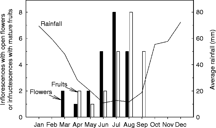
Inflorescences and fruits at all maturity stages were observed in P. remota trees from March through September. Blooming began in March near the end of the rainy season and flushed in June through August, the driest months in Hawaii (Fig. 1). Fruits matured in 13 months regardless of the flowering date; hence a flush of mature fruit was shed in July (peak dry season) through September (onset of rainy season) (Fig. 1). We can infer from this phenological pattern that histo-differentiation of newly formed embryos and maturation of the previous year's fruits were concurrent during the dry season and that fruits and seeds accumulated dry matter mostly during the rainy season.
Phenology of flowering and fruiting of Pritchardia remota growing in stands at the University of Hawaii at Manoa (Honolulu, Hawaii, USA) in 2004–2005. Number of inflorescences with open flowers and infructescences with mature fruits are represented by closed and open bars, respectively. Solid curve depicts average rainfall in Honolulu over a 30-year period (data provided by United States National Weather Service).

P. remota fruits are single-seeded drupes and maturity was assessed by size and colour. The exocarps of mature fruits (400 DPA) were black and leathery, the mesocarp was fibrous, the endocarp was thick and rigid. At maturity, dry mass of the pericarp (i.e. exo-, meso-, endocarp) was 1.9 ± 0.03 g (mean ± SE). Fruits were smooth when shed (about 400 DPA); the exocarp and mesocarp became brittle when fruits were left on the ground during the peak dry season. Seeds are endospermic with a relatively thick testa. At maturity, the dense, hard seed separated slightly from the endocarp except at the hilum, where it usually stayed attached. An operculum, derived from the testa, occurred opposite to the hilum (Pérez et al., Reference Pérez, Criley and Baskin2008). The embryo was located under the operculum. At shedding, embryos had a conical cotyledon that enveloped a rudimentary embryonic axis (Fig. 2A, B).
Morphology of developing Pritchardia remota embryos. (A) Embryo excised from seeds at shedding [400 d post-anthesis (DPA)] comprised mostly of a cotyledonary petiole that envelops an embryonic axis. The embryonic axis is contained within the apical portion of the cotyledonary petiole (ac) while the distal portion of the cotyledonary petiole (dc) remains within the seed and enlarges as endosperm is digested. Scale bar = 1.0 mm; magnified × 200. (B) Free-hand section of 400 DPA embryo showing close-up of embryonic axis (ax). The radicle emerges from the apical portion of the cotyledon (white arrowhead). The shoot emerges from transparent region of cotyledonary petiole (black circle). Scale bar = 0.5 mm; magnified × 450. (C) Embryo (em) at 70 DPA. Scale bar = 0.25 mm; magnified × 500.

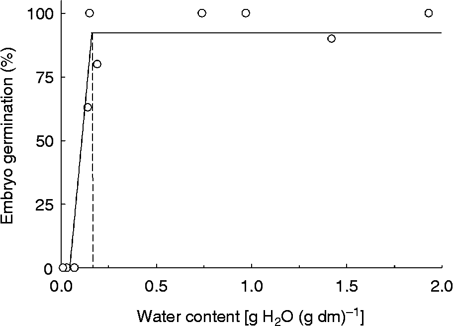
Between 80 and 100% of embryos harvested at 340 DPA germinated, and this proportion was relatively constant with water content until embryos were dried to less than 0.16 g g− 1 (Fig. 3). Drying embryos to less than 0.10 g g− 1 was lethal. Tolerance to drying could not be assessed in embryos harvested at 370 and 400 DPA, because of the general tendency for lower germination in undried samples, possibly as a result of induced dormancy.
Germination of Pritchardia remota embryos harvested 340 d post-anthesis (DPA) and dried to various water contents using a stream of N2 or exposure to different RH. The horizontal line represents the average germination of embryos dried to 0.3 [g H2O (g dry mass)− 1 (g g− 1)] and the sloped line represents the regression (r 2 = 0.91) between germination percentage and water content for embryos between 0 and 0.2 g g− 1. The two lines intersect at 0.16 g g− 1.

Changes in fruits and seeds during development
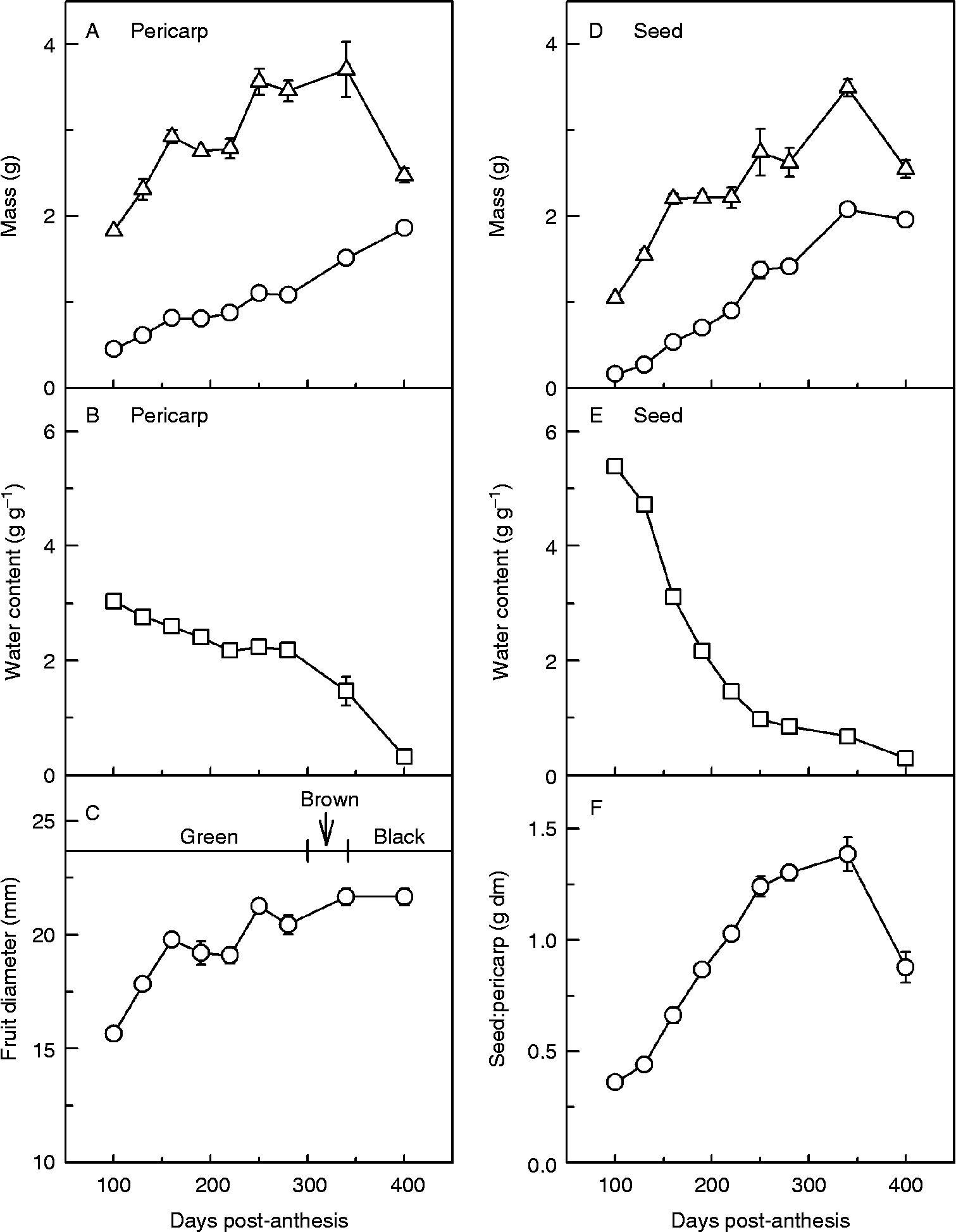
At 70 DPA fruits were bright green; a flexible endocarp and testa were tightly appressed. Dry mass of the pericarp increased about twofold, between 100 and 250 DPA (Fig. 4A). At about 300 DPA the exocarp began to brown and at 340 DPA fruit tissues began to dry rapidly, harden and turn black (Fig. 4C). Dry matter of the pericarp continued to increase until shedding at 400 DPA when it reached a final dry mass of about 1.9 ± 0.03 g (Fig. 4A). Dry mass at 250 and 400 d was significantly different for pericarps (F 1,13 = 105.4, P < 0.001) according to one-way ANOVA (α = 0.05).
Accumulation of dry matter and water in fruits (A–C) and seeds (D, E) of Pritchardia remota collected in 2004–2005. Changes in fresh (triangles) and dry (circles) mass of (A) pericarps with seeds removed and (D) seeds with embryos removed. Changes in water content [g H2O (g dry mass)− 1 (g g− 1)] of (B) pericarps and (E) seeds. (C) Change in diameter of P. remota fruits and (F) dry mass ratio of seeds:pericarps. Error bars denote SE.

The dry mass of the seed increased about eightfold between 100 and 250 DPA and continued to increase until 340 DPA when maximum dry mass of the seed was about 2.1 ± 0.1 g (Fig. 4D). The endosperm thickened from a clear liquid with water content >5 g g− 1 at 70 DPA to a cloudy gel with water content about 1.5 g g− 1 at 190 DPA (Fig. 4E). Dry matter continued to accumulate and water content decreased, and by 340 DPA, the endosperm had solidified to an extremely hard substance that required a hammer to break it. The seed gained dry matter faster than the fruit between 70 and 340 DPA, eventually filling the fruit with dense material (Fig. 4F). Seed dry mass was significantly different between 250 and 400 DPA (F 1,13 = 31.1, P < 0.001), demonstrating that dry matter accumulated throughout most of the developmental period.
Changes in embryo during development
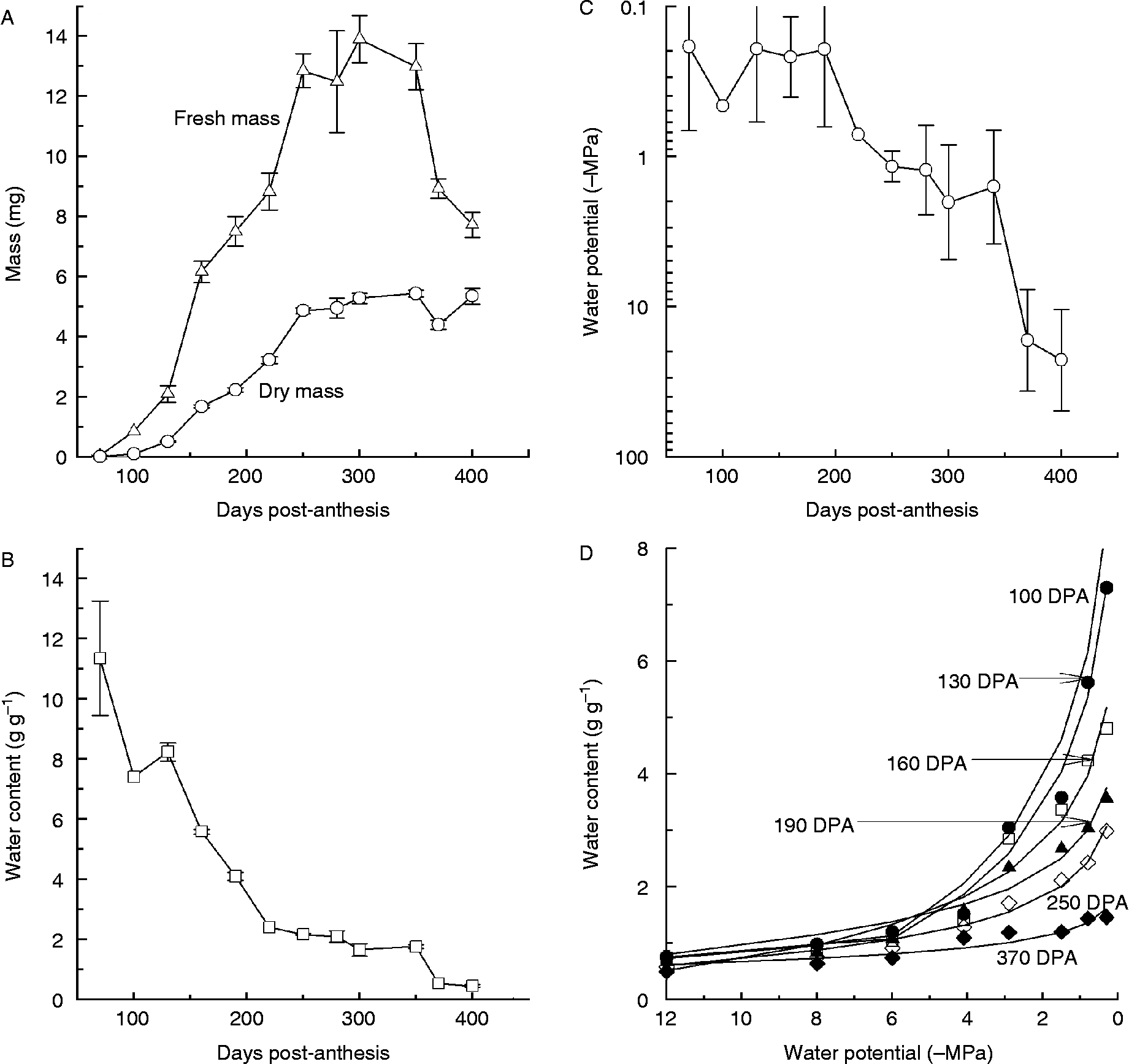
At 70 DPA, the embryo was a microscopic globular mass of cells attached to the operculum, and had the appearance typical of angiosperm embryos before histo-differentiation (Fig. 2C). The dry mass of the embryo increased 50-fold by 250 DPA to a maximum of 4.9 ± 0.1 mg (Fig. 5A). Dry mass of the embryo at 250 and 400 DPA was not significantly different (F 1,17 = 0.23, P = 0.6), suggesting that embryos had attained maximum dry mass before the fruit and seed. By 340 DPA, the hardened endosperm tissues allowed the embryo to separate from the operculum. At 400 DPA, the embryo occupied about 0.3% of the total dry mass of the fruit.
Dry matter accumulation and water relations of developing Pritchardia remota embryos collected in 2004–2005. Changes in (A) fresh mass (triangles) and dry mass (circles), (B) water content [g H2O (g dry mass)− 1 (g g− 1)] and (C) water potential. Error bars denote standard error of mean. (D) Pressure–volume (PV) curves for developing P. remota embryos. Data points represent water content–water potential relationships of embryos measured using PEG solutions and curves were calculated from the regression (r 2 range 0.92–0.98) of water content with ln(water potential) for Ψw ≤ − 8 MPa.

Embryo water content at 70 DPA was 11.3 ± 3.8 g g− 1 (Fig. 5B), higher than fruit or seed tissues at the same maturity stage (Fig. 4B, E). Water content decreased rapidly in embryo cells until 250 DPA, and then slowly until fruits were shed at 400 DPA (Fig. 5B). Despite large changes in water content in embryo cells between 70 and 250 DPA, the water potential of embryos was relatively constant at about − 0.2 MPa (range of − 0.05 to − 0.5 MPa) throughout most of the dry matter accumulation phase (Fig. 5C). Embryo water potential decreased to about − 1.2 MPa (range from − 0.9 to − 1.5 MPa) after 250 DPA, coincident with embryos attaining maximum dry mass. Water potential abruptly decreased to between − 10 and − 50 MPa when the fruits were shed (Fig. 5C).
PV curves constructed for embryos excised from fruits at different DPA allowed us to quantify relationships between dry mass accumulation, water content and water potential (Fig. 5D). At a given water potential, embryo water content decreased as the dry matter of embryos increased, suggesting that water mass was displaced by osmotically inactive materials. Difference in water content–water potential relationships among embryos at different maturities is most pronounced at Ψw>–3 MPa and can be visualized by the difference in water contents (Fig. 5D). According to the PV curve for embryos at 340 DPA (data not shown, but similar to embryos at 370 DPA), the critical water content of 0.16 g g− 1 below which embryos are killed by desiccation (Fig. 3) corresponds to − 49 MPa (~70% RH).
Patterns of dry matter deposition and organelle modifications during seed maturation were visualized from ultrastructure studies of developing embryos. At the early stages of dry matter accumulation (100 DPA), cells with a large central nucleus and thin cell wall were dominated by large, irregularly shaped vacuoles that were mostly electron-transparent (Fig. 6A). Vacuoles became more spherical and filled with flocculent electron-dense material between 100 and 250 DPA, as embryo dry mass increased from 0.1 to 4.9 mg. Cytoplasm became denser as embryos matured; the nucleus, a large central vacuole and smaller vacuoles containing reserves, dense mitochondria and thickened cell walls could be visualized at 400 DPA (Fig. 6B, C).
Ultrastructural changes during development in embryo cells of Pritchardia remota collected in 2004–2005: (A) 100 d post-anthesis (DPA), (B) 400 DPA, (C) detail of mitochondria from cells of 400 DPA embryos. Scale bars in (A), (B) and (C) are 5 μm, 2 μm and 200 nm, respectively. Black arrowhead, lipid droplet; cr, cristae; cw, cell wall; mi and white arrowhead, mitochondria; nu, nucleus; s, starch granule; t, tonoplast; va, vacuole.

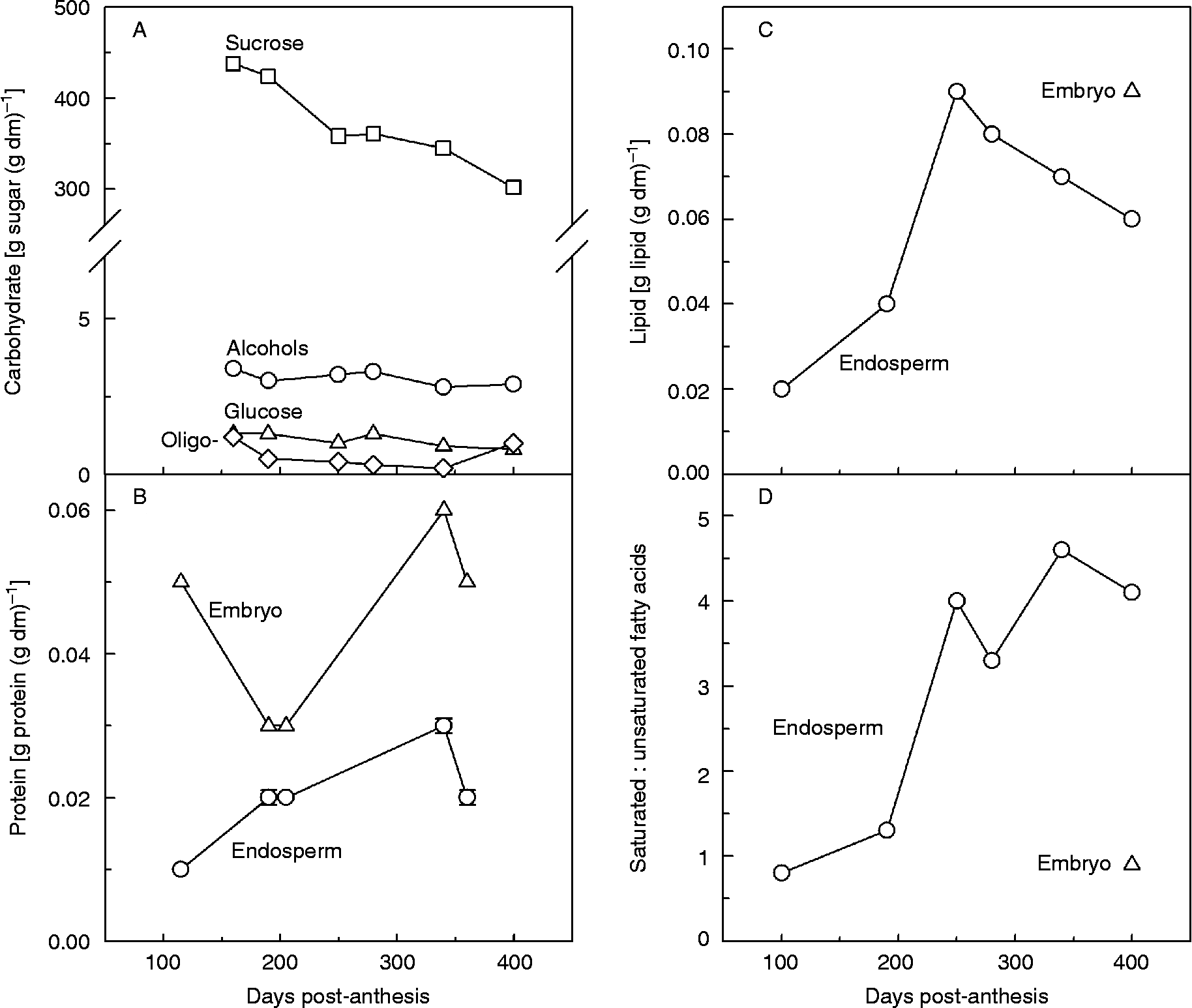
The composition of dry matter deposited into embryo and endosperm cells during seed development and maturation was evaluated by quantitative assessments of soluble carbohydrate, lipids and proteins. At 160 DPA, about 44% of embryo dry matter was soluble carbohydrate, with sucrose accounting for about 98% of the sugars and glucose and oligosaccharides accounting for about 1% each of the embryo dry mass (Fig. 7A). Soluble carbohydrate content decreased as embryos matured, and sucrose content comprised 30 ± 2% of the total dry mass of embryos at 400 DPA when fruits were shed. Proteins comprised a lower proportion of P. remota dry mass compared to carbohydrates, accounting for about 5% and 2% of the dry mass of the embryo and endosperm, respectively, throughout development (Fig. 7B). Lipids were also a minor reserve comprising about 9% of the accumulated dry mass in fully mature embryos of P. remota (Fig. 7C, triangle). Lipid content of the endosperm increased initially to nearly 9% of the dry mass at 250 DPA and then decreased (Fig. 7C). Both embryo and endosperm tissues contained high proportions of saturated fatty acids, as is typical for palms (Fig. 7D). Oleic (18:1), palmitic (16:0) and linoleic (18:2) were the predominant fatty acids in the embryo, comprising about 32, 23 and 21% of the total lipid, respectively. Minor fatty acids in the embryo were stearic (18:0), myristic (14:0) and lauric (12:0). In the endosperm, oleic (18:1) and linoleic (18:2) were the predominant fatty acids in immature seeds, and lauric (12:0) and myristic (14:0) were the predominant fatty acids in mature seeds (Fig. 7D).
Chemical composition of developing Pritchardia remota seeds. (A) Content of sucrose (squares), glucose (triangles), oligosaccharides (raffinose, stachyose) (diamonds) and sugar alcohols (myo-inositol, arabitol, and sorbitol) (circles). (B) Protein content within embryos (triangles) and endosperm (circles). (C) Lipid content of embryo (triangle) and endosperm (circles). (D) Ratios of saturated fatty acids (C8:0+C10:0+C12:0+C14:0+C16:0+C18:0) to unsaturated fatty acids (C18:1+C18:2) in embryos (triangle) and endosperm (circles).

Calculation of cell shrinkage in developing embryos in response to water stress
Accumulated dry matter, initial water potential and PV relationships were used to predict the extent to which cells shrink during water stress. Water occupied about 89% of the volume of cells from embryos at 100 DPA (Fig. 5A, B), which were highly vacuolated (Fig. 6A). Water potential of freshly harvested embryos at 100 DPA was close to − 0.5 MPa (Fig. 5C). Exposure to − 5 MPa and − 15 MPa reduced the water content of these immature embryos to 1.58 and about 0.55 g g− 1, respectively (Fig. 5D), causing a 68 and 81% loss of the cell volume (Fig. 8A). The same degree of water stress resulted in much less cell shrinkage in embryos that had accumulated dry matter reserves. For example, water in embryos 340 DPA occupied about 70% of cell volume at Ψw = − 0.5 MPa, and volume decreased by approximately 32 and 47% when Ψw was reduced to − 5 and − 15 MPa, respectively (Fig. 8B). The critical water content for 340 DPA embryos was 0.16 g g− 1 (Fig. 3) or − 49 MPa (Fig. 5D), which would lead to 63% decrease in cell volume (calculations not shown).
Schematic illustration of osmotic shrinkage of cells with dry mass contents similar to Pritchardia remota embryos that are (A) 100 d post-anthesis (DPA) and (B) 340 DPA and exposed to − 5 or − 15 MPa. Various organelles are represented: nucleus (black circles), vacuoles (white circles), and vacuoles with accumulated dry matter reserves (grey circles). Loss of volume resulting from loss of water was calculated from PV curves (Fig. 5D) assuming initial volume at Ψw = − 0.5 MPa and that dry matter occupied 11 and 30% of the volume in cells of 100 and 340 DPA embryos. Cells were assumed to be initially spherical and the surface area was assumed constant during shrinkage. Water density of 1 g ml− 1 was used to convert mass changes to volume changes.

Discussion
The patterns of dry matter accumulation and water relations in developing embryos of the endangered palm, Pritchardia remota, were investigated with the goal of identifying factors that contribute to the enigmatic physiology of seeds in Arecaceae. Flowering and fruiting patterns reported here for P. remota trees in Honolulu are similar to those reported for trees in situ (Conant, Reference Conant1985). Embryo development of P. remota is prolonged compared to other species of the Coryphoid group (Henderson, Reference Henderson2002) where gestation typically lasts 180–270 d (Chapin, Reference Chapin1999; Henderson, Reference Henderson2002). Duration of seed development period or shedding of fruit during the dry season has been positively correlated with seed tolerance to water stress (Dussert et al., Reference Dussert, Chabrillange, Engelmann, Anthony, Louran and Hamon2000; Daws et al., Reference Daws, Lydall, Chmielarz, Leprince, Matthews, Thanos and Pritchard2004, Reference Daws, Cleland, Chmielarz, Gorian, Leprince, Mullins, Thanos, Vandvik and Pritchard2006; Pritchard et al., Reference Pritchard, Daws, Fletcher, Gaméné, Msanga and Omondi2004a) and, in combination with a lengthy gestation period, may suggest that P. remota seeds acquire tolerance to desiccation. During gestation, fruits of P. remota grow to an average diameter of about 22.0 ± 4.0 mm (Fig. 4C) which is within the range of 4–100 mm diameter reported for Coryphoids, but larger than the median diameter of 14 mm reported for this group (Chapin, Reference Chapin1999; Henderson, Reference Henderson2002). The endocarp and endosperm become extremely hard, and these tissues impede but do not prevent water uptake (Pérez et al., Reference Pérez, Criley and Baskin2008). This may contribute to morpho-physiological dormancy that delays germination (Pérez et al., Reference Pérez, Criley and Baskin2008) but also protects the embryo from severe dehydration during the driest part of the season (Tweddle et al., Reference Tweddle, Dickie, Baskin and Baskin2003; Pritchard et al., Reference Pritchard, Daws, Fletcher, Gaméné, Msanga and Omondi2004a; Daws et al., Reference Daws, Garwood and Pritchard2005).
In contrast to the dry mass of the whole seed, the embryo of P. remota accumulates only about 5 mg dry mass (Fig. 5A) and tissues are underdeveloped (Baskin and Baskin, Reference Baskin and Baskin2001) (Fig. 2A, B). The developmental pattern of a rudimentary embryo and massive endosperm appears to be archetypal of palms (Uhl and Dransfield, Reference Uhl and Dransfield1987; Baskin and Baskin, Reference Baskin and Baskin2001; Henderson, Reference Henderson2002; Orozco-Segovia et al., Reference Orozco-Segovia, Batis, Rojas-Aréchiga and Mendoza2003; Pérez et al., Reference Pérez, Criley and Baskin2008). In P. remota, the small embryo may be, in part, an effect of the truncated period for dry matter accumulation which occurs mostly during the rainy season and ends at 250 DPA (Figs 1, 5A), about 90 d before the endosperm achieves maximum dry mass and 150 d before the fruit is shed. The pattern of seed and embryo development in P. remota superficially resembles seeds that acquire desiccation tolerance, in that there is a period at which maximum dry matter is achieved (i.e. physiological maturity) followed by a period of apparent quiescence during maturation drying (Kermode and Finch-Savage, Reference Kermode, Finch-Savage, Black and Pritchard2002). This pattern contrasts with desiccation-sensitive seeds that continue development until they are shed from the plant (Kermode and Finch-Savage, Reference Kermode, Finch-Savage, Black and Pritchard2002; Berjak and Pammenter, Reference Berjak and Pammenter2008). The curious early cessation of growth of embryos relative to endosperm is atypical of other angiosperm seeds, but has been reported in other palm species (Aberlenc-Bertossi et al., Reference Aberlenc-Bertossi, Chabrillange, Corbineau and Duval2003; von Fintel et al., Reference von Fintel, Berjak and Pammenter2004; Wen and Song, Reference Wen and Song2007).
The extent to which embryos accumulate dry matter varies among palm species, and this may be an important factor in acquiring desiccation tolerance (Kermode and Finch-Savage, Reference Kermode, Finch-Savage, Black and Pritchard2002; Berjak and Pammenter, Reference Berjak and Pammenter2008). P. remota embryos at the onset of dry matter accumulation follow the general pattern of having highly vacuolated cells (Fig. 6A) with a high water potential (Fig. 5C) (Farrant et al., Reference Farrant, Pammenter, Berjak and Walters1997; Farrant and Walters, Reference Farrant and Walters1998; Berjak and Pammenter, Reference Berjak and Pammenter2008). As with most species, food reserves displace water in P. remota embryos during dry matter accumulation resulting in a general decrease in water content (Fig. 5B). When dry matter accumulation had finished, embryos of Phoenix dactylifera L. and Washingtonia filifera (Linden ex Andre) H. Wendl. had acquired the typical appearance of desiccation-tolerant cells, with cytoplasm filled with insoluble reserves deposited into protein and lipid bodies (DeMason and Thomson, Reference DeMason and Thomson1981; Chandra Sekhar and DeMason, Reference Chandra Sekhar and DeMason1988; DeMason, Reference DeMason1988). In contrast, cells of desiccation sensitive embryos of Euterpe edulis Mart. retained large, fluid-filled vacuoles and did not accumulate insoluble reserves (Panza et al., Reference Panza, Láinez and Maldonado2004). Development of P. remota embryos was somewhat intermediate between these extremes, with lipid and protein comprising a small proportion of dry matter reserves (9 and 5%, respectively), and between 33 and 44% of the dry mass consisting of low molecular weight sugars, mostly sucrose (Fig. 7A). The unusually high sugar content of P. remota embryos is comparable to reports for embryos of Cocos nucifera L. and E. guineensis, which are considered sensitive and sufficiently tolerant of desiccation, respectively (Steadman et al., Reference Steadman, Pritchard and Dey1996; Aberlenc-Bertossi et al., Reference Aberlenc-Bertossi, Chabrillange, Corbineau and Duval2003). The water potential within P. remota embryos remained high and nearly constant at about − 0.2 MPa during the first 200 d of embryo growth (Fig. 5C), indicating that sugars did not behave as an osmoticum. Hence, we suggest that sugars and other soluble reserves are initially sequestered in the vacuole, which remained large (Fig. 6A) until embryos reached maximum mass (Fig. 5A).
Part way through seed development, between 250 and 340 DPA, the embryo sustained water stress conditions in which water potential declined from − 1 to about − 2 MPa (Fig. 5C). Mild water stress at water potentials between − 1 and − 2 MPa can induce metabolic shifts towards protection from desiccation stress in many plant systems (Alpert and Oliver, Reference Alpert, Oliver, Black and Pritchard2002; Walters et al., Reference Walters, Farrant, Pammenter, Berjak, Black and Pritchard2002). At this stage, the embryo does not accumulate dry matter, nor does the water content change within the embryo, and this, in combination with declining water potential, suggests an increasing concentration of osmolytes in the cytoplasm, either through catabolism or repartitioning from the vacuole. This period is also marked by a loss of well-developed cristae in mitochondria (Fig. 6C). These metabolic responses and structural changes are considered protective against desiccation damage (Farrant et al., Reference Farrant, Pammenter, Berjak and Walters1997; Farrant and Walters, Reference Farrant and Walters1998) and appear analogous to maturation drying observed in desiccation tolerant seeds (Reference Baskin and BaskinFig. 5A–C; Kermode and Finch-Savage, Reference Kermode, Finch-Savage, Black and Pritchard2002). De-differentiation of mitochondria was also observed in desiccation tolerant embryos of P. dactylifera, W. filifera and Phoenix reclinata Jacq. (DeMason and Thomson, Reference DeMason and Thomson1981; DeMason, Reference DeMason1988; von Fintel et al., Reference von Fintel, Berjak and Pammenter2004), but not in desiccation sensitive embryos of E. edulis (Panza et al., Reference Panza, Láinez and Maldonado2004). Despite apparent symptoms of water stress in P. remota embryos, the endosperm continued to accumulate dry matter reserves, indicating ongoing carbon transport between seed and parent plant.
Near 340 DPA, the water potential of P. remota fruit and seed tissues decreased towards equilibrium with the outside environment, indicating vascular separation from the parent plant. The extent of drying in P. remota embryos to about 0.45 g g− 1 ( − 26 MPa) (Fig. 5C) was comparable to other palm seed tissues having a range of physiologies (Pritchard et al., Reference Pritchard, Wood, Hodges and Vautier2004b; von Fintel et al., Reference von Fintel, Berjak and Pammenter2004; González-Benito et al., Reference González-Benito, Huertas-Micó and Pérez-García2006). P. remota embryos, harvested at 340 DPA, exhibited considerable tolerance to desiccation, surviving water contents as low as 0.16 g g− 1 ( − 49 MPa). However, embryos were killed if dried to less than 0.10 g g− 1. Clearly, embryos of P. remota are more tolerant to desiccation than palm seeds classified as recalcitrant, but they do not acquire full tolerance of desiccation exhibited by orthodox species (Grout et al., Reference Grout, Shelton and Pritchard1983; Ellis et al., Reference Ellis, Hong, Roberts and Soestisna1991b; Davies and Pritchard, Reference Davies and Pritchard1998). The range of critical water contents and water potentials observed in Arecaceae explains ambiguous assessments of desiccation tolerance and lends support to the idea that critical water contents or water potentials used to quantify desiccation tolerance are not discrete, but occur on a continuum.
One factor contributing to the range of critical water contents or water potentials among cells is the variation in contribution of water to the stabilization of cell structure. Highly vacuolated cells require water to fill spaces and will experience greater compression when water is removed compared to cells that are loaded with dry matter and have much less void volume to compress (Walters and Koster, Reference Walters, Koster, Jenks and Wood2007). Benchmarks for lethal mechanical stress during cell compression are reductions in volume of over 50% (Meryman, Reference Meryman1974) or 65% (Steponkus et al., Reference Steponkus, Uemura, Webb, Disalvo and Simon1995). General changes in cell geometry during water stress were calculated using PV curves (Fig. 5D) (Walters et al., Reference Walters, Farrant, Pammenter, Berjak, Black and Pritchard2002; Walters and Koster, Reference Walters, Koster, Jenks and Wood2007) and are illustrated schematically for immature and more mature P. remota embryos (Fig. 8). Predicted benchmarks for lethal mechanical stress (50 and 65% volume loss) occur between − 2.7 and − 4.4 MPa (3.1 and 1.9 g g− 1), respectively, in the 100 DPA embryos. The greater structural support conferred by accumulated food reserves in the 340 DPA embryos allow for significantly greater water stress before lethal shrinkage: 50% and 65% volume loss occurred at about − 22 MPa (0.55 g g− 1) and − 52 MPa (0.13 g g− 1), respectively. In other words, while the milder water stress of − 5 MPa is predicted to be lethal to the 100 DPA embryos (Fig. 8A), the 340 DPA embryos are expected to survive the more severe water stress of − 15 MPa (Fig. 8B). Survival of 340 DPA embryos to 0.16 g g− 1 (Fig. 3) ( − 49 MPa and 63% volume loss) is consistent with these predictions. The analysis leads to the conclusion that dry matter accumulation into embryo cells protects them from mechanical strain during desiccation, and that loading more than 35% of cell volume with dry matter is required to survive complete drying. About 30% of the cell volume of P. remota embryos used in this study was occupied by dry matter, which suggests that even fully mature embryos will be lethally damaged by drying to less than − 75 MPa (about 58% RH).
P. remota embryo cells may survive osmotic contraction at ambient temperatures, but may succumb to an abrupt loss of cell volume when lipids contract during crystallization at lower temperatures (Walters et al., Reference Walters, Ballesteros and Vertucci2010). Palm seeds contain oils with high proportions of saturated fatty acids (Fig. 7D; López-Villalobos et al., Reference López-Villalobos, Dodds and Hornung2001; Al-Shahib and Marshall, Reference Al-Shahib and Marshall2003), and these so-called ‘tropical oils’ crystallize between 10 and − 10°C causing damage to some seeds categorized as having ‘intermediate’ storage physiology (Crane et al., Reference Crane, Miller, Van Roekel and Walters2003, Reference Crane, Kovach and Gardner2006).
Conclusions
Loading dry matter into cells decreases mechanical strain during cell shrinkage incumbent with water loss. Cells with a high percentage of the cell volume occupied by food reserves will, therefore, survive to lower water potentials. P. remota embryos initially accumulate high amounts of sugars, but dry matter accumulation appears arrested before the majority of the cell volume is filled and before germination metabolism begins. Hence, despite the large seed size and relatively long gestation period, mature embryos of P. remota exhibit stress responses typical of immature seeds (Farrant and Walters, Reference Farrant and Walters1998; Corbineau et al., Reference Corbineau, Picard, Fougereux, Ladonne and Côme2000; Bailly et al., Reference Bailly, Audigier, Ladonne, Wagner, Coste, Corbineau and Côme2001; Kermode and Finch-Savage, Reference Kermode, Finch-Savage, Black and Pritchard2002; Walters et al., Reference Walters, Farrant, Pammenter, Berjak, Black and Pritchard2002; Probert et al., Reference Probert, Adams, Coneybear, Crawford and Hay2007; Walters and Koster, Reference Walters, Koster, Jenks and Wood2007). The tendency to stop dry matter accumulation midway through gestation places P. remota embryos on the cusp between orthodox and recalcitrant physiology. The elusive physiology of Pritchardia spp. seeds, and perhaps palm seeds in general, may be attributed to carbon allocation to embryos, in which lethal mechanical strain from extreme desiccation rests upon the ability to deposit sufficient reserves into embryo cells during the rainy season.
Acknowledgements
We thank T. Carvalho, M. Chow, B. Schutzman, J. Waddell, D. Woodside and A. Yoshinaga for technical and logistical assistance. R. Criley, C. Baskin, D. Drake, M. Maunder, K. Kobayashi, A. Powell and anonymous reviewers who provided constructive comments. Financial support was provided to H.E.P. from the National Science Foundation Grades K-12 Teaching Fellowship; Ecology, Evolution, and Conservation Biology Interdisciplinary Specialization at the University of Hawaii; and University of Florida – Institute of Food and Agricultural Sciences Innovative Projects Initiative.