Introduction
The apicomplexan genera Karyolysus Labbé, 1894 (Adeleorina) and Schellackia Reichenow, Reference Reichenow1919 (Eimeriorina) are parasites with heteroxenous life cycles that parasitize saurian hosts, both genera being transmitted by the ingestion of infected invertebrate vectors. In the life cycle of Karyolysus, the mite Ophionyssus Mégnin, 1884, is considered as the main invertebrate host (Reichenow, Reference Reichenow1913; Svahn, Reference Svahn1975; Haklová-Kočíková et al., Reference Haklová-Kočíková, Hižňanová, Majláth, Račka, Harris, Földvári, Tryjanowski, Kokošová, Malčeková and Majláthová2014). After blood feeding on the infected lizard, gamogony and sporogony occur in the associated mites. Merogony takes place in the endothelial cells of the liver, lungs, spleen and heart of the vertebrate host (Reichenow, Reference Reichenow1913; Svahn, Reference Svahn1975; Telford, Reference Telford2009). Schellackia parasites use a variety of invertebrates (leeches and arthropods) as paratenic hosts for the transmission of sporozoites, whereas all development takes place in the gut of vertebrates. In the peripheral blood of vertebrate hosts, the intraerythrocytic stages most frequently found are trophozoites or gamonts for Karyolysus and sporozoites for Schellackia.
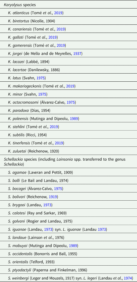
Altogether, 18 species of the genus Karyolysus have been described and named so far (Table 1). They have been considered to be parasites of Palearctic lizards, mainly reported from European countries such as Poland, Hungary, Romania, Slovakia and Spain (Álvarez-Calvo, Reference Álvarez-Calvo1975; Haklová-Kočíková et al., Reference Haklová-Kočíková, Hižňanová, Majláth, Račka, Harris, Földvári, Tryjanowski, Kokošová, Malčeková and Majláthová2014), but also from Scandinavia (Svahn, Reference Svahn1974, Reference Svahn1975) and Russia (Beyer and Sidorenko, Reference Beyer and Sidorenko1984). Newly, seven species have been found in the Canary Islands (Tomé et al., Reference Tomé, Pereira, Harris, Carretero and Perera2019). Nevertheless, reports from India (de Mello and de Meyrelles, Reference de Mello and de Meyrelles1937), Kenya (Mutinga and Dipeolu, Reference Mutinga and Dipeolu1989) and recently also from South Africa (Cook et al., Reference Cook, Netherlands and Smit2016) are changing this view. The prevailing vertebrate hosts are lacertid lizards of the genera Gallotia, Lacerta, Podarcis and Zootoca. However, records of Karyolysus from varanid, scincid and geckonid lizards (Mutinga and Dipeolu, Reference Mutinga and Dipeolu1989; Cook et al., Reference Cook, Netherlands and Smit2016; Tomé et al., Reference Tomé, Pereira, Harris, Carretero and Perera2019) have broadened its known host range. Furthermore, based on molecular surveys, there are many probably incorrectly assigned Hepatozoon isolates from lizards and snakes, which might resemble species of Karyolysus (Maia et al., Reference Maia, Harris and Perera2011; Tomé et al., Reference Tomé, Maia and Harris2012, Reference Tomé, Maia and Harris2013; Karadjian et al., Reference Karadjian, Chavatte and Landau2015). For the genus Schellackia, there are 11 described and named species parasitizing lizards worldwide. Eight species have been reported from the Old World, two from North America and one in Brazil (Table 1). Additionally, two species from South America are reported infecting frogs: Schellackia balli Le Bail and Landau, Reference Le Bail and Landau1974 from the Cane toad (Rhinella marina) and an unnamed Schellackia from a Brazilian tree-frog (Phrynohyas venulosa) (Le Bail and Landau, Reference Le Bail and Landau1974; Paperna and Lainson, Reference Paperna and Lainson1995). Furthermore, two Lainsonia Landau, Reference Landau1973 species (Lainsonia iguana Landau, Reference Landau1973 and Lainsonia legeri Landau et al., Reference Landau, Lainson, Boulard and Shaw1974), described also from Brazil (Landau, Reference Landau1973; Landau et al., Reference Landau, Lainson, Boulard and Shaw1974) are considered by some authors to be Schellackia (Levine, 1980; Upton, Reference Upton2000). Thus, according to their distribution data, the two genera, Karyolysus and Schellackia, potentially overlap over a large geographic area. Specifically, from the Iberian Peninsula, Schellackia bolivari Reichenow, Reference Reichenow1919, Schellackia bocagei Álvarez-Calvo, Reference Álvarez-Calvo1975, Karyolysus lacertae (Danilewsky, 1886) and Karyolysus octocromosomi Álvarez-Calvo, Reference Álvarez-Calvo1975, have been reported (Reichenow, Reference Reichenow1919; Álvarez-Calvo, Reference Álvarez-Calvo1975).
List of described and named Karyolysus and Schellackia species

Former descriptions of these parasites are based on the morphology of intraerythrocytic stages, often supplied with detailed life history (Reichenow, Reference Reichenow1919; Landau, Reference Landau1973; Álvarez-Calvo, Reference Álvarez-Calvo1975; Svahn, Reference Svahn1975; Mutinga and Dipeolu, Reference Mutinga and Dipeolu1989; Bristovetzky and Paperna, Reference Bristovetzky and Paperna1990; Finkelman and Paperna, Reference Finkelman and Paperna1998). Increasingly, traditional morphological approaches are being supported by molecular genetic tools, which clarify the phylogenetic position and correct taxonomic placement, for example, the polyphyly of the genus Hepatozoon Miller, 1908, and family Lankesterellidae, or the change of generic allocation of Karyolysus paradoxa (Dias, 1954) from Hepatozoon to Karyolysus (Megía-Palma et al., Reference Megía-Palma, Martínez and Merino2013, Reference Megía-Palma, Martínez and Merino2014, Reference Megía-Palma, Martínez, Cuervo, Belliure, Jiménez-Robles, Gomes, Cabido, Pausas, Fitze, Martín and Merino2018; Haklová-Kočíková et al., Reference Haklová-Kočíková, Hižňanová, Majláth, Račka, Harris, Földvári, Tryjanowski, Kokošová, Malčeková and Majláthová2014; Karadjian et al., Reference Karadjian, Chavatte and Landau2015; Cook et al., Reference Cook, Netherlands and Smit2016).
In a previous study, Stuart-Fox et al. (Reference Stuart-Fox, Godinho, de Bellocq, Irwin, Brito, Moussalli, Široký, Hugall and Baird2009) detected unspecified haemogregarines from blood smears of Lacerta schreiberi Bedriaga, 1878, from the Iberian Peninsula. Here, we work with material from that study to identify these haemoparasites by combining both microscopy and PCR-based methods. Since no blood other than the blood smears was available, we use engorged ticks collected from the lizards as an alternative source of host blood and also of haemoparasite DNA. Hence, a secondary aim of this study was to test the possibility of using this alternative non-invasive source of blood to genetically characterize the haemoparasites of these lizards.
Materials and methods
Sampling
Ticks were collected from L. schreiberi lizards in several localities in the Central System mountains of the Iberian Peninsula around the Portuguese–Spanish border during April and May 2006 in the framework of a previous study, and during an additional sampling season in May 2007 (see Stuart-Fox et al., Reference Stuart-Fox, Godinho, de Bellocq, Irwin, Brito, Moussalli, Široký, Hugall and Baird2009 and Table S1). A subset of the ticks found on the lizards was collected and preserved in 70% ethanol (see Kubelová et al., Reference Kubelová, Papoušek, Bělohlávek, De Bellocq, Baird and Široký2015). After clipping the tail tip, blood smears were made of a drop of blood (Stuart-Fox et al., Reference Stuart-Fox, Godinho, de Bellocq, Irwin, Brito, Moussalli, Široký, Hugall and Baird2009). Tick species and life-stage determination was done using available keys (Nosek and Sixl, Reference Nosek and Sixl1972; Estrada-Peña et al., Reference Estrada-Peña, Bouattour, Camicas and Walker2004). The ticks and blood smears used in the present study are from 48 lizards (see details in Table S1).
Morphological diagnosis of blood protists
All 48 blood smears were fixed in absolute methanol for 5 min, stained with Giemsa for 15 min and examined with an Olympus BX53 microscope using the 100× magnification lens equipped with immersion oil (Široký et al., Reference Široký, Kamler and Modrý2004). The intensity of parasitaemia was estimated for each infected lizard as the percentage of infected red blood cells found in approximately 104 cells (Široký et al., Reference Široký, Kamler, Frye, Fictum and Modrý2007). Images of parasites were captured using an Olympus DP 73 digital camera and QuickPhoto Micro 3.0 software. Basic morphometric characteristics were measured: maximum length and width of distinguished developmental stages of the parasite, and maximum length and width of nuclei for gamonts where possible; LW (length × width) and L/W (length/width) values were calculated. All measurements are given in micrometres (μm) as the mean followed by standard deviation and range in parentheses. Morphological identification of blood parasites was based on data from the literature (Álvarez-Calvo, Reference Álvarez-Calvo1975; Svahn, Reference Svahn1975; Telford, Reference Telford2009; Megía-Palma et al., Reference Megía-Palma, Martínez and Merino2013, Reference Megía-Palma, Martínez and Merino2014; Haklová-Kočíková et al., Reference Haklová-Kočíková, Hižňanová, Majláth, Račka, Harris, Földvári, Tryjanowski, Kokošová, Malčeková and Majláthová2014).
DNA extraction, amplification and sequencing
A total of 264 DNA samples of Ixodes ricinus (Linneaus, 1758) ticks were used. Each DNA sample represents a single nymph of I. ricinus. Nymphs were collected from 48 lizards with 1–23 (5.5 on average) nymphs per lizard. These samples correspond to a subset of the nymph DNA samples used in Kubelová et al. (Reference Kubelová, Papoušek, Bělohlávek, De Bellocq, Baird and Široký2015) and obtained by alkaline hydrolysis (Rijpkema et al., Reference Rijpkema, Golubić, Molkenboer, Verbeek-De Kruif and Schellekens1996), following the modified protocol as described in Kubelová et al. (Reference Kubelová, Tkadlec, Bednář, Roubalová and Široký2011).
For PCR detection of apicomplexan DNA, we used five pairs of primers targeting part of the 18S rDNA (for the primer sequences and PCR conditions, see Table S2). Primers EF/ER were designed for Eimeria spp. and they also amplify both haemogregarines and haemococcidia (Kvičerová et al. Reference Kvičerová, Pakandl and Hypša2008). To distinguish between haemogregarines and haemococcidia, and for diagnostic purposes, the other primers specific for haemococcidia were also used, which amplify shorter portions of the same gene (BT-F1/EimRodR, Hep600F1/Hep1600R, Sbol2F/EimRodR and BT-F1/SbolR) (Megía-Palma et al. Reference Megía-Palma, Martínez and Merino2013, Reference Megía-Palma, Martínez and Merino2014). All PCR reactions were carried out in 25 µl volumes, including 1 µl each of 10 µ m primer, 12.5 µl of Combi PPP Master Mix (Top-Bio, Czech Republic), 9.5 µl of water and 1 µl of extracted DNA. Morphologic determination of ticks was completed by molecular analysis because of the similar morphologies of preimaginal stages of ixodid ticks, especially between I. ricinus and the newly described species from the Mediterranean – Ixodes inopinatus (Estrada-Peña et al., Reference Estrada-Peña, Nava and Petney2014). This molecular determination was based on the part of the 16S rRNA gene (Table S2, Mangold et al. Reference Mangold, Bargues and Mas-Coma1998). PCR reactions for species diagnosis of ticks were carried out in a final volume of 23 µl including 0.25 µl each of 10 µ m primer, 12.5 µl of Combi PPP Master Mix, 9.5 µl of water and 0.5 µl of extracted DNA. Products were separated by electrophoresis on a 1.5% agarose gel with nucleic acid staining solution Midori Green (Elisabeth Pharmacon, Brno, Czech Republic) and visualized using a UV transilluminator. All parasite positive PCR products were cut off as gel slices and purified using a Gel/PCR DNA Fragments Extraction Kit (Geneaid Biotech, New Taipei City, Taiwan) and Sanger sequenced on an automatic 3730XL DNA analyser (Macrogen Inc., Amsterdam, the Netherlands).
Phylogenetic analyses
The sequences generated in this study were edited in Geneious 9.1.5 (Kearse et al., Reference Kearse, Moir, Wilson, Stones-Havas, Cheung, Sturrock, Buxton, Cooper, Markowitz, Duran, Thierer, Ashton, Meintjes and Drummond2012) and compared with the GenBank database (www.ncbi.nlm.nih.gov/genbank/) using the BLAST algorithm (blast.ncbi.nlm.nih.gov/Blast.cgi). The following analyses were carried out for haemogregarines and Schellackia-like parasites separately. Our sequences together with selected sequences available from GenBank (see Tables S3 and S4 for accession numbers) were aligned using the MUSCLE algorithm (Edgar, Reference Edgar2004). The final alignment of haemogregarines with a length of 1850 bp consisted of 45 sequences. The final alignment for haemococcidians with 1618 bp consisted of 24 sequences. A substitution model GTR + G + I was selected by the Smart model selection software (Lefort et al., Reference Lefort, Longueville and Gascuel2017) as the best model. Bayesian inference (BI) analysis was performed in Geneious 9.1.5 (Ronquist and Huelsenbeck, Reference Ronquist and Huelsenbeck2003) using Mr. Bayes 3.2.6 plugin with the GTR + G + I model for 107 generations, with trees and parameters sampled every 200 generations. The trees were summarized after a ‘burn-in’ of 25%. Maximum likelihood (ML) phylogenetic analysis was performed by PhyML 2.2.0 plugin in Geneious 9.1.5 (Guindon and Gascuel, Reference Guindon and Gascuel2003) with the GTR + G + I model and parameters estimated from the data. Nodal supports were generated with 1000 bootstrap replicates. Haemogregarina stepanowi Danilewsky, 1885, was set as an outgroup for the haemogregarines and Acroeimeria tropidura (Aquino-Shuster, Duszynski and Snell, 1990) for the haemococcidia. Trees were visualized using Tree-Graph 2.12.0 (Stöver and Müller, Reference Stöver and Müller2010) and graphically edited in Adobe Illustrator CS6.
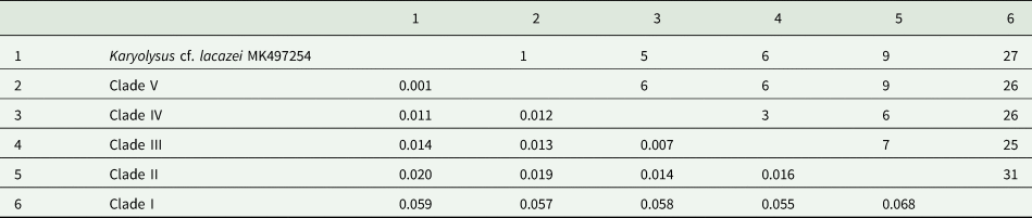
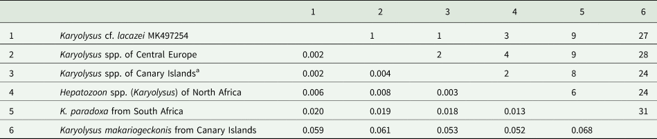
Genetic distances as p-distances and number of nucleotide differences were calculated in Mega 5.0 (Tamura et al., Reference Tamura, Peterson, Peterson, Stecher, Nei and Kumar2011). Our haemogregarine sequence was compared with the groups corresponding to clades obtained in the phylogenetic analyses (Table 2) and then to groups created within the Karyolysus clade on the base of the phylogeny (Table 3) and their geographical origin (Table 4). Pairwise distances between our haemogregarine sequence and other sequences from Karyolysus clade were also calculated (Table S5).
p-distances (lower-left) and number of nucleotide differences (upper-right), between our Karyolysus cf. lacazei sequence and haemogregarine sequences in three groups composed according to phylogenetic tree in Fig. 2

The number of base differences per site from estimation of net average between groups of sequences in the 455 bp long fragment are shown. All positions containing gaps and missing data were eliminated.
p-distances (lower-left) and number of nucleotide differences (upper-right) between our Karyolysus cf. lacazei sequence and clades I–V in the Karyolysus clade

The number of base differences per site from estimation of net average between groups of sequences in the 455 bp long fragment are shown. All positions containing gaps and missing data were eliminated.
p-distances (lower-left) and number of nucleotide differences (upper-right) between our Karyolysus cf. lacazei sequence and four geographic groups from Karyolysus clade sequences

K. makariogeckonis is separated from Canary Island group due to its significant genetic difference. The number of base differences per site from estimation of net average between groups of sequences in the 455 bp long fragment are shown. All positions containing gaps and missing data were eliminated.
a Without K. makariogeckonis MG787251.
Results
Parasite identification
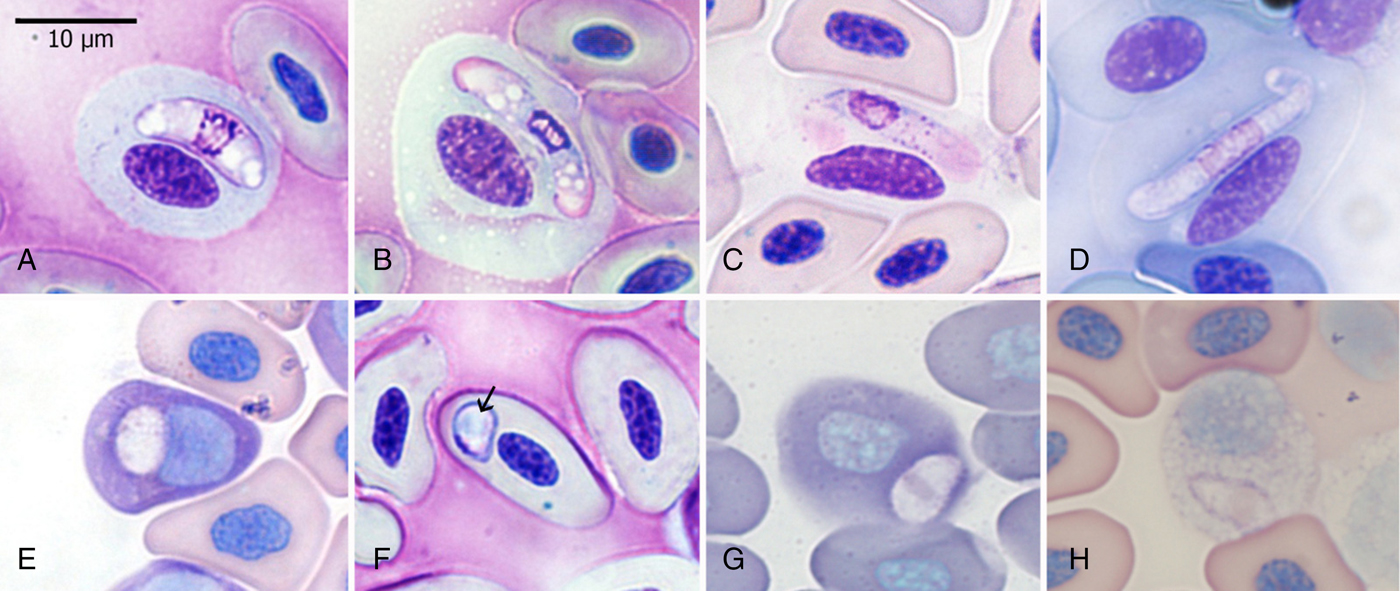
Altogether, 31 out of the 48 examined lizards (prevalence 64.6%) were diagnosed to be infected by blood protists. Two types of parasites were observed: trophozoites and gamonts similar to those previously described as Karyolysus lacazei (Labbé, 1894) in 24 smears (50%; Fig. 1A–D); and Schellackia-like sporozoites in 20 blood smears (41.7%; Fig. 1E–H). Double infection by both parasites occurred in 13 cases (27.1%). Parasitaemia of infected individuals ranged between 0.02 and 4.24% (mean 0.62%) for Karyolysus cf. lacazei; and between 0.01 and 0.12% (mean 0.04%) for Schellackia. Parasites were localized in red blood cells or occasionally seen freely in plasma. We also found objects similar to the sporozoites of Schellackia, rarely found in leucocytes (Fig. 1H).
Haemoparasites of L. schreiberi. K. cf. lacazei: trophozoites (A, B) and gamonts (C, D). Sporozoites of Schellackia sp. (E–F). The arrow indicates refractile body.

Two developmental stages of Karyolysus were distinguished. Trophozoites showed a variety of shapes, from reniform (Fig. 1A) to oval or slender (Fig. 1B). Pale blue or pinkish stained cytoplasm was markedly vacuolated. A granular or reticular nucleus spread across the whole width of the parasite was placed centrally. Trophozoites measured 14.6 ± 1.6 (12–18) × 3.9 ± 1.0 (3–6) (n = 35). Gamonts (Fig. 1C, D) appeared uniform with an elongate slender shape, and one end recurved as described by Reichenow (Reference Reichenow1919), or bent according to Svahn (Reference Svahn1975); usually, they were placed parallel to the host cell nucleus. Cytoplasm stained pale blue and no vacuoles were present. The nuclei of gamonts were elongated and filled the entire width of the parasite. Although sometimes clearly demarcated, they were usually diffuse making their exact measurements impossible. Nuclei were located in the centre, or more often shifted closer to the bend. Macro- and micro-gamonts were indistinguishable. Gamonts measured: 20.3 ± 1.6 (17–26) × 3.1 ± 0.4 (2–4) (n = 118), LW = 63.3 ± 9.2 (38–96) μm2, L/W = 6.6 ± 0.9 (4.3–10.5). Nuclei measured: 5.8 ± 1.0 (4–8) × 3.3 ± 0.3 (3–4) (n = 32), LW = 17.9 ± 3.2 (12–24) μm2, L/W = 1.9 ± 0.3 (1.3–2.7).
Gamonts and trophozoites had an evident impact on the host cell (Fig. 1A-D). Hypertrophy and loss of the staining properties of cytoplasm and nucleus appeared in all infected cells. In some cases, erythrocyte cytoplasm was completely absent. The host cell nucleus stained paler in infected than uninfected cells, and was swollen and displaced laterally.
The shape of Schellackia sporozoites ranged from round (in 11% of cases) and oval (23%) (Fig. 1E) to broadly pyriform (66%) (Fig. 1F–H). Cytoplasm stained light pinkish. A single round refractile body with diameter 2 µm located in the centre of the sporozoite was clearly seen in only one case (Fig. 1F). The nuclei showed variable forms and placements: narrow bands touching the parasite membrane (Fig. 1F), a band-like nucleus usually situated centrally (Fig. 1G, H); a couple of chromatin granules, or diffuse areas of chromatin (Fig. 1E). Sporozoites were situated in a polar or lateropolar position within erythrocytes and under light microscopy generated no visible changes to the infected cells. Sporozoites measured 5.89 ± 0.70 (5.0–7.0) × 3.49 ± 0.52 (3.0–5.0) (n = 81).
All ticks were identified by microscopic examination as nymphs of I. ricinus. All analysed ticks showed 100% molecular identity with I. ricinus.
Phylogenetic analyses
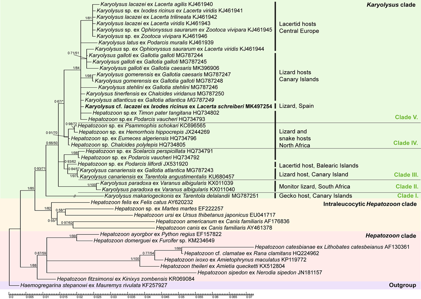
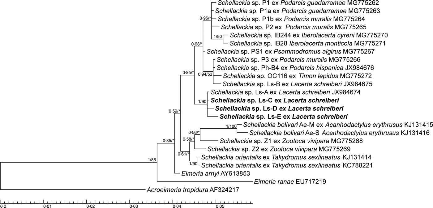
We obtained 24 18S rDNA sequences: four sequences (929–1442 bp), all identical, belonged to haemogregarines, and 20 sequences corresponding to four haplotypes belonging to haemococcidia (926–1497 bp). For the phylogenetic analysis, our longest haemogregarine sequence (1442 bp) and three new haplotypes of haemococcidia (1025 bp for Ls-E, 1006 bp for Ls-D and 1497 bp for Ls-C) were used (see Tables S3 and S4 for accession numbers). Both BI and ML analyses provided phylogenies with identical topologies. The phylogenetic tree for haemogregarines is shown in Fig. 2 and the one for Schellackia-like sequences in Fig. 3.
Phylogenetic tree of Karyolysus and related haemogregarines based on Bayesian inference (BI) analyses of 18S rDNA. Numbers at the nodes show posterior probabilities under BI/bootstrap values for maximum likelihood (ML) higher than 0.50 or 50%, respectively. Nodal supports (BI/ML) lower than 0.50 or 50% are marked with an asterisk (*). Branch with K. makariogeckonis is shortened in half. New sequence obtained in this study is marked in bold.

Phylogenetic tree of Schellackia sp. based on Bayesian inference (BI) analyses of 18S rDNA. Numbers at the nodes show posterior probabilities under BI/bootstrap values for maximum likelihood (ML) higher than 0.50 or 50%, respectively. Nodal supports (BI/ML) lower than 0.50 or 50% are marked with an asterisk (*). The three new Schellackia haplotypes (Ls-C, Ls-D, Ls-E) are marked in bold.

Our haemogregarine sequence (MK497254) clustered together with known Karyolysus species from lacertid, geckonid and varanid hosts and Hepatozoon species from snakes (Colubridae, Lamprophiidae), skinks (Scincidae), lizards (Lacertidae) and geckos (Phyllodactylidae) (Fig. 2). Genetic distances are shown in Tables 2, 3, 4 and Table S5. From three groups of sequences according to the clades in the haemogregarine phylogenetic tree (Karyolysus clade, intraleucocytic Hepatozoon clade and Hepatozoon clade) all of our haemogregarine sequences were most closely related to the Karyolysus clade with a p-distance of 0.002, whereas the genetic distance to the intraleucocytic Hepatozoon species was 0.020 and to other Hepatozoon species 0.030 (Table 2). Five main clades (clades I–V in Fig. 2) are in the Karyolysus clade: I. K. makariogeckonis; II. K. paradoxa; III. K. canariensis and haemogregarines from other lacertids; IV. haemogregarines from North African snakes and skinks; V. haemogregarines from lizards including the rest of the Canarian Karyolysus species and the sequence identified in this study (for their p-distances see Table 3). Three species in the fifth clade, K. lacazei, K. galloti and K. gomerensis, formed separated clusters while other sequences remained in the polytomy. Within the Karyolysus clade, our sequence was most closely related to the Karyolysus species of Central Europe and Canary Islands (p-distance 0.002 for both) (Table 4). According to the pairwise distances, our haemogregarine was most similar to Karyolysus atlanticus (MG787249) from the Canary Islands and Karyolysus sp. (KJ461944) from Central Europe (Hungary) (Table S5).
Three out of 20 obtained sequences of Schellackia were identical to haplotype Ls-A (JX984674) previously found in L. schreiberi (Megía-Palma et al., Reference Megía-Palma, Martínez and Merino2013). Three other new haplotypes, Ls-C (MK512382), Ls-D (MK512383) and Ls-E (MK512384), formed a monophyletic cluster with this Schellackia sp. haplotype Ls-A (Fig. 3). All Schellackia sequences form a monophyletic clade but with low support with Eimeria arnyi (AY613853) from the ringneck snake and E. ranae (EU717219) from terrestrial frogs at basal positions. Our new haplotypes came from 17 ticks collected from 9 individual lizards. Haplotype Ls-E was the most common (9/17), followed by haplotype Ls-C (7/17), while haplotype Ls-D occurred only in one tick. One lizard was infested by ticks containing all four haplotypes; two lizards were infested by ticks containing two haplotypes, Ls-C and Ls-E, and another one with Ls-A and Ls-E. Each tick contained only one haplotype.
Usability of engorged ticks as an alternative source of haemoparasite DNA
Primer pair EF/ER confirms positive microscopic findings in 71% of cases, but notably, the primer pair also diagnoses infection in 41% of microscopically negative samples. Primers Sbol2F/EimRodR confirm microscopic findings of haemococcidia in 61% of cases; another 15% were discovered in specimens microscopically haemococcidia negative. Hence, primers EF/ER had a sensitivity of 71%, a specificity of 59% and an accuracy of 67%, whereas primers Sbol2F/EimRodR had a sensitivity of 61%, a specificity of 85% and an accuracy of 71% (Fig. S1).
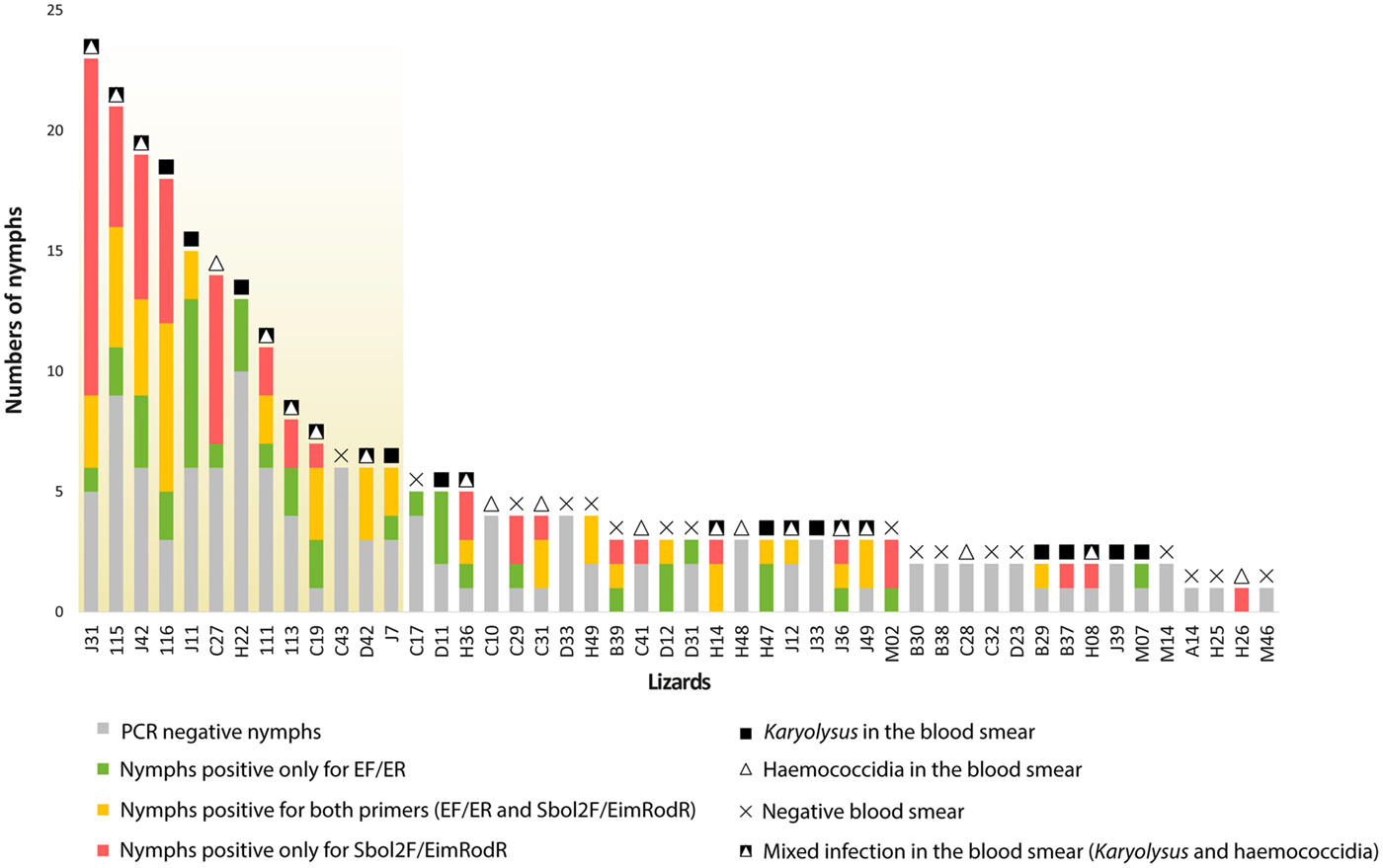
The correlation between PCR detection of haemoparasites from engorged nymphs and microscopic findings is high for lizards with six and more collected nymphs (6–23 nymphs). Three exceptions included lizards that were PCR positive for Schellackia, but negative for haemococcidia in microscopy. Results were markedly contrasted in lizards carrying 1–5 nymphs. Among 35 lizards, for 18 lizards their PCR and microscopy findings correlated fully (microscopy and PCR for both haemogregarine and haemococcidia), for four lizards only partially, whereas for the remaining 13 lizards they did not correlate. Negative microscopy with positive PCR results accounted for most of the miscorrelations (8/13 lizards). The remaining five lizards were microscopically positive with negative results from PCR analyses. For a comparison of the microscopy and PCR based analyses for each particular lizard see Fig. 4.
Summary view on PCR and microscopic results of individual lizards. Lizards are ordered in descending number of collected nymphs. Symbols above columns refer to microscopic findings. Correlating results between PCR detection of haemoparasites from engorged nymphs and microscopic findings on blood smears in the lizards with six and more collected nymphs are marked with a yellowish background.

Discussion
We detected two types of apicomplexan parasites, the genera Karyolysus and Schellackia, infecting the Iberian lizard L. schreiberi using a combination of microscopy and PCR based methods. The genetic diversity of Schellackia was higher than that of Karyolysus, with four haplotypes detected in our tick samples vs a single haplotype, respectively, although haemogregarines were more prevalent.
Morphological uniformity of reptilian blood apicomplexans makes their microscopic differentiation on the generic level difficult. Recently, the combination of microscopy with the use of molecular genetic markers has become standard in species diagnosis and taxonomy of apicomplexans. Nevertheless, even such an approach cannot avoid all risk of error, such as mismatches between the results of microscopy and molecular genetic data from mixed infections. Such mismatch can occur, for example, when one of the co-infecting parasitic species dominates on blood smears, whereas PCR based diagnosis can be more sensitive to the other one, e.g. due to the use of better matching primers. Also, the use of engorged nymphs itself as the source of host blood brings uncertainty into the results. The use of I. ricinus, a species of three-host tick, introduces the possibility that blood parasites from a previous host become included in the analysis. Thus far, ticks have not been reported as part of the Karyolysus life cycle. Gamonts and sporokinetes have been found in mites (Svahn, Reference Svahn1975; Haklová-Kočíková et al., Reference Haklová-Kočíková, Hižňanová, Majláth, Račka, Harris, Földvári, Tryjanowski, Kokošová, Malčeková and Majláthová2014), while they have not been observed in tick squashes (Haklová-Kočíková et al., Reference Haklová-Kočíková, Hižňanová, Majláth, Račka, Harris, Földvári, Tryjanowski, Kokošová, Malčeková and Majláthová2014; Cook et al., Reference Cook, Netherlands and Smit2016). For haemococcidia, haematophagous invertebrates including ticks are merely passive vectors. Sporozoites accumulate in the wall of the digestive tract of invertebrates but there is no development (Telford, Reference Telford2009). We are not aware of transstadial or transovarial transmission of these haemoparasites in I. ricinus having been recorded. Based on the available information, the infection of the ectoparasites before blood feeding seems to be unlikely. Thus, we assume that the examined ticks were merely acting as paratenic or temporary transport hosts for Schellackia and Karyolysus respectively by feeding on the sampled lizards. Nevertheless, haematophagous ectoparasites can be used as a valuable non-invasive source of host and/or haemoparasite genetic information (Mathew et al., Reference Mathew, Van Den Bussche, Ewing, Malayer, Latha and Panciera2000; Sloboda et al., Reference Sloboda, Kamler, Bulantová, Votýpka and Modrý2007; Vilcins et al., Reference Vilcins, Ujvari, Old and Deane2009; Bataille et al., Reference Bataille, Fournié, Cruz, Cedeño, Parker, Cunningham and Goodman2012; Harris et al., Reference Harris, Graciá, Jorge, Maia, Perera, Carretero and Giménez2013; Haklová-Kočíková et al., Reference Haklová-Kočíková, Hižňanová, Majláth, Račka, Harris, Földvári, Tryjanowski, Kokošová, Malčeková and Majláthová2014; Hamšíková et al., Reference Hamšíková, Silaghi, Rudolf, Venclíková, Mahríková, Slovák, Mendel, Blažejová, Berthová, Kocianová, Hubálek, Schnittger and Kazimírová2016). This method may be suitable especially for small-sized hosts where manipulation and blood collection can be difficult, particularly in threatened or endangered species.
The application of engorged nymphs as the source of host/parasite DNA is possible, but its reliability remains uncertain. Our dataset was unbalanced regarding nymph collection (1–23 nymphs per lizard, mean 5.5, s.d. 5.4). The used primers possess various specificity and sensitivity, and amplicons of different length. Individual lizards were set as PCR positive if carrying at least one positive nymph. These factors may all have distorted the results provided in Fig. 4, and Fig. S1, where correlations between lizards, PCR results, and microscopy are shown. Nevertheless, the values of sensitivity, specificity and accuracy according to Fig. S1 indicate our method to be suitable. According to this study, six and more collected nymphs per lizard could provide reliable PCR analysis comparing with microscopic findings. PCR detection is generally more sensitive than microscopy. Nevertheless, the volumes of blood meal of nymphs are small and depend on feeding time. Thus the rate of nymphs' engorgement also influences the results and should be adjusted for. This might be so in the case of microscopically positive, but PCR negative lizards in our study.
The observed morphology of available life stages of haemogregarines was similar or identical to those of K. lacazei (Svahn, Reference Svahn1975; Haklová-Kočíková et al., Reference Haklová-Kočíková, Hižňanová, Majláth, Račka, Harris, Földvári, Tryjanowski, Kokošová, Malčeková and Majláthová2014). Both species reported from Spain, K. octocromosomi and K. lacertae, differ from our haemogregarine in morphology (see Table S6 for comparison of Karyolysus species). The phylogenetic analysis places our sequence within a clade containing ten morphologically described and named species of Karyolysus (see Fig. 2) along with Hepatozoon isolates lacking morphological descriptions. These Hepatozoon isolates originate mostly from lacertid hosts. However, a few sequences are from other squamates from the Mediterranean region and North Africa (snakes and skinks), and are clearly separated from the clade of intraerythrocytic Hepatozoon from amphibians and reptiles. Hepatozoon isolates in the Karyolysus clade are obviously wrongly assigned members of the genus Karyolysus (Fig. 2). This statement is in concordance with generic transfers of K. paradoxa and K. canariensis on the basis of both molecular and morphological traits. According to phylogeny and nomenclature development on the generic level, including generic allocations of haemogregarines discussed thoroughly during the last decade (Maia et al., Reference Maia, Perera and Harris2012; Haklová-Kočíková et al., Reference Haklová-Kočíková, Hižňanová, Majláth, Račka, Harris, Földvári, Tryjanowski, Kokošová, Malčeková and Majláthová2014; Kvičerová et al., Reference Kvičerová, Hypša, Dvořáková, Mikulíček, Jandzik, Gardner, Javanbakht, Tiar and Široký2014; Karadjian et al., Reference Karadjian, Chavatte and Landau2015; Cook et al., Reference Cook, Netherlands and Smit2016), we consider the placement of our isolate into the genus Karyolysus to be correct. The alignment of 18S rDNA did not solve the phylogeny of our Karyolysus isolate, which together with other available sequences of different species of Karyolysus form a poorly resolved clade with considerable polytomy. Genetic distances revealed that Karyolysus species from Central Europe (Karyolysus sp. from Hungary) and K. atlanticus from the Canary Islands are the most closely related to our isolate, having almost equal p-distance values. Overall, genetic distances based on available data for Karyolysus clade are low and these organisms are genetically very similar regardless of their geographic origin.
Along with molecular data, the morphology and biology of the parasites may help their diagnosis. Karyolysus gamonts cause typical destruction of the host cell with some exceptions (Telford, Reference Telford2009); e.g. Karyolysus minor Svahn, Reference Svahn1975, does not seem to have any effects on host cells (Svahn, Reference Svahn1975). Haklová-Kočíková et al. (Reference Haklová-Kočíková, Hižňanová, Majláth, Račka, Harris, Földvári, Tryjanowski, Kokošová, Malčeková and Majláthová2014) considered that the Karyolysus clade includes both forms – with and without an effect on the host cell. Maia et al. (Reference Maia, Perera and Harris2012) did not provide morphological descriptions; however, in the presented figures typical changes of host cells are visible. Other studies (Maia et al., Reference Maia, Harris and Perera2011; Tomé et al., Reference Tomé, Maia and Harris2012, Reference Tomé, Maia and Harris2013, Reference Tomé, Rato, Perera and Harris2016; Damas-Moreira et al., Reference Damas-Moreira, Harris, Rosado, Tavares, Maia, Salvi and Perera2014) provided no information about morphology. However, morphological changes of host cells analogous to those induced by Karyolysus have been reported in Hepatozoon theileri (Laveran, 1905) from frog hosts that are grouped within the Hepatozoon clade (Fig. 2) (Netherlands et al., Reference Netherlands, Cook, Smit and du Preez2014; Conradie et al., Reference Conradie, Cook, du Preez, Jordaan and Netherlands2017). Hepatozoon curvirostris (Billet, Reference Billet1904) from the lacertid host Timon pater (Lataste, 1880) also causes karyolysis, but an early study of this by Billet (Reference Billet1904) naturally lacks molecular data. The effect on host cells is not always uniform among species. For example, various levels of erythrocyte distortion were observed between seven Canarian species of Karyolysus (Tomé et al., Reference Tomé, Pereira, Harris, Carretero and Perera2019). The recently redescribed K. paradoxa (Cook et al., Reference Cook, Netherlands and Smit2016) shows specific effects on host cells such as the shrinkage of erythrocytes and vacuolization of its nucleus, different from the effects of other species of Karyolysus. Additionally, the hypertrophy of the host cell observed with light microscopy may look different under electron microscopy. While for Hepatozoon ixoxo Netherlands et al., Reference Netherlands, Cook, Smit and du Preez2014, hypertrophy was confirmed, H. theileri can be observed causing extensive flattening of the host cell, with small excrescences covering the surface of the infected erythrocyte; this is not observable at the light microscopy level (Conradie et al., Reference Conradie, Cook, du Preez, Jordaan and Netherlands2017). According to the morphology of available blood stages, phylogenetic analysis and the distortion effect on host cells, we refer to this haemogregarine as Karyolysus cf. lacazei. Our finding also represents the first detection of Karyolysus in L. schreiberi.
It seems that the Schellackia-like parasites show some genetic variability within the 18S rRNA. A recent study by Megía-Palma et al. (Reference Megía-Palma, Martínez, Cuervo, Belliure, Jiménez-Robles, Gomes, Cabido, Pausas, Fitze, Martín and Merino2018) found a high haplotype diversity for Schellackia in lacertid lizards with 11 new haplotypes, which mostly showed high host-specificity. To date, two Schellackia haplotypes have been found in L. schreiberi and have been shown to be host specific (Megía-Palma et al., Reference Megía-Palma, Martínez and Merino2013, Reference Megía-Palma, Martínez, Cuervo, Belliure, Jiménez-Robles, Gomes, Cabido, Pausas, Fitze, Martín and Merino2018). We obtained four Schellackia haplotypes, one identical to haplotype Ls-A, the first genetically characterized Schellackia species (Megía-Palma et al., Reference Megía-Palma, Martínez and Merino2013), and three new haplotypes closely related to haplotype Ls-A. Haplotype Ls-A was previously found in the same host species in Segovia (Spain) 300 km away from our sampling area. The morphology of our isolates is very similar to the description of Schellackia haplotype Ls-A (Megía-Palma et al., Reference Megía-Palma, Martínez and Merino2013). Additionally, another Schellackia sp. with similar morphology but genetically different, haplotype Ls-B, was found in L. schreiberi in the same locality as haplotype Ls-A (Megía-Palma et al., Reference Megía-Palma, Martínez and Merino2013). Within named Schellackia species, two were described from the Iberian Peninsula – S. bocagei and S. bolivari (the type species of the genus). However, both differ from our isolates in morphological traits, and S. bolivari also by the available sequence data (Álvarez-Calvo, Reference Álvarez-Calvo1975; Reichenow, Reference Reichenow1919; Megía-Palma et al., Reference Megía-Palma, Martínez and Merino2014). We observed no correlation between the shape of sporozoites and the haplotypes. The variety of shapes of sporozoites was seen in each of the positive blood smears. Also, various haplotypes were found in several ticks collected from one lizard. Phylogenetic analysis placed our isolates among a group of unnamed Schellackia (Fig. 3), and available morphological traits obtained from blood smears were insufficient to make thorough comparisons with already described species and/or a proper description of new species, respectively.
Proper identification of the haemoprotozoa requires a complex approach. Molecular tools, parasite morphology and its effect on the host cell, life cycle patterns, host associations and geographic distribution should be used together to reach the most accurate result. Even applied molecular genetic methods are not always specific enough for species determination. The 18S rDNA is handy for comparison with data from public databases but offers insufficient information for studies on the phylogeny of lower taxa, namely species. This situation may improve with the application of a multigene approach.
Supplementary material
The supplementary material for this article can be found at https://doi.org/10.1017/S0031182019001112.
Acknowledgements
The sampling of L. schreiberi and their ectoparasites was organized by S.J.E. Baird and funded by INRA, France through the CBGP and by the Fundacão para a Ciência e Tecnologia (FCT, Portugal). We thank the Malcata Nature Reserve for providing access to their field station. We thank H. Freitas and R. Vasconcelos for their assistance in the field, and A. Moussalli, D. Stuart Fox, A. F. Hugall, J. C. Brito, R. Godinho and N. R. Irvin for help both in the field and in processing lizard ectoparasites. We are grateful to the Instituto de Conservacão da Natureza in Portugal and Junta de Castilla y León in Spain for permits and support. The authors thank Christopher Steer for language correction and three anonymous reviewers for their effort as well.
Financial support
The sampling of L. schreiberi and their ectoparasites was funded by INRA, France through the CBGP and by the Fundacão para a Ciência e Tecnologia (FCT, Portugal). Pavel Široký has been financially supported by the Ministry of Education, Youth, and Sports of the Czech Republic under the project CEITEC 2020 (LQ1601).
Conflict of interest
None.
Ethical standards
Not applicable.