




No CrossRef data available.
Article contents
Tetratrichomonads from the oral cavity and respiratory tract of humans
Published online by Cambridge University Press: 10 June 2005
Abstract
To clarify the taxonomy of trichomonads associated with human respiratory diseases, we examined a collection of axenic trichomonad strains isolated from the oral cavity and bronchi of patients from pulmonary diseases clinics in Tallin, Estonia. The oral and bronchial strains were compared mutually as well as with a reference strain of Trichomonas tenax, a common inhabitant of the human oral cavity, and other trichomonad species from humans and animals. Unexpectedly, the morphological studies, as well as DNA sequencing of ITS1-5.8S rRNA-ITS2 regions revealed that the Estonian strains belong to the genus Tetratrichomonas, with a high similarity to the avian species Tetratrichomonas gallinarum. None of the strains belonged to Trichomonas tenax. DNA fingerprinting using the RAPD method separated Estonian strains into 2 distinct groups: ‘bronchial’ consisting of 5 and 2 strains isolated from bronchi and ‘oral’ cavity, respectively, and oral consisting of 3 oral strains. Consistent differences between ‘bronchial’ and ‘oral’ groups were confirmed by analysis of ITS1-5.8S rRNA-ITS2 sequences. Our results have revealed novel trichomonad species of the human oral cavity and bronchi.
Keywords
Information
- Type
- Research Article
- Information
- Copyright
- © 2005 Cambridge University Press
INTRODUCTION
The common parabasalid flagellate Trichomonas tenax frequently inhabits the oral cavity of humans. It has a world-wide distribution with high prevalence in persons with periodontal diseases (reviewed by Honigberg and Burgess, 1994), although the contribution of T. tenax to pathological changes in the periodontium is debated (Feki et al. 1981; Honigberg, 1990). More importantly, a number of cases of pulmonary trichomoniasis ascribed to T. tenax have been reported (reviewed by Hersh, 1985 and Honigberg, 1990). Besides broncho-pulmonary exudates the trichomonads have also been found in pleural fluid of predisposed patients (Memik, 1968; Walzer, Rutherford and East, 1978; Osborne, Giltman and Uthman, 1984; Ohkura, Suzuki and Hashiguchi, 1985; Shiota et al. 1998; Porcheret et al. 2002; Lewis et al. 2003), submaxillary gland (Duboucher, Mogenet and Perie, 1995), and infra-auricular lymph node (Duboucher et al. 2000).
The generally accepted view is that trichomonads occurring in the oral cavity and the respiratory tract of humans are T. tenax (Honigberg and Burgess, 1994). However, some authors considered the trichomonads found in the respiratory tract to be independent species and named them on the basis of their location Trichomonas pulmonalis (Schmidt, 1895) or Trichomonas broncho-pulmonalis (Kazakova, Roigas and Teras, 1977). In addition, reports have been published on symptomatic infections of respiratory passages and oral mucosa by T. vaginalis in adults (Rebhun, 1964; Terezhalmy, 1983) including a case of an immunocompromised patient infected with HIV (Duboucher et al. 2003), or newborns infected during delivery by mothers with trichomonad vaginitis (McLaren et al. 1983; Hiemstra, Van Bel and Berger, 1984). Although a method for specific detection of T. tenax by polymerase chain reaction is available (Kikuta et al. 1997), most reports of the identification of parasites, if attempted at all, have relied on microscopic examination using unreliable methods for dependable classification of trichomonad species.
Trichomonas tenax infections in patients with respiratory diseases were extensively studied in Estonia. In the prospective study by Tumka (1956), trichomonads were found in 19 of 111 patients with chronic pneumonia. Kazakova et al. (1980) reported T. tenax in the bronchi of 37 out of 370 patients with chronic pneumonia and bronchitis. Sardis et al. (1983) found trichomonads in the bronchi of 16 out of 30 patients with various pulmonary diseases. These investigators successfully established several axenic isolates of oral trichomonads from asymptomatic patients (Teras et al. 1970; Teras, Kumm and Kallas, 1972), as well as from bronchi, sputum and oral cavity of patients suffering from different chronic pulmonary diseases (Kazakova et al. 1980, 1985). These isolates were used in comparative serological studies involving both sera of patients with pulmonary trichomoniasis (Roigas et al. 1980; Kazakova et al. 1985) and polyclonal rabbit antibodies raised against representative isolates. The results obtained with the aid of complement fixation reaction, immunofluorescence, and agglutination revealed distinct serotypes among these organisms and showed considerable differences between the strains isolated from the bronchi and those from the oral cavity. Interestingly, antigenic differences were demonstrated even between bronchial and oral isolates obtained from a single patient. On the basis of these results the authors suggested that bronchial and oral trichomonads belonging to different serotypes may represent different subspecies (Kazakova et al. 1980, 1985) if not species (Kazakova et al. 1977).
In the present study, the oral and bronchial strains of human trichomonads isolated and axenized in Estonia by Teras and colleagues (1980) were compared mutually as well as with a T. tenax reference strain and other trichomonad species from humans and animals with the aim being to clarify the taxonomy of trichomonads associated with human respiratory diseases. Surprisingly, genetic analysis as well as morphological observations revealed that the archival Estonian strains, of both oral and bronchial origin, are distinct from T. tenax and belong to the genus Tetratrichomonas that has not previously been reported as a human parasite.
MATERIALS AND METHODS
Organisms and cultivation
The Estonian strains were isolated from patients suffering from different chronic pulmonary diseases at the State Antituberculosis Dispensary and the Pulmonary Disease Department, Institute of Experimental and Clinical Medicine, Tallin, Estonia between 1964 and 1977 (Teras et al. 1980) (Table 1). The strains TXB1–TXB5 were isolated by means of bronchoscopy, and strains TXO6–TXO10 were isolated from oral cavity or sputum. Samples containing trichomonads were initially inoculated to Locke-egg-serum double phase medium LSM-2 (Wantland, Wantland and Winquist, 1963) without antibiotics, and maintained at 37 °C. The cultures were axenized using TT1 medium (Teras et al. 1970) as described by Kazakova et al. (1985). After the transfer to the Prague laboratory, the strains were adapted to the TYM medium (Clark and Diamond, 2002), cryopreserved and stored in liquid nitrogen. Other trichomonad strains (Table 1) were grown in axenic culture except for Trichomonas canistomae, Tetratrichomonas limacis and Tetratrichomonas prowazeki that were cultivated xenically. The axenic strains were grown in TYM medium (Clark and Diamond, 2002) at pH 6·2 (Trichomonas vaginalis), or pH 7·2 (all other strains) at 37 °C. The TYSGM medium (Clark and Diamond, 2002) without mucin and with the addition of rice starch was used for xenic cultures that were maintained at 37 °C (T. canistomae) or 26 °C (T. limacis, and T. prowazeki). These strains are available at the Department of Parasitology, Faculty of Science, Charles University in Prague.

Light microscopy
Moist films of trichomonads were spread onto cover-slips that were fixed in Hollande's fluid and stained with protargol as described previously (Tachezy et al. 2002).
DNA isolation
Genomic DNA was isolated from trichomonads using the method of guanidium thiocyanate or guanidium chloride extraction (Pramanick, Forstová and Pivec, 1976; Tachezy et al. 2002). DNA isolated from strain numbers 12–15 and 18 (see Table 1) was kindly provided by Vladimír Hampl (Charles University, Prague).
Random amplified polymorphic DNA (RAPD) analysis
Random oligonucleotides (10-mers) (RAPD Primer set #2, http://www.michaelsmith.ucb.ca/services/NAPSPrimer_Sets/Primers.pdf) were obtained from the University of British Columbia, Vancouver, Canada. Each primer (UBC157, UBC173, UBC174, UBC 175, UBC177, UBC178, UBC188, UBC190, UBC194) provided distinct patterns of amplified DNA fragments. DNA amplifications were performed in PTC-200 thermocycler (MJ Research) using the following parameters: initial denaturation at 94 °C, 5 min; 35 cycles of 40 s at 92 °C, 40 s at 32 °C, and 90 s at 72 °C; 9 min. final step at 72 °C. Reaction mixtures consisted of 50 ng DNA, 10 pmol of primer, 5 mM MgCl2, 400 μM of each of dNTPs, PCR reaction buffer, and 5 units of Taq Polymerase (Promega) in a total volume of 50 μl. PCR products were separated in a 2% (w/v) agarose gel at 80 V for 6 h. Electrophoretic patterns were scored according to Tachezy et al. (2002). FreeTree program (Hampl, Pavlicek and Flegr, 2001) was used for construction of trees and for computation of bootstrap values. Genetic distances of samples were computed from Nei-Li's coefficient of similarity (Nei and Li, 1979) and the tree was constructed by the neighbor-joining method (Saitou and Nei, 1987). Bootstrap values were computed for every node of the dendrogram based on 1000 resamplings.
Amplification and cloning of internal transcribed spacer (ITS) regions and 5.8S ribosomal RNA genes
Primers and conditions for DNA amplification of the ITS1-5.8S rRNA-ITS2 region were adapted from Felleisen (1997). DNA was amplified using primers complementary to conserved regions near to the 3′-end of the small subunit of the ribosomal RNA gene (ITSR: 5′-TTC AGT TCA GCG GGT CTT CC), and at the 5′-end of the large subunit of the ribosomal RNA gene (ITSF: 5′-GTA GGT GAA CCT GCC GTT GG). The PCR products of 370–500 bp were separated in a 2% (w/v) agarose gel, extracted from the gel by means of a QIAquick Gel Extraction Kit (Qiagen, Hilden, Germany), and subcloned using an Original TA Cloning Kit (Invitrogen, Carlsbad, CA, USA). Selected clones were sequenced in both directions using T3/T7α and M13 primers. ITS regions of T. gallinarum strain numbers 20–22 were sequenced directly from PCR products using the ITSF and ITSR primers. The sequences of strain numbers 3, 5–9, 20–25, were submitted to GenBank (National Center of Biotechnology Information) under accession numbers: Estonian strains – AY244641 (TXB3), AY244642 (TXB5), AY244644 (TXO6), AY244645 (TXO7), AY244646 (TXO8), AY244643 (TXO9); Tetratrichomonas gallinarum – AY244647 (ZPA2), AY244648 (Z26/7), AY244649 (1-11-M2); Tetratrichomonas prowazeki AY244650; Tetratrichomonas limacis AY244651; and Trichomonas canistomae AY244652.
Sequence analysis
Trichomonad sequences of the ITS1-5.8S rRNA-ITS2 regions were obtained directly in this study or from existing sequences in GenBank (Tetratrichomonas sp. DS, AF236105; Tetratrichomonas 1998-5003, AF340154; Tetratrichomonas 1999-5000, AF342742; Tetratrichomonas 2000-0017, AF342740; Tetratrichomonas tenax, U86615; Trichomonas canistomae, AY244652; Trichomonas gallinae, U86614; Trichomonas vaginalis, U86613; Pentatrichomonas hominis, U86616; Tritrichomonas foetus, U85967; Tritrichomonas suis, U85966; Tritrichomonas mobilensis, U86612). The sequences were aligned using the ClustalX, v. 1.81 program (Thompson et al. 2000) and manually edited using the Bioedit program v. 5.0.0. (Hall, 1999) and MUST program v. 1.0 (Philippe, 1993). All positions containing gaps were removed from the alignment. Phylogenetic trees and distance matrix were constructed using the PHYLIP program, v. 3.6 alpha (Felsenstein, 1997). Bootstrap analyses were performed with 1000 replicates for each method. Dendrograms were constructed using the methods of neighbor-joining, Maximum Likelihood and Maximum Parsimony.
Experimental infections
Protozoan-free turkey poults were used for experimental infections as previously described (Kulda, Suchankova and Svoboda, 1974). Three groups, each containing eight 10-day-old poults, were intercloacally infected with the Estonian strains TXB5 (bronchial) and TXO6 (oral), and by Tetratrichomonas gallinarum strain 1-11-M2 of turkey origin. Infection doses were 2×250 μl of trichomonads (4×10⁶ cells/ml) in TYM medium, pH 7·2. A control group of non-infected animals consisted of 6 poults. Each group of poults was maintained separately and fed with coccidiostat-free diet. Birds were sacrificed 7 days after inoculation and their coecal content was examined by microscopy (fresh mounts) and cultivation using TYM medium supplemented with antibiotics (penicillin 2000 I.U./ml and amicacin 300 μg/ml). Animal experiments were performed according to Animal Welfare and Animal Protection Protocols of the Ministry of Agriculture of the Czech Republic.
RESULTS
Morphology
Estonian strains isolated either from the respiratory tract (strains TXB1 – TXB5) or from the oral cavity and sputum (strain TXO6 – TXO10) of humans were of similar morphology. The cells were ellipsoidal or ovoid in shape measuring 6·5–8·0 μm×5·0–8·0 μm. Non-dividing organisms possessed 4 subequal anterior flagella and 1 recurrent flagellum associated with the undulating membrane and continuing as a free posterior flagellum beyond its end. A well-developed undulating membrane supported by a relatively stout costa extended for the entire length of the body. The capitulum of the axostyle was spatulate and relatively wide, with the axostylar trunk of uniform diameter along its length protruding posteriorly in a short cone-shaped projection (Fig. 1A). The ellipsoidal nucleus measured 1·5–1·8 μm×1·0–1·5 μm. The parabasal apparatus was discoid (Fig. 1B), typically with a central granule. From this description it is apparent that the Estonian strains do not belong to the T. tenax species. Their morphological characters conform to the genus Tetratrichomonas, morphologically similar to the avian species Tetratrichomonas gallinarum (Martin and Robertson, 1911), but significantly differ from those of the genus Trichomonas (Table 2). The gross morphology of the Estonian tetratrichomonads and T. tenax is documented in Fig. 1.

Fig. 1. Morphological comparison of Tetratrichomonas sp. and Trichomonas tenax. (A) Cell of Tetratrichomonas sp. – oral strain TXO7. (B–C) Detail of parabasal apparatus of Tetratrichomonas sp. – bronchial strain TXB3. (D) Detail of parabasal apparatus of Trichomonas tenax Hs-4:NIH. (E) Cell of Trichomonas tenax Hs-4:NIH. AF, anterior flagella; Ax, axostyl; C, costa; FPF, free posterior flagellum; N, nucleus; P, pelta; PA, parabasal apparatus; UM, undulating membrane.

Comparative genetic analysis of bronchial and oral strains by RAPD
As the study of the cell morphology did not reveal any differences between bronchial and oral isolates, analysis of PCR-amplified anonymous DNA fragments was chosen as a more sensitive method. The RAPD analysis separated Estonian strains into 2 distinct groups, each providing characteristic patterns of DNA fragments (Fig. 2). The ‘bronchial’ group included all 5 bronchial isolates (TXB1 – TXB5) as well as 2 isolates collected from oral cavities (TXO9 – TXO10). The ‘oral’ group consisted of 3 oral isolates (TXO6 – TXO8). Distinct species-specific patterns were observed for Trichomonas vaginalis, Trichomonas gallinae, Pentatrichomonas hominis and Tritrichomonas foetus (Fig. 2). The data from RAPD analyses using all 9 primers, which provided altogether 838 DNA fragments were used for construction of a dendrogram (Fig. 3). The ‘bronchial’ and ‘oral’ groups were separated into distinct clades, each with high bootstrap support (100%). The dendrogram also confirmed separation of the Estonian isolates from all other trichomonad species involved in the RAPD analysis including 2 representatives of the genus Trichomonas (T. vaginalis and T. gallinae).

Fig. 2. Results of RAPD amplification with primer UBC 174. Strains TXB1 – TXB5 and TXO 9 – TXO10 represent the ‘bronchial’ group, strains TXO6 – TXO8 the ‘oral’ group. For specification of other used strains see Table 1.

Fig. 3. Tree based on RAPD PCR amplification. ‘Oral’ and ‘bronchial’ groups of Tetratrichomonas sp. form separate branches supported with high bootstraps, distinct from Trichomonas vaginalis group and other included strains.
Analysis of ITS1-5.8S rRNA-ITS2 regions
To establish the taxonomic position of the Estonian strains within the Trichomonadinae subfamily, sequences of 5.8S rRNA genes and the flanking internal transcribed ITS1 and ITS2 regions were analysed. Complete sequences of ITS1-5.8S rRNA-ITS2 regions of 328 and 330 bp were determined for 3 strains of the ‘bronchial’ (TXB3, TXB5, TXO9) and the ‘oral’ (TXO6, TXO7, TXO8) groups, respectively. As the morphological data indicated, these strains belong to the genus Tetratrichomonas. We also determined nucleotide sequences of 3 strains of Tetratrichomonas gallinarum isolated from duck (Anas platyrhynchos), turkey (Meleagris gallopavo), and fowl (Gallus gallus), Tetratrichomonas limacis from garden slug (Limax maximus) and Tetratrichomonas prowazeki from slow worm (Anguis fragilis), the type species of the genus. The set of data further included Tetratrichomonas sp. isolated from duck by Crespo et al. (2001) and 3 tetratrichomonad strains isolated from the bovine preputial cavity (Walker et al. 2003). In addition to tetratrichomonads, the analysis included 4 species of the genus Trichomonas including oral species from a human and a dog, Trichomonas tenax and Trichomonas canistomae, respectively (sequence determined in this study), and Pentatrichomonas hominis. Tritrichomonas species were used as the outgroup. The Estonian strains formed 2 closely related subgroups of tetratrichomonad clade corresponding to the ‘oral’ and ‘bronchial’ groups revealed by RAPD analysis (Fig. 4). In addition to strains TXB3, TXB5 and TXO9, the ‘bronchial’ subgroup also included 3 strains of Tetratrichomonas gallinarum (ZPA2, Z26/7, 1-11-M2). The complete nucleotide sequences of all members of this subgroup were 100% identical. Also, no sequence differences were observed among 3 strains of the ‘oral’ subgroup, which differed from the bronchial group at 9 positions: 2 deletions and 3 transversions (A-C) in the ITS1 region, 2 transitions (C-T and A-G) in the 5.8S rRNA gene, and 1 transition (A-G) and 1 transversion (G-T) in the ITS 2 region (Fig. 5). Interestingly, Tetratrichomonas sp. from a duck was distinct from 3 strains of Tetratrichomonas gallinarum and it was placed as an outgroup to ‘oral’ and ‘bronchial’ subgroups. Tetratrichomonads from the bovine preputial cavity formed a separate subgroup that appeared at the sister position of the ‘bronchial’ and ‘oral’ subgroups. T. limacis and T. prowazeki formed the most divergent group of all tetratrichomonads containing 2 and 3 insertions, respectively, which were not present in other trichomonad species. As expected, Trichomonas tenax was placed within a separate Trichomonas clade with Trichomonas canistomae as a sister species. These results confirmed that all Estonian strains belong to the genus Tetratrichomonas and indicated that these organisms are closely related to, if not identical with the intestinal parasite of birds, Tetratrichomonas gallinarum.

Fig. 4. Trichomonad tree based on comparison of the ITS1-5.8S rRNA-ITS2 region sequences. The tree was constructed using the neighbor-joining/Maximum Likelihood/Maximum Parsimony methods. Asterisks designate nodes with bootstrap values below 50%.

Fig. 5. Alignment of sequences of the ITS1-5.8SrRNA-ITS2 region of Tetratrichomonas sp. strain TXB3 (bronchial group) and TXO6 (oral group), Tetratrichomonas gallinarum strain 1-11-M2 and T. canistomae (strain BRIXI). Differences between oral and bronchial isolates are marked with boxes.
Experimental infections
To examine the infectivity of the human tetratrichomonad strains for avian hosts, the bronchial TXB5 and oral TXO6 strains were inoculated intracloacally into turkey poults. Tetratrichomonas gallinarum strain 1-11-M2 of turkey origin was used as a positive control. The animals were sacrificed 10 days after inoculation and examined at autopsy. All poults inoculated with the control T. gallinarum strain became infected. They showed watery caecal content containing numerous motile trichomonads. In contrast, both TXB5 and TXO6 strains failed to establish an infection. No trichomonads were observed in a formed caecal contents at microscopic examination, and attempts to re-isolate trichomonads in TYM medium were negative from all birds of these latter groups.
DISCUSSION
Genetic analysis as well as morphological examination of axenic trichomonad strains isolated in Estonia from patients with chronic respiratory diseases revealed that these organisms represent novel trichomonad species. All examined Estonian strains showed typical morphology of the genus Tetratrichomonas. They significantly differ from T. tenax, and they were unequivocally separated by RAPD analysis from Trichomonas vaginalis and Pentatrichomonas hominis as well as from 2 pathogenic species from animals Tritrichomonas foetus and Trichomonas gallinae. In a dendrogram based on sequence analysis of the ITS1-5.8S rRNA-ITS2 region, these strains clustered with other members of the genus Tetratrichomonas to form a monophyletic clade of the genus. Sequences of 3 strains of the ‘bronchial’ group and 3 strains of the ‘oral’ group showed 100% and 95% identity, respectively, with the 3 strains of Tetratrichomonas gallinarum (ZPA2, Z26/7, 1-11-M2) isolated from duck, fowl and turkey. The members of the genus Trichomonas including T. vaginalis and oral parasites of humans (T. tenax) and dog (T. canistomae) formed a distinct, statistically well-supported clade.
Why were only tetratrichomonads found among the archival axenic strains isolated in Tallin? According to Teras et al. (1980), numerous xenic trichomonad isolates were obtained during the Tallin study that was aimed at revealing the aetiological role of Trichomonas tenax in human lung pathologies. All were assumed to be T. tenax and it is likely that the true T. tenax isolates were present among them. Axenization of some of these cultures was later attempted with variable success and axenic cultures only were further maintained for use in laboratory experiments. The authors employed their original technique of axenization using a complex TT-1 medium with antibiotics, gradual transition from biphasic to monophasic culture and U-tube migration (Teras et al. 1970). It is generally known that unlike some other trichomonad species, T. tenax is rather difficult to axenize. A critical step in T. tenax axenization is pre-adaptation of the trichomonads to bacteria-free conditions in monoxenic culture with a kinetoplastid flagellate such as Trypanosoma cruzi (Diamond, 1962) or Crithidia sp. (Feki and Molet, 1982). It can be assumed that the method employed by the Estonian workers, that did not involve the monoxenic cultivation step, might exert a negative selection pressure against T. tenax, while permitting axenic growth of tetratrichomonads. It should be noted in this context that attempts to establish axenic cultures directly from the bacterial xenic stage by us as well as by others were unsuccessful (Diamond, 1962; Ribaux, 1979). Repeated attempts in the Prague laboratory to axenize local isolates of T. tenax by adhering strictly to the protocol provided by Teras were also unsuccessful (Urbánková and Kulda, unpublished observations).
Differences between the oral and bronchial strains
Roigas et al. (1980) and Kazakova et al. (1985) reported differences in antigenic properties between their isolates from the oral cavity and bronchi of humans. Although we found that all the examined Estonian strains are morphologically indistinguishable, our DNA analyses confirmed the presence of 2 subsets of organisms assigned as the ‘oral’ and the ‘bronchial’ groups. The RAPD technique has previously been shown to provide species and/or strain-specific ‘fingerprints’ of trichomonads (Vanacova et al. 1997; Tachezy et al. 2002). This technique separated the ‘oral’ group (consisting of oral isolates only) from the ‘bronchial’ group that included all bronchial isolates and 2 oral isolates. This separation was confirmed by sequencing of ITS1-5.8S rRNA-ITS2 regions.
Relationships of the Estonian strains to Tetratrichomonas gallinarum
Our morphological observations and genetic analyses have shown a very close relationship of the human tetratrichomonad strains with a common avian species T. gallinarum. There is strong support for the placement of these strains into subgroups of T. gallinarum isolates from domesticated galliform or anseriform birds as confirmed by more extensive genetic analyses involving additional Tetratrichomonas species and strains (Cepicka et al. 2005).
To test whether these birds might be natural hosts of tetratrichomonads isolated from humans, transmission of the bronchial strain TXB5 and the oral strain TXO6 to turkey poults was attempted by intracloacal inoculation. Susceptibility of turkey poults to experimental infection by this route has been reported previously for various axenic strains of T. gallinarum isolated from a ducks, geese and hens (Kulda et al. 1974). Failure of our attempts to transmit tetratrichomonads of human origin, both bronchial (TXB5) and oral (TXO6), to turkey poults, while the turkey T. gallinarum strain (1-11-M2) infected all tested animals, indicates biological separation of the human-host-adapted T. gallinarum-like trichomonads. Accordingly a current study on T. gallinarum-like trichomonads (Cepicka et al. 2005) showed that human strains pertinent to a subgroup that includes also trichomonads from galliform and anseriform birds, form an independent, host-specific branch within this subgroup. It cannot be completely ruled out that the failure to infect avian hosts by the human tetratrichomonads resulted from a loss of their infectivity due to prolonged in vitro cultivation. Such an explanation, however, seems unlikely in light of findings on virulence attenuation of related bird pathogens Trichomonas gallinae and Histomonas meleagridis that maintained their infectivity for avian hosts after prolonged in vitro cultivation (Stabler, Honigberg and King, 1964; Dwyer and Honigberg, 1970, 1972).
The question of pathogenicity of the Estonian strains
All Estonian strains examined in this study were isolated from patients with chronic pulmonary disorders. According to Estonian workers, patients with pulmonary infection by these parasites presented specific humoral response at the serological examination (Kazakova et al. 1979, 1980; Roigas et al. 1980) and reacted positively in intradermal test for delayed hypersensitivity with corpuscular antigen from axenic Estonian isolates but not with those from Trichomonas vaginalis or Pentatrichomonas hominis (Sardis et al. 1983). Intraperitoneal inoculation of the axenic isolates into laboratory mice induced self-healing fibrinous peritonitis with transient exudative period and localized inflammatory foci that were eventually encapsulated (Sardis et al. 1982). No invasions of internal organs typical for infections with virulent strains of T. vaginalis or T. gallinae were observed. However, the possibility of virulence attenuation by prolonged in vitro cultivation should be considered. More recently, Bozner and Demes (1991a) characterized cell-associated as well as secreted proteolytic activities of the Estonian TXB1 strain (reported as Trichomonas tenax). The major proteolytic activity was ascribed to cysteine proteinases. These authors also showed activity of secreted proteinases against 4 native types of collagens (Bozner and Demes, 1991b), a property that may further support presumable pathogenic potential of the tetratrichomonad strains for humans. It is tempting to speculate that trichomonads isolated in Tallin that were identified in this study to be members of the Tetratrichomonas genus might be responsible for pulmonary pathologies ascribed to T. tenax. However, equivocal evidence on their primary pathogenicity is not yet available.
Conclusions
Results presented in this paper identify a novel trichomonad flagellate from lungs and the oral cavity of humans that has not previously been recognized as a human parasite. The organism belongs to the genus Tetratrichomonas being closely related to the avian species Tetratrichomonas gallinarum. We refrain, at present, from naming the species as the taxonomy of the Tetratrichomonas genus is unsettled. Current data indicate that T. gallinarum apparently is a species complex comprising several major groups, each with additional cryptic taxons (Cepicka et al. 2004). The human isolates clearly belong to the T. gallinarum complex. Further studies are required to clarify (i) whether the human infections are zoonotic and if so, which avian host may serve as a source of infection (ii) how frequent the human infections by tetratrichomonads are (iii) what their geographical distribution is, and (iv) and what is the pathogenic potential of these parasites.
This work was supported by grant 264/1999 (J.T.) from the Grant Agency of the Charles University in Prague. The authors wish to thank to Dr E. M. Roigas, Dr I. I. Kazakova, Dr H. Sardis-Tompel and other workers of the former Department of Protozoology, Institute of Experimental Biology, Estonian Academy of Science in Tallin, who were involved in projects that lead to isolation of the trichomonad strains employed in this study.
References
REFERENCES
Bozner, P. and
Demes, P. (
1991 a).
Cell-associated and extracellular proteolytic activity of an oral flagellate, Trichomonas tenax.
Archives of Oral Biology
36,
77–83.Google Scholar
Bozner, P. and
Demes, P. (
1991 b).
Degradation of collagen types I, III, IV and V by extracellular proteinases of an oral flagellate Trichomonas tenax.
Archives of Oral Biology
36,
765–770.Google Scholar
Cepicka, I.,
Kutisova, K.,
Tachezy, J.,
Kulda, J. and
Flegr, J. (
2005).
Cryptic species within the Tetratrichomonas gallinarum species complex revealed by molecular polymorphism.
Veterinary Parasitology
28,
11–21.CrossRefGoogle Scholar
Clark, G. C. and
Diamond, L. S. (
2002).
Methods for cultivation of luminal parasitic protists of clinical importance.
Clinical Microbiology Reviews
15,
329–341.CrossRefGoogle Scholar
Crespo, R.,
Walker, R. L.,
Nordhausen, R.,
Sawyer, S. J. and
Manalac, R. B. (
2001).
Salpingitis in Pekin ducks associated with concurrent infection with Tetratrichomonas sp. and Escherichia coli.
Journal of Veterinary Diagnostic Investigation
13,
240–245.CrossRefGoogle Scholar
Diamond, L. S. (
1962).
Axenic cultivation of Trichomonas tenax, the oral flagellate of man. I. Establishment of cultures.
Journal of Protozoology
9,
442–444.Google Scholar
Duboucher, C.,
Farto-Bensasson, F.,
Cheron, M.,
Peltier, J. Y.,
Beaufils, F. and
Perie, G. (
2000).
Lymph node infection by Trichomonas tenax: Report of a case with co-infection by Mycobacterium tuberculosis.
Human Pathology
31,
1317–1321.CrossRefGoogle Scholar
Duboucher, C.,
Mogenet, M. and
Perie, G. (
1995).
Salivary trichomoniasis – A case report of infestation of a submaxillary gland by Trichomonas tenax.
Archives of Pathology and Laboratory Medicine
119,
277–279.Google Scholar
Duboucher, C.,
Noel, C.,
Durand-Joly, I.,
Gerbod, D.,
Delgado-Viscogliosi, P.,
Jouveshomme, S.,
Leclerc, C.,
Cartolano, G. L.,
Dei-Cas, E.,
Capron, M. and
Viscogliosi, E. (
2003).
Pulmonary coinfection by Trichomonas vaginalis and Pneumocystis sp. as a novel manifestation of aids.
Human Pathology
34,
508–511.Google Scholar
Dwyer, D. M. and
Honigberg, B. M. (
1970).
Effects of certain laboratory procedures on the virulence of Histomonas meleagridis for turkeys and chickens.
Journal of Parasitology
56,
694–700.CrossRefGoogle Scholar
Dwyer, D. M. and
Honigberg, B. M. (
1972).
Immunologic analysis by quantitative fluorescent antibody methods of effects of prolonged cultivation of Histomonas meleagridis for turkeys and chickens.
Journal of Parasitology
39,
39–52.Google Scholar
Feki, A. and
Molet, B. (
1982).
Trichomonas tenax: axenization of another strain.
Transactions of the Royal Society of Tropical Medicine and Hygiene
76,
425.CrossRefGoogle Scholar
Feki, A.,
Molet, B.,
Haag, R. and
Kremer, M. (
1981).
Les protozoaires de la cavité buccale humaine: Corrélations épidémiologiques et possibilités pathogéniques.
Journal de Biologie Buccale
9,
155–161.Google Scholar
Felleisen, R. (
1997).
Comparative sequence analysis of 5.8S rRNA genes and internal transcribed spacer (ITS) regions of trichomonadid protozoa.
Parasitology
115,
111–119.Google Scholar
Felsenstein, J. (
1997).
PHYLIP – phylogeny interface package (version 3.2).
Cladistics
5,
164–166.Google Scholar
Hall, T. A. (
1999).
BioEdit: a user-friendly biological sequence alignment editor and analysis program for Windows 95/98/NT.
Nucleic Acids Symposium Series
41,
95–98.Google Scholar
Hampl, V.,
Pavlicek, A. and
Flegr, J. (
2001).
Construction and bootstrap analysis of DNA fingerprinting-based phylogenetic tree with the freeware program FreeTree: application to trichomonad parasites.
International Journal of Systematic and Evolutionary Microbiology
51,
731–735.CrossRefGoogle Scholar
Hersh, S. M. (
1985).
Pulmonary trichomoniasis and Trichomonas tenax.
Journal of Medical Microbiology
20,
1–10.CrossRefGoogle Scholar
Hiemstra, J.,
Van Bel, F. and
Berger, H. M. (
1984).
Can Trichomonas vaginalis cause pneumonia in newborn babies?
British Medical Journal
289,
355–356.Google Scholar
Honigberg, B. M. (
1990).
Trichomonads found outside the urogenital tract of humans. In
Trichomonads Parasitic in Humans (
ed. Honigberg, B. M.), pp.
342–393.
Springer-Verlag,
New York.CrossRef
Honigberg, B. M. and
Burgess, D. E. (
1994).
Trichomonads of importance in human medicine including Dietamoeba fragilis. In
Parasitic Protozoa (
ed. Kreier, J. P.), pp.
1–109.
Academic Press,
San Diego.
Kazakova, I.,
Roigas, E. and
Teras, J. (
1977).
Comparative investigations of the antigenic properties of trichomonads isolated from the human oral cavity and bronchial tubes. In
5th International Congress of Protozoology, p.
177.
New York City, 26 June–2 July 1977.
Kazakova, I.,
Teras, J.,
Roigas, E.,
Sardis, C. H.,
Kumm, R. and
Kaal, V. (
1980).
Trichomonad invasion in respiratory tract and specific agglutinins in the blood sera of patients suffering from lung diseases.
Izvestii Akademii Nauk Estonskoi SSR (Biologiia)
29,
85–94. In Russian, English summary.Google Scholar
Kazakova, I. I.,
Roigas, E.,
Teras, J. and
Kumm, R. (
1979).
Specific antibodies in the blood sera of patients harbouring trichomonads in their auditory passages. In
Theoretical and Practical Problems of Parasitology, pp.
39–40.
Tartu.
In Russian.
Kazakova, I. I.,
Teras, J.,
Roigas, E. M. and
Sardis, H. (
1985).
Intraspecific antigenic differences between Trichomonas strains isolated from the human respiratory tract and oral cavity.
Medicinskaia Parazitologia Moscow
4,
34–38.
In Russian, English summary.Google Scholar
Kikuta, N.,
Yamamoto, A.,
Fukura, K. and
Goto, N. (
1997).
Specific and sensitive detection of Trichomonas tenax by the polymerase chain reaction.
Letters in Applied Microbiology
24,
193–197.CrossRefGoogle Scholar
Kulda, J.,
Suchankova, E. and
Svoboda, S. (
1974).
Studies of the pathogenicity of Tetratrichomonas gallinarum in mice and turkey poults.
Acta Veterinaria (Brno)
43,
53–64.Google Scholar
Kulda, J.,
Vojtechovska, M.,
Tachezy, J.,
Demes, P. and
Kunzova, E. (
1982).
Metronidazole resistance of Trichomonas vaginalis as a course of treatment failure in trichomoniasis.
British Journal of Venereal Diseases
58,
394–399.Google Scholar
Lewis, K. L.,
Doherty, D. E.,
Ribes, J.,
Seabolt, J. P. and
Bensadoun, E. S. (
2003).
Empyema caused by Trichomonas.
Chest
123,
291–292.CrossRefGoogle Scholar
Martin, C. H. and
Robertson, M. (
1911).
Further observations on the caecal parasites of fowls, with some reference to the rectal fauna of other vertebrates.
Quarterly Journal of Microscopical Science
57,
53–81.Google Scholar
McLaren, L. C.,
Davis, L. E.,
Healy, G. R. and
James, C. G. (
1983).
Isolation of Trichomonas vaginalis from the respiratory tract of infants with respiratory disease.
Pediatrics
71,
888–890.Google Scholar
Memik, F. (
1968).
Trichomonads in pleural effusion.
The Journal of the American Medical Association
204,
1145–1146.CrossRefGoogle Scholar
Nei, M. and
Li, W. H. (
1979).
Mathematical model for studying genetic variation in the terms of restriction endonucleases.
Proceedings of the National Academy of Sciences, USA
76,
5269–5273.CrossRefGoogle Scholar
Ohkura, T.,
Suzuki, N. and
Hashiguchi, Y. (
1985).
Invasion of the human respiratory tracts by trichomonads.
American Journal of Tropical Medicine and Hygiene
34,
823.Google Scholar
Osborne, P. T.,
Giltman, L. I. and
Uthman, E. O. (
1984).
Trichomonads in the respiratory tract. A case report and literature review.
Acta Cytologica
28,
136–138.Google Scholar
Philippe, H. (
1993).
MUST, a computer package of Management Utilities for Sequences and Trees.
Nucleic Acids Research
21,
5264–5272.CrossRefGoogle Scholar
Porcheret, H.,
Maisonneuve, L.,
Esteve, V.,
Jagot, J. L. and
Le Pennec, M. P. (
2002).
Pleural trichomoniasis due to Trichomonas tenax.
Revue des Maladies Respiratoires
19,
97–99.Google Scholar
Pramanick, D.,
Forstova, J. and
Pivec, L. (
1976).
4 M guanidine hydrochloride applied to the isolation of DNA from different sources.
FEBS Letters
62,
81–84.CrossRefGoogle Scholar
Rebhun, J. (
1964).
Pulmonary trichomoniasis associated with a fever of unknown origin.
California Medicine
100,
443–444.Google Scholar
Ribaux, C. L. (
1979).
Étude du protozoaire buccal Trichomonas tenax en microscopie électronique a balayage et en transmission
Journal de Biologie Buccale
7,
157–168.Google Scholar
Roigas, E.,
Teras, J.,
Kaal, V.,
Kazakova, I. and
Sardis, CH. (
1980).
Results of quantitative complement fixation and indirect immunofluorescence method with blood sera of persons in cases of trichomonad invasion of lungs.
Izvestii Akademii Nauk Estonskoi SSR (Biologiia)
29,
95–102.
In Russian, English summary.Google Scholar
Saitou, N. and
Nei, M. (
1987).
The neighbor-joining method – a new method for reconstructing phylogenetic trees.
Molecular Biology and Evolution
4,
406–425.Google Scholar
Sardis, C. H.,
Teras, J.,
Roigas, E.,
Kazakova, I. and
Trapido, L. (
1983).
Allergenic properties of trichomonads inhabiting human respiratory tract.
Izvestii Akademii Nauk Estonskoi SSR (Biologiia)
32,
134–141.Google Scholar
Sardis, H.,
Roigas, E. M.,
Teras, J. H. and
Kazakova, I. I. (
1982).
On the virulence of trichomonad strains isolated from respiratory tract and oral cavity of man. In
Problems of Parasitology in Baltic Countries (
ed. Teras, J.), pp.
83–85.
Akademia Nauk Estonskoi SSR,
Vilnius.
In Russian.
Schmidt, A. (
1895).
Ueber parasitäte Protozoen (Trichomonas pulmonalis) im Auswurf.
Medizinische Wochenschrift
42,
1181–1183.Google Scholar
Shiota, T.,
Arizono, N.,
Morimoto, T.,
Shimatsu, A. and
Nakao, K. (
1998).
Trichomonas tenax empyema in an immunocompromised patient with advanced cancer.
Parasite
5,
375–377.CrossRefGoogle Scholar
Stabler, R. M.,
Honigberg, B. M. and
King, V. M. (
1964).
Effect of certain laboratory procedures on virulence of the Jones' Barn strain of Trichomonas gallinae from pigeons.
Journal of Parasitology
50,
36–41.CrossRefGoogle Scholar
Tachezy, J.,
Tachezy, R.,
Hampl, V.,
Sedinova, M.,
Vanacova, S.,
Vrlik, M.,
Van Ranst, M.,
Fleger, J. and
Kulda, J. (
2002).
Cattle pathogen Tritrichomonas foetus (Riedmuller, 1928) and pig commensal Tritrichomonas suis (Gruby and Delafond, 1843) belong to the same species.
Journal of Eukaryotic Microbiology
49,
154–163.CrossRefGoogle Scholar
Teras, J.,
Kumm, R. and
Kallas, E. V. (
1972).
Intraspecific variations of Trichomonas tenax. In
2nd Congress of Epidemiology, Microbiology and Hygiene, pp.
370–372.
Tallin.
In Russian.
Teras, J.,
Tompel, H.,
Mirme, E. and
Kallas, E. (
1970).
Development of media for axenic cultivation and obtaining of axenic cultures of Trichomonas hominis and Trichomonas tenax. In
Problems of Parasitology in Baltic Countries, pp.
251–252.
Riga.
In Russian.
Teras, J. K.,
Roigas, E. M.,
Kazakova, I. I.,
Ranne, H. R.,
Trapido, L. E.,
Sardis, H. J. and
Kaal, V. A. (
1980).
Detection of Trichomonas in the bronchi, sputum and oral cavity in patients with diverse pulmonary abnormalities.
Terapeuticheskii Arkhiv
52,
123–125.
In Russian, English summary.Google Scholar
Terezhalmy, G. T. (
1983).
Oral manifestations of sexually related diseases.
Ear Nose and Throat Journal
62,
287–296.Google Scholar
Thompson, J. D.,
Gibson, T. J.,
Plewniak, F.,
Jeanmougin, F. and
Higgins, D. G. (
2000).
The CLUSTAL_X windows interface: flexible strategies for multiple sequence alignment aided by quality analysis tools.
Nucleic Acids Research
25,
4876–4882.Google Scholar
Tumka, A. F. (
1956).
Trichomonad invasion of the lung.
Klinicheskaia Medicina
34,
35–40.
In Russian.Google Scholar
Vanacova, S.,
Tachezy, J.,
Kulda, J. and
Flegr, J. (
1997).
Characterization of trichomonad species and strains by PCR fingerprinting.
Journal of Eukaryotic Microbiology
44,
545–552.Google Scholar
Walker, R. L.,
Hayes, D. C.,
Sawyer, S. J.,
Nordhausen, R. W.,
Van Hoosear, K. A. and
Bondurant, R. H. (
2003).
Comparison of the 5.8S rRNA gene and internal transcribed spacer regions of trichomonadid protozoa recovered from the bovine preputial cavity.
Journal of Veterinary Diagnostic Investigation
15,
14–20.Google Scholar
Walzer, P. D.,
Rutherford, I. and
East, R. (
1978).
Empyema with Trichomonas species.
American Review of Respiratory Diseases
118,
415–418.Google Scholar
Wantland, W. W.,
Wantland, E. M. and
Winquist, D. L. (
1963).
Collection, identification and cultivation of oral Protozoa.
Journal of Dental Research
49,
1234–1241.CrossRefGoogle Scholar
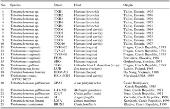
Table 1. List of trichomonad strains

Fig. 1. Morphological comparison of Tetratrichomonas sp. and Trichomonas tenax. (A) Cell of Tetratrichomonas sp. – oral strain TXO7. (B–C) Detail of parabasal apparatus of Tetratrichomonas sp. – bronchial strain TXB3. (D) Detail of parabasal apparatus of Trichomonas tenax Hs-4:NIH. (E) Cell of Trichomonas tenax Hs-4:NIH. AF, anterior flagella; Ax, axostyl; C, costa; FPF, free posterior flagellum; N, nucleus; P, pelta; PA, parabasal apparatus; UM, undulating membrane.

Table 2. Main morphological characters of the genera Trichomonas and Tetratrichomonas
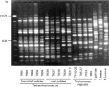
Fig. 2. Results of RAPD amplification with primer UBC 174. Strains TXB1 – TXB5 and TXO 9 – TXO10 represent the ‘bronchial’ group, strains TXO6 – TXO8 the ‘oral’ group. For specification of other used strains see Table 1.

Fig. 3. Tree based on RAPD PCR amplification. ‘Oral’ and ‘bronchial’ groups of Tetratrichomonas sp. form separate branches supported with high bootstraps, distinct from Trichomonas vaginalis group and other included strains.

Fig. 4. Trichomonad tree based on comparison of the ITS1-5.8S rRNA-ITS2 region sequences. The tree was constructed using the neighbor-joining/Maximum Likelihood/Maximum Parsimony methods. Asterisks designate nodes with bootstrap values below 50%.
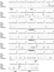
Fig. 5. Alignment of sequences of the ITS1-5.8SrRNA-ITS2 region of Tetratrichomonas sp. strain TXB3 (bronchial group) and TXO6 (oral group), Tetratrichomonas gallinarum strain 1-11-M2 and T. canistomae (strain BRIXI). Differences between oral and bronchial isolates are marked with boxes.
 You have
Access
- 59
- Cited by