

1. Introduction
Ontogenetic modification of skeletal morphology in extinct vertebrates can be hard to detect, especially when isolated holotype remains provide the only basis for evolutionary comparisons. This is particularly problematic for phylogenetic studies, which must assume intraspecific homogeneity for discrete character states and antonymous operational taxonomic units. A prime example of this paradox is Cretaceous ichthyosaurians (Ichthyopterygia: Ichthyosauria), whose interrelationships have been historically plagued by fragmentary fossils and uncertainty surrounding the validity of diagnostic traits (see discussions in McGowan & Motani, Reference McGowan and Motani2003; Fischer, Reference Fischer2012; Zammit, Reference Zammit2012). A compounding issue is the long recognized difficulty in differentiating disassociated juvenile and adult taxon morphotypes, except by inferred proportional criteria (e.g. McGowan, Reference McGowan1973; Deeming et al. Reference Deeming, Halstead, Manabe and Unwin1993; Fernández et al. Reference Fernández, Archuby, Talevi and Ebner2005; Maxwell, Reference Maxwell2012) or relative ossification of the limb bones (e.g. Johnson, Reference Johnson1977; Wade, Reference Wade1984; Caldwell, Reference Caldwell1997a ,Reference Caldwell b ; Motani & You, Reference Motani and You1998). Fortunately, however, all ichthyosaurians appear to have produced free-swimming young (either via ‘ovoviviparity’ or true viviparity: Lee & Shine, Reference Lee and Shine1998; see Blackburn, Reference Blackburn2000 for detailed terminology relating to amniote reproductive strategies); thus, the occasionally preserved skeletons of gravid females provide a direct means of comparing character state expression between conspecific age classes.
In utero ichthyosaurian remains have been reported for only a few Triassic−Cretaceous taxa: Besanosaurus Dal Sasso & Pinna, Reference Dal Sasso and Pinna1996 (Anisian−Landinian, Middle Triassic: Dal Sasso & Pinna, Reference Dal Sasso and Pinna1996), Mixosaurus Baur, Reference Baur1887 (Anisian−Landinian, Middle Triassic: Brinkmann, Reference Brinkmann1996), Shonisaurus Camp, Reference Camp1976 (Carnian, Late Triassic: Camp, Reference Camp1980), Leptonectes McGowan, Reference McGowan1996 (Rhaetian−Sinemurian, Early Jurassic: Lomax & Massare, Reference Lomax and Massare2012), Ichthyosaurus de la Beche & Conybeare, Reference de la Beche and Conybeare1821 (Hettangian, Early Jurassic: Deeming et al. Reference Deeming, Halstead, Manabe and Unwin1993), Stenopterygius Jaekel, Reference Jaekel1904 (Toarcian, Early Jurassic: McGowan, Reference McGowan1979; Böttcher, Reference Böttcher1990; A. Hungerbühler, unpub. M.Sc. thesis, Eberhard-Karls Universität, Tübingen, 1991; Maxwell, Reference Maxwell2012), Maiaspondylus Maxwell & Caldwell, Reference Maxwell and Caldwell2006 (Albian, Early Cretaceous: Maxwell & Caldwell, Reference Maxwell and Caldwell2003) and Platypterygius von Huene, Reference von Huene1922 (Albian, Early Cretaceous: Wade, Reference Wade1990; Kear, Boles & Smith, Reference Kear, Boles and Smith2003). To date, these embryonic materials have not been specifically utilized for assessing character state stability. Consequently, the recovery of exceptionally preserved three-dimensional fossils completely freed of matrix using acetic acid offers an unprecedented opportunity to explore developmental changes affecting phylogenetic traits within a model taxon: Platypterygius australis (McCoy, Reference McCoy1867) from the Early Cretaceous (late Albian) of Australia (see Kear, Reference Kear2003 for stratigraphical distribution). Platypterygius australis is arguably the most complete Cretaceous ichthyosaurian and is the only example represented by multiple adult and juvenile specimens (see Wade, Reference Wade1984; Kear, Reference Kear2005). Moreover, the genus Platypterygius is critical for resolving derived ichthyosaurian (Ophthalmosauria) topologies, since it is the stratigraphically youngest (late Cenomanian: Bardet, Wellnhofer & Herm, Reference Bardet, Wellnhofer and Herm1994), most pandemic and yet phylogenetically nebulous species-level complex (McGowan & Motani, Reference McGowan and Motani2003; Druckenmiller & Maxwell, Reference Maxwell2010; Fischer, Reference Fischer2012; Fischer et al. Reference Fischer, Masure, Arkhangelsky and Godefroit2011, Reference Fischer, Maisch, Naish, Kosma, Liston, Joger, Krüger, Pardo Perez, Tanish and Appleby2012). This paper presents an ontogenetic assessment of the discrete character states used to differentiate Platypterygius spp. (see Maxwell & Kear, Reference Maxwell and Kear2010 for species list). Their contextual implications for other ophthalmosaurian ichthyosaurians are also discussed.
Institutional abbreviations. AM – Australian Museum, Sydney, Australia; BSP – Bayerische Staatssammlung für Paläontologie und historische Geologie, München, Germany; DON – Museo Geológico José Royo y Gómez del Instituto de Investigaciones en Geociencias, Mineria y Química (INGEOMINAS), Bogota, Colombia; GLAHM – The Hunterian Museum, University of Glasgow, Glasgow, Scotland; IRNSB – Royal Belgian Institute of Natural Sciences, Brussels, Belgium; MHNH – Museum Histoire Naturelle Le Havre, Le Havre, France; MV – Museum Victoria, Melbourne, Australia; QM – Queensland Museum, Brisbane, Australia; SMSS – Städtisches Museum Schloss Salder, Salzgitter, Germany; SNHM – Staatliches Naturhistorisches Museum Braunschweig, Braunschweig, Germany; UALVP – University of Alberta, Edmonton, Canada; UW – University of Wyoming, Laramie, USA.
2. Character state survey of the in utero Platypterygius fossils
Although a number of small-bodied (c. < 1.5 m) Platypterygius australis individuals have been recorded in the literature (e.g. Wade, Reference Wade1984; Kear, Reference Kear2005), this study focused on only those that were identifiable as in utero foetuses. The most complete specimen, QM F16812, was found preserved within the body cavity of an articulated adult skeleton (QM F16811), situated adjacent to the ribs in the mid-dorsal region. Wade (Reference Wade1990) first identified QM F16812 as a foetus, and Kear, Boles & Smith (Reference Kear, Boles and Smith2003) considered QM F16812 to be distinct from associated gastric residues because of its large size (estimated minimum skull length ~ 500 mm), articulated condition and cranially oriented position within the parent's ribcage (comparable to other embryonic ichthyosaurians: Böttcher, Reference Böttcher1990; Deeming et al. Reference Deeming, Halstead, Manabe and Unwin1993). A second previously undocumented foetal postcranial skeleton (AM F90830) was also selected for analysis. This individual was partially articulated and occurred in direct contact with two displaced adult dorsal centra and some associated ribs. Size compatibility with QM F16812 (vertebral centrum diameters < 20 mm), coupled with incomplete cortical ossification throughout the axial skeleton and limb elements accords with early ontogeny (sensu Johnson, Reference Johnson1977; Caldwell, Reference Caldwell1997a ,Reference Caldwell b ). In addition, the lack of obvious surface corrosion or edge rounding along breaks indicates that there was no digestive modification of the bones otherwise indicative of gut contents (Andrews, Reference Andrews1990). Both QM F16812 and AM F90830 were recovered from outcrops of the Toolebuc Formation (Rolling Downs Group) in central-western Queensland, Australia (see Kear, Reference Kear2003 for locality maps and stratigraphical disposition). This unit has been correlated with the uppermost middle to upper Albian (Lower Cretaceous) P. ludbrookiae and upper C. paradoxa–P. pannosus zones (Moore, Pitt & Dettmann, Reference Moore, Pitt, Dettmann, Gravestock, Moore and Pitt1986; McMinn & Burger, Reference McMinn, Burger, Gravestock, Moore and Pitt1986).
2.a. Facial skeleton
QM F16812 includes components of the cranium and mandible, but much of the facial and mandibular rostra had broken off prior to collection. The dorsal and left-hand-side of the skull had also been exposed to weathering on the surface, resulting in loss of most of the dermocranial elements. The basicranium and some external surfaces from the right side of the skull have likewise been partially worn away. Despite this, all of the remaining bones from the right orbital and lateral temporal regions were preserved in articulation and cemented together by concretionary limestone matrix. We completely dissolved this rock using acetic acid to examine the internal cranial structures and intricate bone contacts in optimal detail.
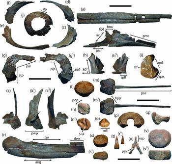
The premaxillae of QM F16812 are incomplete both anteriorly and posteriorly, and the internal walls of the alveolar grooves are broken off (Fig. 1a). This renders cross-sectional shape, structure of the processus supranarialis/processus subnarialis (sensu Maisch & Matzke, Reference Maisch and Matzke1997) and the premaxillary contributions to the bony nasal aperture impossible to reconstruct. Nevertheless, Kear (Reference Kear2005) described well-ossified posterior extremities of the premaxillae in a small-bodied (estimated maximum skull length ~ 700 mm), juvenile individual of Platypterygius australis (AM F98273: listed by Kear, Reference Kear2005 under the junior synonym P. longmani Wade, Reference Wade1990; see Zammit, Reference Zammit2010 for a discussion of taxonomy). This fossil was subjected to high-resolution computed tomography (CT) scans, from which the compiled three-dimensional rendering (see Kear, Reference Kear2005, p. 588, fig. 2A; and accompanying photograph on p. 591, fig. 5A) clearly showed that the posterior extremity of the premaxilla was deeply forked to form tapering projections of sub-equal length. Of these, the dorsalmost processus supranarialis had only minimal contact with the bony nasal aperture, while the ventral processus subnarialis extended below the naris and across the external face of the maxilla (Kear, Reference Kear2005). This arrangement is identical to larger, presumably adult conspecifics (Longman, Reference Longman1922, p. 249, text-fig. 1; Wade, Reference Wade1984, p. 104).
Selected elements of the in utero Platypterygius australis specimen QM F16812: premaxilla (a), maxilla (b), lacrimal (c), prefrontal (d), jugal (e) and postorbital (f) shown in lateral view; articulated parietal and supratemporal in dorsal (g) and ventral (g1) views; quadratojugal in lateral (h) and medial (h1) views; quadrate (i) in medial view; articulated sclerotic ring (j) in external view; pterygoid in medial (k), dorsal (k1) and lateral (k2) views; basioccipital condyle (l) in posterior view; basisphenoid-parasphenoid complex in dorsal (m) and ventral (m1) views; opisthotic in ventral (n) and anterior (n1) views; prootic in posterior view (o); supraoccipital in posterior view (p); stapes in dorsal view (q); posterior mandible in lateral view (r); articular in medial (s) and dorsal (s1) views; functional (t) tooth and replacement tooth crown (t1); cervical neural arch in anterior view (u); and atlas-axis complex in posterior articular (v) and dorsal (v1) views. Abbreviations: ang – angular; bna – bony nasal aperture; bpp – basipterygoid process; den – dentary; juc – jugal contact; lac – lacrimal; mli – membranous labyrinth impression; nsp – neural spine; par – parietal; pas – parasphenoid; plp – posterolateral process of the parietal; pmc – premaxillary contact; pop – paraoccipital process; ppf – paraoccipital process facet; pwp – posterior wing of the pterygoid; pzy – prezygapophysis; qas – quadrate articular surface; qdf – quadrate facet (processus quadratus) of quadratojugal; qol – occipital lamella of quadrate; slp – sclerotic plate; stf – stapes facet on quadrate; stp – supratemporal; s-ui – saccular-utricular impression; sur – surangular. All skeletal components are depicted at relative scale with measurement bars equal to 50 mm.

Druckenmiller & Maxwell (Reference Maxwell2010) asserted that the processus supranarialis was well developed in all species of Platypterygius. However, the form of the posterior premaxilla is ambiguous in most taxa represented by adequate cranial remains. For example, the premaxillae of P. sachicarum Páramo, Reference Páramo1997 (DON 19671: Páramo, Reference Páramo1997) are incomplete. Romer (Reference Romer1968) alternatively described the processus supranarialis in P. americanus (Nace, Reference Nace1939) as bounding most of the dorsal margin of the external naris, while the processus subnarialis extended a short distance below it (based on UW 2421: Romer, Reference Romer1968). Nonetheless, he also reported that the bones surrounding the narial opening in UW 2421 were distorted (Romer, Reference Romer1968, p. 28), thus making their exact relationships unclear. Disposition of the posterior premaxilla (where preserved) is variable in other Cretaceous ichthyosaurian genera. Maxwell & Caldwell (Reference Maxwell and Caldwell2006, p. 1046, text-fig. 1B) depicted a ventrally tapered structure in Maiaspondylus Maxwell & Caldwell, Reference Maxwell and Caldwell2006 that did not contact the external narial opening. Likewise, Druckenmiller & Maxwell (Reference Maxwell2010, pp. 1043–4) inferred that the processus supranarialis was extremely reduced in Athabascasaurus Druckenmiller & Maxwell, Reference Maxwell2010, and that the processus subnarialis terminated on the lateral face of the maxilla, but this was based on facet shape alone. The Jurassic ophthalmosaurians Ophthalmosaurus Seeley, Reference Seeley1874 (A. M. Kirton, unpub. Ph.D. thesis, Univ. Newcastle upon Tyne, 1983), Caypullisaurus Fernández, Reference Fernández1997 (Fernández, Reference Fernández2007), Aegirosaurus Bardet & Fernández, Reference Bardet and Fernandez2000 (Bardet & Fernández, 2000) and Cryopterygius Druckenmiller, Hurum, Knutsen & Nakrem, 2012 (Druckenmiller et al. Reference Druckenmiller, Hurum, Knutsen and Nakrem2012) conversely display a forked outline with the more prominent processus subnarialis interacting with the bony nasal opening.
The right maxilla of QM F16812 is missing only its anteriormost extremity, but was evidently drawn out into an elongate anterior process inset dorsally for receipt of the premaxilla (Fig. 1b). This contact terminates posteriorly in a well-defined lanceolate depression, probably accommodating the processus subnarialis as it extended below the bony external naris (see Kear, Reference Kear2005). The ventral alveolar edge of the maxilla is raised, suggesting considerable external exposure anterior to the nasal opening (a classic defining feature of Platypterygius: McGowan, Reference McGowan1972; McGowan & Motani, Reference McGowan and Motani2003). The maxillary dorsal lamina of QM F16812 divides into two sub-parallel processes that enclose the bony nasal opening (note though that the sloping edge of the anterior process is damaged: see Fig. 1b). Druckenmiller & Maxwell (Reference Maxwell2010) recognized that the bony margins of the nasal opening of adult P. australis diverge at a 90° angle, based upon AM F116940 figured by Kear (Reference Kear2005). Druckenmiller & Maxwell (Reference Maxwell2010) also stated that the maxilla of P. australis (vis-à-vis AM F116940) bore ‘three or more’ dorsal processes. However, the outline of this particular specimen is deceptive because an oblique fracture has broken off a large section of its anterodorsal edge (this is clearly visible in Kear, Reference Kear2005, p. 591, fig. 5D), thus giving a false impression of a third anteriormost process.
The dorsal lamina of the maxilla in QM F16812 forms the entire ventral margin of the bony nasal aperture (Fig. 1b), thus excluding the lacrimal. This is likewise consistent with all other P. australis remains (Kear, Reference Kear2005), as well as some Jurassic ophthalmosaurians such as Cryopterygius (Druckenmiller et al. Reference Druckenmiller, Hurum, Knutsen and Nakrem2012). In contrast, the lacrimal extensively overlaps the maxilla and contacts the premaxilla in Caypullisaurus (Fernández, Reference Fernández2007), Maiaspondylus (Maxwell & Caldwell, Reference Maxwell and Caldwell2006) and purportedly also P. americanus (Romer, Reference Romer1968), although re-examination of the only articulated cranium for this species (UW 2421) indicated that this traditional reconstruction is incorrect (Wade, Reference Wade1984; B. P. Kear, pers. obs.). Interestingly, Maxwell (Reference Maxwell2010) identified a broad contact between the premaxilla and lacrimal as an unambiguous synapomorphy of Ophthalmosauria, but with reversals occurring in Ophthalmosaurus and some species of Platypterygius (this trait might also be intraspecifically variable in other thunnosaurians such as Stenopterygius quadriscissus (Quenstedt, Reference Quenstedt1856) (A. Hungerbühler, unpub. M.Sc. thesis, Eberhard-Karls Universität, Tübingen, 1991). Fischer (Reference Fischer2012) additionally concluded that the lacrimal formed the posteroventral margin of the external naris in a badly fractured skull (MHNH 2010.4.4) attributed to P. hercynicus (Kuhn, Reference Kuhn1946). However, first-hand inspection of this specimen (B. P. Kear, pers. obs.) shows these bone identifications are equivocal, and the original narial shape cannot be recreated accurately.
The location of the anterior maxillary foramen (usually bordered by the dorsal lamina of the maxilla: Wade, Reference Wade1984; Kear, Reference Kear2005) was not distinguishable in QM F16812. However, Kear (Reference Kear2005) documented this structure, as well as accessory nasal foramina (situated posterodorsal to the bony external naris) in both osteologically immature (AM F98273: Kear, Reference Kear2005, p. 591, fig. 5A) and apparently adult P. australis (QM F2453: Wade, Reference Wade1990, p. 122; and the holotype MV P12989: Zammit, Reference Zammit2010, p. 289). Ochev & Efimov (Reference Ochev and Efimov1985), Arkhangelsky (Reference Arkhangelsky1998) and McGowan & Motani (Reference McGowan and Motani2003) described similar foramina in other Platypterygius spp.
Only a tiny fragment of the right nasal was recovered with QM F16812, but this clearly contacted the maxilla via a descending process and contributed to most (if not all) of the dorsal edge of the foreshortened external naris (a ubiquitous trait amongst P. australis specimens: Kear, Reference Kear2005). In contrast, the bony nasal aperture of Cretaceous ichthyosaurians including Athabasacasaurus (Druckenmiller & Maxwell, Reference Maxwell2010) and Sveltonectes Fischer, Masure, Arkhangelsky, & Godefroit, Reference Fischer, Masure, Arkhangelsky and Godefroit2011 (Fischer et al. Reference Fischer, Masure, Arkhangelsky and Godefroit2011) is much more anteroposteriorly elongate. Where discernible, the narial opening of other recognized Platypterygius spp. are either incomplete or distorted, e.g. P. americanus (Romer, Reference Romer1968), P. sachicarum (Páramo, Reference Páramo1997) and remains attributed to P. hercynicus (Fischer, Reference Fischer2012).
Despite its immaturity, QM F16812 maintained remarkably well-formed interdigitating sutures between the maxilla, lacrimal, prefrontal and probably also the nasal (Fig. 1b–d). The maxilla and jugal would likewise have been rigidly interlocked. Based on the condition in other P. australis fossils (Kear, Reference Kear2005), and the well-delineated contact surface on the maxilla of QM F16812, the jugal apparently did not interact with the premaxilla as it does in adult Brachypterygius von Huene, Reference von Huene1922 (A. M. Kirton, unpub. Ph.D. thesis, Univ. Newcastle upon Tyne, 1983; Bardet & Fernández, Reference Bardet and Fernandez2000) and Maiaspondylus (Maxwell & Caldwell, Reference Maxwell and Caldwell2006). There is also a slight differentiation in the anterior process of the jugal (Fig. 1e), which formed a curved ventral orbital margin in P. australis (Longman, Reference Longman1922; Kear, Reference Kear2005), as opposed to a relatively straight and more elongate profile in the holotype of P. hercynicus (SMSS unregistered: Kolb & Sander, Reference Kolb and Sander2009) and indeterminate Platypterygius sp. remains from the late Cenomanian of Bavaria (BSP Nr 1991 I 111: Bardet, Wellnhofer & Herm, 1994).
The prefrontal of QM F16812 (Fig. 1d) has lost most of its dorsal and posterior extremities to erosion. Nevertheless, it closely abutted both the lacrimal and maxilla, and thus probably contacted the nasal as in adult P. australis and all parvipelvians (Motani, Reference Motani1999; McGowan & Motani, Reference McGowan and Motani2003). Kear (Reference Kear2005) remarked that there is a minor internal contribution from the prefrontal to the posterior rim of the bony nasal aperture in at least one P. australis specimen in which this area of the skull is visible (AM F98273). This could not be detected in QM F16812, but it might have been broken off.
2.b. Postorbital region of the skull
The postorbital components of QM F16812, including parts of the parietal, supratemporal, postorbital, quadratojugal and the quadrate were preserved in articulation. Like all other skulls of P. australis, there is no evidence of a squamosal (Kear, Reference Kear2005). Maisch & Matzke (Reference Maisch and Matzke2000, p. 27) remarked that the squamosal might have been ‘entirely lost’ in Platypterygius, and Romer (Reference Romer1968) provided a lengthy discussion on the absence of a corresponding element in P. americanus and other ichthyosaurians (the postorbital region is unknown or badly damaged in most Platypterygius spp.: Páramo, Reference Páramo1997; McGowan & Motani, Reference McGowan and Motani2003: Kolb & Sander, Reference Kolb and Sander2009). Alternatively, Fischer (Reference Fischer2012) extrapolated that a squamosal might have occurred in P. hercynicus, based on a referred left quadratojugal (MHNH 2010.4.9) that possessed an embayed posterolateral surface; note that no squamosal element was actually located with this specimen. Fischer et al. (Reference Fischer, Maisch, Naish, Kosma, Liston, Joger, Krüger, Pardo Perez, Tanish and Appleby2012) similarly suggested that a squamosal facet was present on the supratemporal of Acamptonectes Fischer, Maisch, Naish, Kosma, Liston, Joger, Krüger, Pardo Pérez, Tainsh & Appleby, 2012. Druckenmiller & Maxwell (Reference Maxwell2010) identified a squamosal bone in Athabascasaurus, although they also conceded that this might represent a fractured ventral extension of the supratemporal.
Little remains of the dorsal and medial surfaces of the parietal of QM F16812. However, the ventral surface does incorporate endocranial impressions (see Kear, Reference Kear2005 for detailed description in other P. australis) and a well-preserved posterolateral process that interdigitates with the supratemporal (Fig. 1g, g1). The ventral extremity of the parietal in QM F16812 also contributes to the paraoccipital process facet on the supratemporal – a feature reported in other members of the species (Kear, Reference Kear2005). Fischer et al. (Reference Fischer, Maisch, Naish, Kosma, Liston, Joger, Krüger, Pardo Perez, Tanish and Appleby2012) explicitly stated that the parietal of Sveltonectes, and part of a skull assigned to P. hercynicus (MHNH 2010.4.1), differed from P. australis in displaying a restricted lateral supratemporal contact. Unfortunately, this is not palpable in the original descriptions or illustrations of these fossils (Fischer et al. Reference Fischer, Masure, Arkhangelsky and Godefroit2011, pp. 1011–12, fig. 1; Fischer, Reference Fischer2012. p. 125, fig. 2, p. 128). Moreover, contrary to Fischer et al. (Reference Fischer, Maisch, Naish, Kosma, Liston, Joger, Krüger, Pardo Perez, Tanish and Appleby2012, p. 8) the supratemporal does not strictly ‘[buttress] most of the posterior part of the parietal’ in P. australis. Rather, it articulates primarily with the supratemporal process and partly overlies some of the posterodorsal surface of the parietal (see QM F16812 in Fig. 1g), leaving much of the posteromedial (occipital) face exposed (Kear, Reference Kear2005, p. 145; see also Romer, Reference Romer1968, p. 38, fig. 8A for a comparable condition in Ichthyosaurus).
Maisch & Matzke (Reference Maisch and Matzke2000, p. 81) listed a ‘long postorbital region’ as a defining feature of Platypterygius, although they also recognized its occurrence in a variety of ichthyosaurians. Accordingly, the postorbital and quadratojugal of QM F16812 (Fig. 1f, h, h1), as in other P. australis, are large elements that would have formed a broad postorbital ‘wall’ similar to that of Brachypterygius (A. M. Kirton, unpub. Ph.D. thesis, Univ. Newcastle upon Tyne, 1983) Caypullisaurus (Fernández, Reference Fernández2007), Athabascasaurus (Druckenmiller & Maxwell, Reference Maxwell2010) and Cryopterygius (Druckenmiller et al. Reference Druckenmiller, Hurum, Knutsen and Nakrem2012). Maxwell, Fernández & Schoch (Reference Maxwell, Fernández and Schoch2012, table S3) specifically scored the quadratojugal of P. australis as having a concave ventral edge, ‘resulting in an embayment anterior to the quadrate articulation’ (state ‘0’ for their character 29: see Maxwell, Fernández & Schoch, Reference Maxwell, Fernández and Schoch2012, table S2). This coding seems to have been based upon the element (AM F98273) figured by Kear (Reference Kear2005, p. 596, fig. 7G), which displays a convex ventral profile arching into an ‘embayment’ lateral to the quadrate facet. However, QM F16812 (Fig. 1h) and most larger P. australis specimens (e.g. QM F2453: Wade, Reference Wade1984, p. 101, fig. 1B) conform to the contrasting state option (‘1’): quadratojugal with straight ventral edge. Thus, the outline shape of the quadratojugal appears to be variable.
The prominent quadrate facet (processus quadratus: sensu Maisch & Matzke, Reference Maisch and Matzke1997) of the quadratojugal in QM F16812 is offset from the main body of the bone (Fig. 1h1). This is characteristic of P. australis (Kear, Reference Kear2005), but differs from the quadratojugal (MHNH 2010.4.9) attributed to P. hercynicus by Fischer (Reference Fischer2012). This specimen lacked a distinct projection emanating from the quadratojugal's posteromedial edge (see Fischer, Reference Fischer2012, p. 129, fig. 5A1). In addition, Fischer (Reference Fischer2012, pp. 128–9) remarked that the posterior margin of MHNH 2010.4.9 formed an ‘acute angle’ relative to its ventral border, unlike the quadratojugal of P. australis, P. americanus and Athabascasaurus in which this same angle is ‘obtuse’. Based on Fischer's original photographs (Fischer, Reference Fischer2012, p. 129, fig. 5A1, B1), the axis of his ‘acute angle’ must have been situated at the posteroventral corner of the bone. Perplexingly though, orientation using this same position shows that the posterior margin of the quadratojugal in both QM F16812 (Fig. 1h) and a juvenile P. australis skull (AMF98273) figured by Kear (Reference Kear2005, p. 596, fig. 7G, H) is acute, not obtuse. The same is true for osteologically mature P. australis skeletons (e.g. QM F2453 as illustrated by Wade, Reference Wade1984, p. 101, fig. 1B), in which the quadratojugal has a near perpendicular posterior profile but retains a strongly acute posterodorsal flange. Furthermore, Platypterygius americanus (Romer, Reference Romer1968, p. 29, fig. 2; B. P. Kear, pers. obs.) appears to manifest a similar condition, as does Athabascasaurus based on Druckenmiller & Maxwell (Reference Maxwell2010, p. 1041, fig. 5B).
The right quadrate of QM F16812 is generally compliant with those of other P. australis (Wade, Reference Wade1990; Kear, Reference Kear2005) except in its poorly defined stapedal facet rim and incompletely ossified condylar articular surface (Fig. 1i). Fischer et al. (Reference Fischer, Masure, Arkhangelsky and Godefroit2011, p. 1013) and Fischer et al. (Reference Fischer, Maisch, Naish, Kosma, Liston, Joger, Krüger, Pardo Perez, Tanish and Appleby2012, p. 9) reported that the quadrate articular condyle is particularly massive in some European Platypterygius species ([sic] P. hercynicus: Kolb & Sander, Reference Kolb and Sander2009) and characteristically lacks a ‘saddle-[like shape]’ or ‘saddle-like zone’ separating it from the anterior pterygoid lamella as found in Sveltonectes and Acamptonectes, respectively. In relation to QM F16812 and other P. australis, these terms could possibly describe the embayed posterior margin of the quadrate's occipital lamella, although the precise meaning intended by Fischer et al. (Reference Fischer, Masure, Arkhangelsky and Godefroit2011) and Fischer et al. (Reference Fischer, Maisch, Naish, Kosma, Liston, Joger, Krüger, Pardo Perez, Tanish and Appleby2012) is unclear. Irrespectively, the dorsal extremity of the quadrate figured by Kolb & Sander (Reference Kolb and Sander2009, p. 166, text-fig. 6) is eroded and appears to be incomplete (E. Maxwell, pers. comm. Reference Maxwell2012), making the outline of its extracondylar area difficult to reconstruct.
2.c. Orbit and sclerotic plates
McGowan (Reference McGowan1972) and McGowan & Motani (Reference McGowan and Motani2003) listed reduced size of the orbit as a distinguishing trait of the genus Platypterygius. McGowan (Reference McGowan1973) concluded that orbital growth was isometric in ichthyosaurians, but A. Hungerbühler (unpub. M.Sc. thesis, Eberhard-Karls Universität, Tübingen, 1991) reported negative allometry, and together with Fernández et al. (Reference Fernández, Archuby, Talevi and Ebner2005), also demonstrated that the diameter of the sclerotic ring was proportionately larger in juveniles relative to adults. Certainly, the reconstructed diameter of the orbit in QM F16812 measured against the conservatively estimated minimum foetal skull length (~ 85 mm versus 500 mm/a ratio of 0.17: see also Wade, Reference Wade1984) would negatively scale relative to that of osteologically more mature individuals (260 mm versus 1043 mm/ratio = 0.2 based on QM F2453: see also measurements in Wade, Reference Wade1984, Reference Wade1990). In addition, the sclerotic aperture/sclerotic ring diameter ratio of QM F16812 (Fig. 1j) is slightly larger than those recorded in other specimens (0.49 versus 0.44: see Kear, Reference Kear2005), and the individual sclerotic plates are less well ossified and fewer in number (12 in QM F16812 versus 13–14 based on AM F98273, AM F116393: Kear, Reference Kear2005); this last feature presumably represents intraspecific variability.
2.d. Palatal complex
Parts of the left and right pterygoids, palatines and vomers have been recovered with QM F16812 (Fig. 1k–k2). Aside from their unfinished cortical surfaces, all are virtually indistinguishable from those found in other P. australis (compare with examples in Kear, Reference Kear2005). However, an important difference is that the posteromedial wing of the quadrate ramus of the pterygoid lacks a fully developed shelf underlying the basisphenoid and stapes (see Kear, Reference Kear2005, pp. 598–9, fig. 8E), a structure that becomes thicker and more evenly rounded in adults (see Wade, Reference Wade1990, p. 119, fig. 1B). The outline shape of the medial, lateral and probably also dorsal wings of the quadrate ramus might therefore change with ontogeny; this observation has significant implications for taxa such as Sveltonectes (holotype IRSNB R269), which was distinguished by its distinctively ‘forked’ posterior pterygoid margin (see Fischer et al. Reference Fischer, Masure, Arkhangelsky and Godefroit2011, p. 1014, fig. 2G). Curiously, IRSNB R269 also displayed other features consistent with osteological immaturity (e.g. incompletely ossified humeral bone surfaces: Fischer et al. Reference Fischer, Masure, Arkhangelsky and Godefroit2011), but was considered an adult because of its sclerotic ring proportions and complete fusion of the basisphenoid-parasphenoid complex (a feature reappraised below).
There is no evidence of a septomaxilla in QM F16812; thus, in accordance with Wade (Reference Wade1984) and Kear (Reference Kear2005), the bone would appear to be absent. Kear (Reference Kear2003) followed the character framework of Maisch & Matzke (Reference Maisch and Matzke2000) and incorrectly reported this element as present in P. australis. Romer (Reference Romer1968) and Fischer (Reference Fischer2012) also speculated that septomaxillae might occur in P. americanus and P. hercynicus, respectively, but we do not advocate these interpretations based on our inspections of the original material (UW 2421 and MHNH 2010.4.4: B. P. Kear, pers. obs.).
2.e. Basicranium
The basicranial elements of QM F16812 are well preserved but were disarticulated prior to burial. Erosion has also unfortunately destroyed the exoccipitals and all of the extracondylar area on the basioccipital (Fig. 1l). The remaining basioccipital condyle is incompletely ossified and totally lacks an outer cortical layer (but a dorsally offset notochordal pit is still visible). Wade (Reference Wade1990) and Kear (Reference Kear2005) described a greatly reduced extracondylar area in P. australis, as well as a hemispherical condylar outline, and a flat anteroventrally sloped basisphenoid contact that lacked a basioccipital peg. These states have been used to define the genus Platypterygius (Maxwell, Reference Maxwell2010; Maisch & Matzke, Reference Maisch and Matzke2000; Sander, Reference Sander2000; McGowan & Motani, Reference McGowan and Motani2003). However, they also occur in various ophthalmosaurians including Brachypterygius (McGowan, Reference McGowan1976; A. M. Kirton, unpub. Ph.D. thesis, Univ. Newcastle upon Tyne, 1983), Athabascasaurus (Druckenmiller & Maxwell, Reference Maxwell2010), Sveltonectes (Fischer et al. Reference Fischer, Masure, Arkhangelsky and Godefroit2011), Palvennia Druckenmiller, Hurum, Knutsen & Nakrem, Reference Druckenmiller, Hurum, Knutsen and Nakrem2012 (Druckenmiller et al. Reference Druckenmiller, Hurum, Knutsen and Nakrem2012) and Arthropterygius Maxwell, Reference Maxwell2010, which additionally retained an incipient basioccipital peg (Maxwell, Reference Maxwell2010). The basioccipital of Ophthalmosaurus (Andrews, Reference Andrews1910; Appleby, Reference Appleby1956; A. M. Kirton, unpub. Ph.D. thesis, Univ. Newcastle upon Tyne, 1983; McGowan & Motani, Reference McGowan and Motani2003; Druckenmiller & Maxwell, Reference Maxwell2010), together with Caypullisaurus (described as being ‘similar to Ophthalmosaurus’ by Fernández, Reference Fernández2007, p. 370; Maxwell, Reference Maxwell2010), Mollesaurus Fernández, Reference Fernández1999 (further distinguished by a conspicuous ventral notch: Fernández, Reference Fernández1999) and Acamptonectes (Fischer et al. Reference Fischer, Maisch, Naish, Kosma, Liston, Joger, Krüger, Pardo Perez, Tanish and Appleby2012) alternatively maintain distinctly offset extracondylar areas. Thus, this trait does not unanimously define Ophthalmosauria (Ophthalmosauridae sensu Fischer et al. Reference Fischer, Masure, Arkhangelsky and Godefroit2011, Reference Fischer, Maisch, Naish, Kosma, Liston, Joger, Krüger, Pardo Perez, Tanish and Appleby2012), or the constituent clade Platypteryginae, which was established using the poorly supported (bootstrap values < 50%) tree topology of Fischer et al. (Reference Fischer, Maisch, Naish, Kosma, Liston, Joger, Krüger, Pardo Perez, Tanish and Appleby2012).
Druckenmiller & Maxwell (Reference Maxwell2010, p. 1049) noted that the basioccipital condyle in a juvenile P. australis (AM F98273), as well as the osteologically immature holotype (destroyed during World War II) of ‘Ichthyosaurus’ brunsvicensis Broili, Reference Broili1909 (referred to Platypterygius sp. by McGowan & Motani, Reference McGowan and Motani2003, and subsequently cf. Acamptonectes by Fischer et al. Reference Fischer, Maisch, Naish, Kosma, Liston, Joger, Krüger, Pardo Perez, Tanish and Appleby2012), was conspicuously compressed, oval in outline and lacked a circumscribing ‘waist’. Fischer et al. (Reference Fischer, Maisch, Naish, Kosma, Liston, Joger, Krüger, Pardo Perez, Tanish and Appleby2012, p. 16) also envisaged a ‘concave extracondylar band’ in the latter taxon, and coined the term ‘peripheral ring’ to describe the depressed area delineating the occipital condyle in the type material of Acamptonectes (GLAHM 132588 and SNHM 1284-R: see Fischer et al. Reference Fischer, Maisch, Naish, Kosma, Liston, Joger, Krüger, Pardo Perez, Tanish and Appleby2012, p. 13). Our examination of AM F98273 confirmed that a constricted ‘waist’ is indeed absent. However, the condyle is demarcated by a sloping peripheral area (see Kear, Reference Kear2005, p. 589, fig. 3B) compatible with the ‘extracondylar band’ depicted in ‘I.’ brunsvicensis (see Broili, Reference Broili1909, tafel XXVII, fig. 5) and reminiscent of an isolated basioccipital (CAMSM B57962) attributed to Acamptonectes sp. by Fischer et al. (Reference Fischer, Maisch, Naish, Kosma, Liston, Joger, Krüger, Pardo Perez, Tanish and Appleby2012, p. 17, fig. 11A–C; in a possible lapsus calami this fossil was not listed with the other formally referred specimens). We also found the apparently unique ‘bilobed’ median cavity of the foramen magnum in Acamptonectes (Fischer et al. Reference Fischer, Maisch, Naish, Kosma, Liston, Joger, Krüger, Pardo Perez, Tanish and Appleby2012, p. 7, fig. 4B) to be difficult to discriminate from the divergent medial edges of the exoccipital facets in P. australis (compare with Kear, Reference Kear2005, p. 602, fig. 10C). This area is highly irregular in AM F98273 (and in the P. australis type specimen MV P12989: Wade, Reference Wade1990, p. 126, fig. 6A; Zammit, Reference Zammit2010, p. 286, fig. 2B), and would have been shaped by relative ossification of the cartilaginous braincase floor (this part of the endocranium was also seemingly modified during ontogeny in Stenopterygius: A. Hungerbühler, unpub. M.Sc. thesis, Eberhard-Karls Universität, Tübingen, 1991).
The basisphenoid and parasphenoid of QM F16812 are fully fused with only a weak trace of suture (Fig. 1m1). This resembles the typical adult condition in P. australis; although, the basisphenoid-parasphenoid can remain completely separate in some smaller individuals (AM F98273: Kear, Reference Kear2005). Co-ossification of the ‘parabasisphenoid’ complex is thus not specifically indicative of osteological maturity, and should be cautiously employed as an ontogenetic determinant for ichthyosaurian fossils (e.g. Fischer et al. Reference Fischer, Masure, Arkhangelsky and Godefroit2011).
The dorsal outline of the basisphenoid QM F16812 (Fig. 1m) conforms to the ‘hexagonal’ shape conceived for P. australis by Fischer et al. (Reference Fischer, Masure, Arkhangelsky and Godefroit2011, p. 1013). However, the basipterygoid processes are minimally developed, giving the basisphenoid a much narrower anterior profile when compared with those of adults (see Kear, Reference Kear2005, p. 603, fig. 11A). Critically, Maxwell (Reference Maxwell2010) and Fischer et al. (Reference Fischer, Masure, Arkhangelsky and Godefroit2011) utilized basipterygoid process size/basisphenoid outline as discriminating characters for Ophthalmosauria: parabasisphenoid gradually narrowing (0), or rapidly tapering posteriorly (1) when viewed in ventral aspect (character 11 of Maxwell, Reference Maxwell2010); and, basipterygoid processes short imparting a square basisphenoid outline (0), or markedly expanded laterally to give a pentagonal basisphenoid outline in dorsal view (character 17 of Fischer et al. Reference Fischer, Masure, Arkhangelsky and Godefroit2011; character 16 of Fischer et al. Reference Fischer, Maisch, Naish, Kosma, Liston, Joger, Krüger, Pardo Perez, Tanish and Appleby2012). Both of these analyses coded P. australis based on its adult morphotype − i.e. state ‘1’ for character 11 of Maxwell (Reference Maxwell2010); state ‘1’ for characters 17/16 of Fischer et al. (Reference Fischer, Masure, Arkhangelsky and Godefroit2011) and Fischer et al. (Reference Fischer, Maisch, Naish, Kosma, Liston, Joger, Krüger, Pardo Perez, Tanish and Appleby2012). In contrast, our foetal individual scores differently − state ‘0’ for both matrices. We also found that the ‘square’ versus ‘pentagonal’ outlines (sensu Fischer et al. Reference Fischer, Masure, Arkhangelsky and Godefroit2011, Reference Fischer, Maisch, Naish, Kosma, Liston, Joger, Krüger, Pardo Perez, Tanish and Appleby2012) could not be delineated on original fossils, and some of the scores for these traits were anomalous − e.g. Maiaspondylus was coded ‘0’ in Fischer et al. (Reference Fischer, Maisch, Naish, Kosma, Liston, Joger, Krüger, Pardo Perez, Tanish and Appleby2012), but the only described basisphenoid of this taxon is incomplete and partly obscured by overlying bones (Maxwell & Caldwell, Reference Maxwell and Caldwell2006, p. 1045, pl. 1, fig. 4).
The large internal carotid foramen piercing the basisphenoid of QM F16812 is posteromedially positioned (like most ophthalmosaurians: Maxwell, Reference Maxwell2010), and bordered anterolaterally by the sloping contact surfaces for the medial lamellae of the pterygoids (Fig. 1m1). The ‘posterolateral depressions’ purportedly evident on the ventral surface of the basisphenoid in P. australis (sensu Fischer et al. Reference Fischer, Maisch, Naish, Kosma, Liston, Joger, Krüger, Pardo Perez, Tanish and Appleby2012, p. 11) could not be detected, nor were they recorded in the comprehensive description of Kear (Reference Kear2005).
The opisthotic and prootic of QM F16812 are poorly ossified, and the paraoccipital process appears to have been mostly cartilaginous (Fig. 1n). In contrast, the structure of the paraoccipital process in adult P. australis is typically short and rounded, similar to that of other Platypterygius spp. in which the opisthotic is known (e.g. P. hercynicus: Kuhn, Reference Kuhn1946; Kolb & Sander, Reference Kolb and Sander2009; and remains referred to P. americanus: Nace, Reference Nace1941, pl. 1, figs 11, 12). The paraoccipital processes of Mollesaurus (Fernández, Reference Fernández1999) and Ophthalmosaurus (Andrews, Reference Andrews1910; Appleby, Reference Appleby1956) apparently also manifested comparable morphologies. However, A. M. Kirton (unpub. Ph.D. thesis, Univ. Newcastle upon Tyne, 1983, p. 26) described the paraoccipital process of Ophthalmosaurus as ‘slender’, and frequently not ossified towards its distal tip. Fischer et al. (Reference Fischer, Maisch, Naish, Kosma, Liston, Joger, Krüger, Pardo Perez, Tanish and Appleby2012) commented that like Ophthalmosaurus (sensu A. M. Kirton, unpub. Ph.D. thesis, Univ. Newcastle upon Tyne, 1983), the paraoccipital process of Acamptonectes forms a proportionately elongate rod, but is subject to intraspecific variability including incomplete ossification of the distal extremity in some osteologically mature individuals (GLAHM 132588). Fischer et al. (Reference Fischer, Maisch, Naish, Kosma, Liston, Joger, Krüger, Pardo Perez, Tanish and Appleby2012, pp. 13–14) additionally mentioned that the otic capsule impression on the opisthotic, and especially prootic, of Acamptonectes was ‘somewhat V-shaped’ as in Mollesaurus and specimens of Ophthalmosaurus, respectively. Platypterygius australis on the other hand purportedly had a more ‘T-shaped’ outline (Fischer et al. Reference Fischer, Maisch, Naish, Kosma, Liston, Joger, Krüger, Pardo Perez, Tanish and Appleby2012), but we found this definition to be subjective and entirely dependent upon how the variably shaped saccular-utricular impression was visualized. Moreover, our examination of QM F16812 demonstrated that both the peripheral margin and relative depth of the otic capsule impression (Fig. 1n1, o) changed shape with ontogeny (an observation also noted in Acamptonectes by Fischer et al. Reference Fischer, Maisch, Naish, Kosma, Liston, Joger, Krüger, Pardo Perez, Tanish and Appleby2012, p. 14, table 3), more closely resembling those outlines depicted for Acamptonectes, Ophthalmosaurus and Mollesaurus in the diagrams of Fischer et al. (Reference Fischer, Maisch, Naish, Kosma, Liston, Joger, Krüger, Pardo Perez, Tanish and Appleby2012, pp. 8, 9, figs 5, 6).
Like all of the recovered basicranial elements of QM F16812, the supraoccipital is incompletely formed and lacks finished bone on all of its articular surfaces (Fig. 1p). The classic U-shaped outline described for P. australis (Wade, Reference Wade1990; Kear, Reference Kear2005) and other Platypterygius species (e.g. P. hercynicus: Kuhn, Reference Kuhn1946; Kolb & Sander, Reference Kolb and Sander2009; and the referred specimen MHNH 2010.4.1: Fischer, Reference Fischer2012) is present, but shallower, and there is no strong midline constriction of the foramen magnum (imparted via advanced ossification of the ventral exoccipital facets). Druckenmiller & Maxwell (Reference Maxwell2010) and Fischer et al. (Reference Fischer, Maisch, Naish, Kosma, Liston, Joger, Krüger, Pardo Perez, Tanish and Appleby2012) reported that midline constriction of the foramina magna is noticeably absent in Athabascasaurus and Acamptonectes.
2.f. Stapes
The stapes of QM F16812 is characteristic of P. australis (Longman, Reference Longman1922; McGowan, Reference McGowan1972; Wade, Reference Wade1990; Kear, Reference Kear2005) and many other derived ophthalmosaurians (see state scores in the data matrix of Motani, Reference Motani1999; Sander, Reference Sander2000; Fischer et al. Reference Fischer, Masure, Arkhangelsky and Godefroit2011) in possessing a short shaft and greatly expanded proximal contact for the basioccipital (Fig. 1q). Conversely, the stapedal shafts of Acamptonectes (Fischer et al. Reference Fischer, Maisch, Naish, Kosma, Liston, Joger, Krüger, Pardo Perez, Tanish and Appleby2012) and Palvennia (Druckenmiller et al. Reference Druckenmiller, Hurum, Knutsen and Nakrem2012) appear to be more elongate. Both the proximal and distal articular surfaces of the stapes in QM F16812 are rounded and highly cancellous, as would be expected for foetal remains. In addition, the shaft lacks the putative hyoid process that intermittently occurs in larger individuals (Kear, Reference Kear2005), as well as other Platypterygius spp. (Fischer, Reference Fischer2012), Ophthalmosaurus (A. M. Kirton, unpub. Ph.D. thesis, Univ. Newcastle upon Tyne, 1983) and Acamptonectes (Fischer et al. Reference Fischer, Maisch, Naish, Kosma, Liston, Joger, Krüger, Pardo Perez, Tanish and Appleby2012).
2.g. Mandible
Large sections of the left and right dentaries were recovered with QM F16812, together with bones from the right posterior mandibular ramus, which are still in articulation (Fig. 1r). The anteriormost tips of the dentaries are missing; nevertheless, Kear (Reference Kear2005, p. 610) described these as being ‘upturned’ in P. australis, not ‘downturned’ as stated by Fischer et al. (Reference Fischer, Maisch, Naish, Kosma, Liston, Joger, Krüger, Pardo Perez, Tanish and Appleby2012, p. 14). Also contrary to Fischer et al. (Reference Fischer, Maisch, Naish, Kosma, Liston, Joger, Krüger, Pardo Perez, Tanish and Appleby2012, p. 14), the anterior ends of the dentaries were purportedly broken off in the skull of P. americanus (UW 2421: Romer, Reference Romer1968, p. 32). Personal inspection by B. P. Kear, however, has found that they are in fact preserved, and correspond in form with the anterior ends of the premaxillae (based on UW 5545: Nace, Reference Nace1941) in being rounded and lacking any ‘downturned’ component.
The surangular and angular of QM F16812 both have large posterolateral exposure. The surangular is comparatively restricted in Athabascasaurus (Druckenmiller & Maxwell, Reference Maxwell2010). Motani (Reference Motani1999) additionally designated extensive external exposure of the angular to be a key synapomorphy for Ophthalmosauria (this trait is expressed in all P. australis specimens: Kear, Reference Kear2005). Maxwell, Fernández & Schoch (2012, table S2) reiterated this character definition but added that the surangular exposure was ‘reduced to a thin strip on the lateral surface of the retroarticular process’. Nonetheless, this is not readily evident in QM F16812, in which the visible retroarticular contribution is extensive (see Fig. 1r).
Unlike more mature P. australis individuals (Kear, Reference Kear2005), the dorsal surface of the angular in QM F16812 forms a smooth groove that lacks a longitudinal ridge separating the surangular contact from the floor of the Meckelian canal. Fischer et al. (Reference Fischer, Maisch, Naish, Kosma, Liston, Joger, Krüger, Pardo Perez, Tanish and Appleby2012) previously recognized this as an ontogenetic feature in their comparisons of ‘sub-adult’ and ‘adult’ Acamptonectes (SNHM 1284-R and GLAHM 132588, respectively).
The prearticular of QM F16812 forms the medial wall of the glenoid fossa, and in accordance with Kear (Reference Kear2005), there is no evidence of a coronoid. The articular (Fig. 1s, s1) lacks cortical bone deposition on its peripheral surfaces, and the whole element is proportionately compact (anteroposterior length ~ 1.6 times maximum breadth: compare with Kear, Reference Kear2005, p. 614, fig. 15D–F). Fernández (Reference Fernández1999) recorded an extremely short articular in Mollesaurus (anteroposterior length ~ 1.15 times breadth), and Maxwell (Reference Maxwell2010, p. 406) remarked that the articular of Arthropterygius had ‘much less rounded’ dorsal and ventral margins than that of Platypterygius (based on P. australis as a generic exemplar). Fischer et al. (Reference Fischer, Maisch, Naish, Kosma, Liston, Joger, Krüger, Pardo Perez, Tanish and Appleby2012, p. 14) reported that the articular of Acamptonectes could be distinguished from other ophthalmosaurians by being ‘nearly as thick as it is long’, but did not provide more detailed comparisons.
2.h. Dentition
None of the teeth of QM F16812 were found in situ, although, numerous loose tooth crowns with either fully or partially ossified roots were recovered (Fig. 1t, t1). Their general morphology is typical of P. australis and other Platypterygius spp. in having a conical enamel cap ornamented by longitudinal ridges, and an exposed cementum ring surmounting an expanded root with squared profile (Bardet, Reference Bardet1990; Maxwell, Caldwell & Lamoureux, Reference Maxwell, Caldwell and Lamoureux2011). Druckenmiller & Maxwell (Reference Maxwell2010) suggested that ichthyosaurian teeth might have scaled isometrically during growth, being less robust in immature individuals; this appears to be the case in QM F16812 (compare with Kear, Reference Kear2005, p. 615, fig. 16).
McGowan (Reference McGowan1976, p. 667) introduced a ‘tooth length index’ to discriminate between the dentitions of various ichthyosaurians. This proportional relationship was elaborated into a phylogenetic character by Fischer et al. (Reference Fischer, Masure, Arkhangelsky and Godefroit2011, p. 1023), who defined their ‘tooth size ratio [TSR]’ as tooth size relative to basioccipital width, with an arbitrary state segregation of ‘large’ ≥ 3 (scored ‘0’ based on outgroup polarity) versus ‘small’ ≤ 1.5 (‘1’). The TSR formula was given as ‘10 × largest crown height/basioccipital width’, and P. australis subsequently scored ‘0’. However, we found this designation to be ambiguous since it is impossible to discriminate the ‘largest crown’ without a complete dentition, which is unknown in this taxon (Kear, Reference Kear2005). Moreover, enamel crown height (vis-à-vis total height of the tooth) varies markedly along the jaw (Kear, Reference Kear2005), and interdependent growth of the dentition relative to the intrinsically disparate components of the basicranium cannot be adequately demonstrated. In accordance, QM F16812 produces discordant TSRs of 6.4 or 2.4 based on crown versus total height of its most fully developed tooth; equivalent parameters taken from the juvenile P. australis AM F98273 ranged between 5.9 and 3.9 for the largest enamel crowns/teeth, respectively.
2.i. Postcranial skeleton
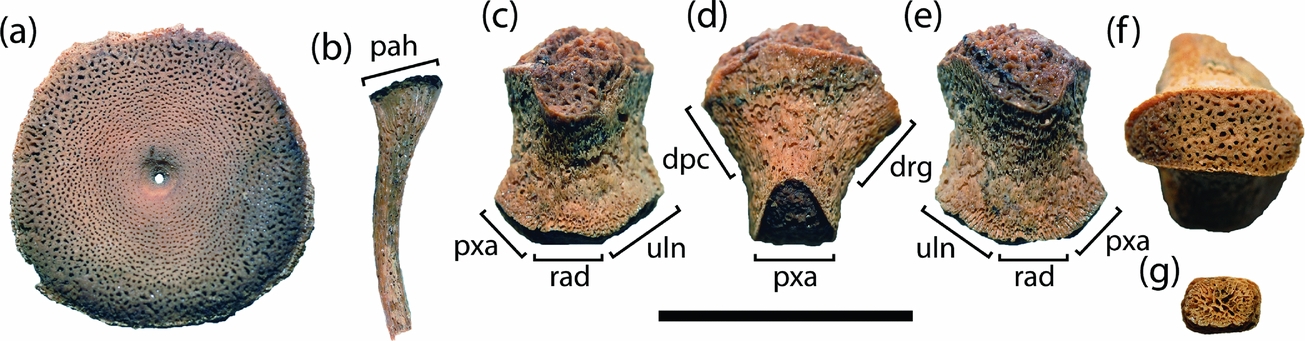
QM F16812 incorporates 16 vertebral centra including the atlas-axis complex and a continuous postaxial series with associated neural arches. The second foetal postcranial skeleton, AM F90830, comprises ten vertebral centra, a few dorsal ribs and some disarticulated limb bones. All of the recovered axial elements are highly cancellous and the centrum peripheries lack features traditionally used to differentiate the cervical and dorsal series, e.g. ossified rib articulations and ventral keels (Fig. 2a). The notochordal pits on the postaxial centra remain perforated, which is similar to in utero specimens of Stenopterygius (A. Hungerbühler, unpub. M.Sc. thesis, Eberhard-Karls Universität, Tübingen, 1991), Maiaspondylus (Maxwell & Caldwell, Reference Maxwell and Caldwell2003) and other possible ophthalmosaurian embryos (O'Keefe et al. Reference O'Keefe, Street, Cavigelli, Socha and O'Keefe2009). The atlas-axis complex (Fig. 1v, v1) completely fused during earliest ontogeny (confirming the hypotheses of A. Hungerbühler, unpub. M.Sc. thesis, Eberhard-Karls Universität, Tübingen, 1991; Maxwell & Kear, Reference Maxwell and Kear2010), but there is no co-ossification with the third cervical as in the immature holotype of P. platydactylus (Broili, Reference Broili1907, p. 19). Maxwell & Kear (Reference Maxwell and Kear2010) and Zammit, Norris & Kear (Reference Zammit, Norris and Kear2010) reported that a dorsal trace of the atlas-axis suture is usually evident in P. australis, but this could not be detected in QM F16812 and thus seems to be intraspecifically variable (it is likewise absent in the holotypes of P. hercynicus and P. americanus: Kolb & Sander, Reference Kolb and Sander2009; Maxwell & Kear, Reference Maxwell and Kear2010).
Selected elements of the in utero Platypterygius australis specimen AM F90830: vertebral centrum in articular view (a); dorsal rib in anteroposterior view (b); humerus in dorsal (c), anterior (d), ventral (e) and distal articular (f) views; distal epipodial or mesopodial element (g) in dorsoventral view. Abbreviations: dpc – deltopectoral crest; drg – dorsal ridge; pah – proximal articular head of rib; pxa – preaxial articular surface; rad – radial articular surface; uln – ulnar articular surface. All skeletal components are depicted at relative scale with measurement bar equal to 20 mm.

The neural arches of AM F90830 are very poorly formed, lacking both the distal articular facets and most of the neural spines. This accords with A. Hungerbühler (unpub. M.Sc. thesis, Eberhard-Karls Universität, Tübingen, 1991), who described increasingly limited ossification throughout the posterior dorsal regions of foetal Stenopterygius. In compliance, the cervical-anterior dorsal series of QM F16812 is much better defined, with ossified neural spines that have flattened dorsal apices (Fig. 1u). Wade (Reference Wade1990) alternatively reported that the neural spines of adult P. australis bore an inset apical notch. Similar excavations have also been described on the presacral neural spines of Ophthalmosaurus (Andrews, Reference Andrews1910; A. M. Kirton, unpub. Ph.D. thesis, Univ. Newcastle upon Tyne, 1983), Acamptonectes (Fischer et al. Reference Fischer, Maisch, Naish, Kosma, Liston, Joger, Krüger, Pardo Perez, Tanish and Appleby2012) and Cryopterygius (Druckenmiller et al. Reference Druckenmiller, Hurum, Knutsen and Nakrem2012), and might have supported either a ligamentous or cartilaginous attachment (Druckenmiller et al. Reference Druckenmiller, Hurum, Knutsen and Nakrem2012).
Despite being partly developed, the neural arches of QM F16812 were taller than their accompanying centra, and thus conform to the diagnostic proportions of Platypterygius spp. (height cervical vertebra centrum/neural arch < 1: Kolb & Sander, Reference Kolb and Sander2009). The zygapophyseal articulations were also separated, unlike in adults (e.g. QM F2453: Wade, Reference Wade1990), which have medially fused zygapophyses in at least the presacral half of the vertebral column. Fischer et al. (Reference Fischer, Maisch, Naish, Kosma, Liston, Joger, Krüger, Pardo Perez, Tanish and Appleby2012, p. 15) listed the retention of separated or ‘unpaired’ zygapophyseal surfaces in the cervicals and anterior dorsals as a distinguishing feature of Acamptonectes. Other ophthalmosaurians such as Ophthalmosaurus (Andrews, Reference Andrews1910; A. M. Kirton, unpub. Ph.D. thesis, Univ. Newcastle upon Tyne, 1983), P. hercynicus (Kolb & Sander, Reference Kolb and Sander2009) and P. americanus (juvenile UW 5547: B. P. Kear, pers. obs.) alternatively display fused zygapophyses. Fischer et al. (Reference Fischer, Masure, Arkhangelsky and Godefroit2011, p. 1015) additionally stated that, ‘as in P. hercynicus’, Sveltonectes has pre- and postzygapophyses that remain ‘separated anteriorly in the cervical and anterior dorsal regions, [but] are usually completely fused in the posterior dorsal and anterior caudal regions’. Puzzlingly, Kolb & Sander (Reference Kolb and Sander2009, p. 172) noted that the zygapophyses of P. hercynicus were ‘paired in the dorsal region and unpaired in the rest of the vertebral column’; thus, the comparisons of Fischer et al. (Reference Fischer, Masure, Arkhangelsky and Godefroit2011) are incongruous.
The dorsal ribs of AM F90830 have a circular cross-section over most of their length and lack the outer compact bone layer otherwise evident in adult ichthyosaurian specimens; Talevi & Fernández (Reference Talevi and Fernández2012) reported that cortical bone deposition was especially thick in Mollesaurus. AM F90830 also shows no evidence of longitudinal costal grooves incising the anterior and posterior surfaces of the ribs, although anteroposterior compression is observable on the proximal head (Fig. 2b). Maxwell & Kear (Reference Maxwell and Kear2010) recognized that the cross-sectional shape of the ribs in P. americanus changed along their length, being circular distally but becoming anteroposteriorly compressed proximally with distinct costal grooves on the proximal head. Fischer et al. (Reference Fischer, Maisch, Naish, Kosma, Liston, Joger, Krüger, Pardo Perez, Tanish and Appleby2012) conversely stated that the rib cross-section was circular in Acamptonectes (but where this occurred along the length of the rib was not explained) and that the costal groves were only weakly developed on one side.
None of the distal limb bones of AM F90830 were found in articulation. Nonetheless, their association with a tiny humerus (16.4 mm maximum proximodistal length) could indicate derivation from the forefins. The humerus of AM F90830 has poorly ossified articular surfaces and the glenoid head is convex (Fig. 2c–f). The humeral shaft also has a ‘sandpaper-like’ texturing consistent with the ‘juvenile’ ichthyosaurian morphotype identified by Johnson (Reference Johnson1977). The humeral proportions of AM F90830 are generally similar to those of adults (see Wade, Reference Wade1984; Zammit, Norris & Kear, Reference Zammit2010) in being longer than wide with the distal extremity anteroposteriorly expanded relative to the proximal glenoid end (exact measurements = 13.1 mm and 11.4 mm, respectively). Motani (Reference Motani1999) identified humeral length/width dimensions (his character 54), together with distal/proximal humeral widths (character 55) to be differential traits amongst ichthyopterygians. Motani (Reference Motani1999, p. 493) scored Platypterygius as ‘0’: humerus ‘much longer than wide’ and ‘nearly equal’ in its distal/proximal articular surface widths. Fernández (Reference Fernández2007) and Druckenmiller & Maxwell (Reference Maxwell2010) followed this state coding. Fischer et al. (Reference Fischer, Masure, Arkhangelsky and Godefroit2011, p. 1024), however, elaborated on the latter character by scoring both P. australis and P. hercynicus as ‘1’: humerus distal/proximal end widths in dorsal view being ‘nearly equal or [with] proximal end slightly wider than distal end’. AM F90830 conversely conforms to the outgroup score of ‘0’: ‘distal end wider than proximal’. We therefore consider the descriptive term ‘nearly equal’ to be ambiguous because it could feasibly incorporate either state option. Furthermore, Motani (Reference Motani1999, p. 493) noted that the dorsal ridge of ophthalmosaurians was pronounced to form a ‘plate-like eminence’, a feature appended by Maxwell (Reference Maxwell2010), Druckenmiller & Maxwell (Reference Maxwell2010) and later Fischer et al. (Reference Fischer, Masure, Arkhangelsky and Godefroit2011, p. 1024) with the deltopectoral crest being of ‘matching’ height and ‘bordered by concave areas’. Both these conditions were considered present (‘1’) in Platypterygius spp. Critically, though, the dorsal ridge and deltopectoral crest are very poorly formed in AM F90830, being distinguishable only as flared protrusions adjacent to the proximal articular head (Fig. 2c–e). This failure to develop distinct structures confounds accurate reconstruction of the dorsal ridge/deltopectoral crest, and subsequently negates any meaningful diagnostic application of these states for ontogenetically indeterminate ophthalmosaurian fossils (e.g. Fischer et al. Reference Fischer, Maisch, Naish, Kosma, Liston, Joger, Krüger, Pardo Perez, Tanish and Appleby2012, p. 15).
The outline shape and number of the distal articular facets on the humerus has also been used to discriminate between ophthalmosaurians (Motani, Reference Motani1999; Maisch & Matzke, Reference Maisch and Matzke2000; Sander, Reference Sander2000; McGowan & Motani, Reference McGowan and Motani2003; Druckenmiller & Maxwell, Reference Maxwell2010; Maxwell, Reference Maxwell2010; Fischer et al. Reference Fischer, Masure, Arkhangelsky and Godefroit2011, Reference Fischer, Maisch, Naish, Kosma, Liston, Joger, Krüger, Pardo Perez, Tanish and Appleby2012), and is variably developed in Platypterygius spp. with P. australis usually displaying three, or more rarely four, concave distal articular surfaces (Maxwell & Kear, Reference Maxwell and Kear2010; Zammit, Norris & Kear, Reference Zammit2010). The distal humeral facets are incompletely ossified in AM F90830; thus, their precise outline shape and relative concavity is difficult to reconstruct. Nevertheless, the distal edges of the humerus are clearly offset (Fig. 2c–f) such that the corresponding contact positions of the ulna, radius and a large preaxial accessory element can be inferred based on comparisons with other P. australis remains (Wade, Reference Wade1984; Zammit, Norris & Kear, Reference Zammit2010). Fisher et al. (Reference Fischer, Masure, Arkhangelsky and Godefroit2011) and Fisher et al. (Reference Fischer, Maisch, Naish, Kosma, Liston, Joger, Krüger, Pardo Perez, Tanish and Appleby2012, character 34 description on p. 2 of supplementary text S1) used relative orientation of the distal humeral facets to designate P. australis and other platypterygiine taxa (Aegirosaurus, Brachypterygius, Caypullisaurus, Maiaspondylus, P. hercynicus, Sveltonectes) as lacking a ‘posterodistally deflected ulnar facet and distally facing radial facet’. Certainly, the radial facet does face distally in the P. australis specimens that we observed, but the ulnar facet is usually posterodistally oriented (see Wade, Reference Wade1984, p. 102, fig. 2B). The degree of this posterodistal ‘deflection’ is also noticeably more marked in AM F90830 where the contact position of the ulna is inclined by as much as 40° relative to a horizontal plane through the radial facet. Posterodistal angling of the ulnar facet would therefore seem to change as ossification progresses, and indeed, is in keeping with ontogenetic modification of the glenoid documented in other juvenile ichthyopterygians (Johnson, Reference Johnson1977; Motani & You, Reference Motani and You1998).
Little of the distal forelimb structure can be discerned from AM F90830 because the individual elements were disarticulated prior to burial. Nevertheless, the phalanges were obviously polygonal (Fig. 2g) as is characteristic of Platypterygius spp. (McGowan, Reference McGowan1972; Maisch & Matzke, Reference Maisch and Matzke2000) and some other ophthalmosaurian taxa including Caypullisaurus (Fernández, Reference Fernández1997), Maiaspondylus (Maxwell & Caldwell, Reference Maxwell and Caldwell2006) and Sveltonectes (Fisher et al. Reference Fischer2011). Maxwell & Kear (Reference Maxwell and Kear2010) also reported that immature individuals of P. americanus, P. australis and P. campylodon (the latter based on Kiprijanoff, Reference Kiprijanoff1881) displayed incised proximal and distal articular surfaces on their phalanges, perhaps indicative of tendinous insertion or fusion from paired ossification centres during early ontogeny. AM F90830, on the other hand, lacks similar incisions and was clearly formed as a single unit (see Caldwell, Reference Caldwell1997a ). The proximal and distal incisions in Platypterygius spp., therefore, presumably relate to later ontogenetic development, possibly the progressive deposition of endochondral bone to produce synarthrotic joints.
3. Conclusions
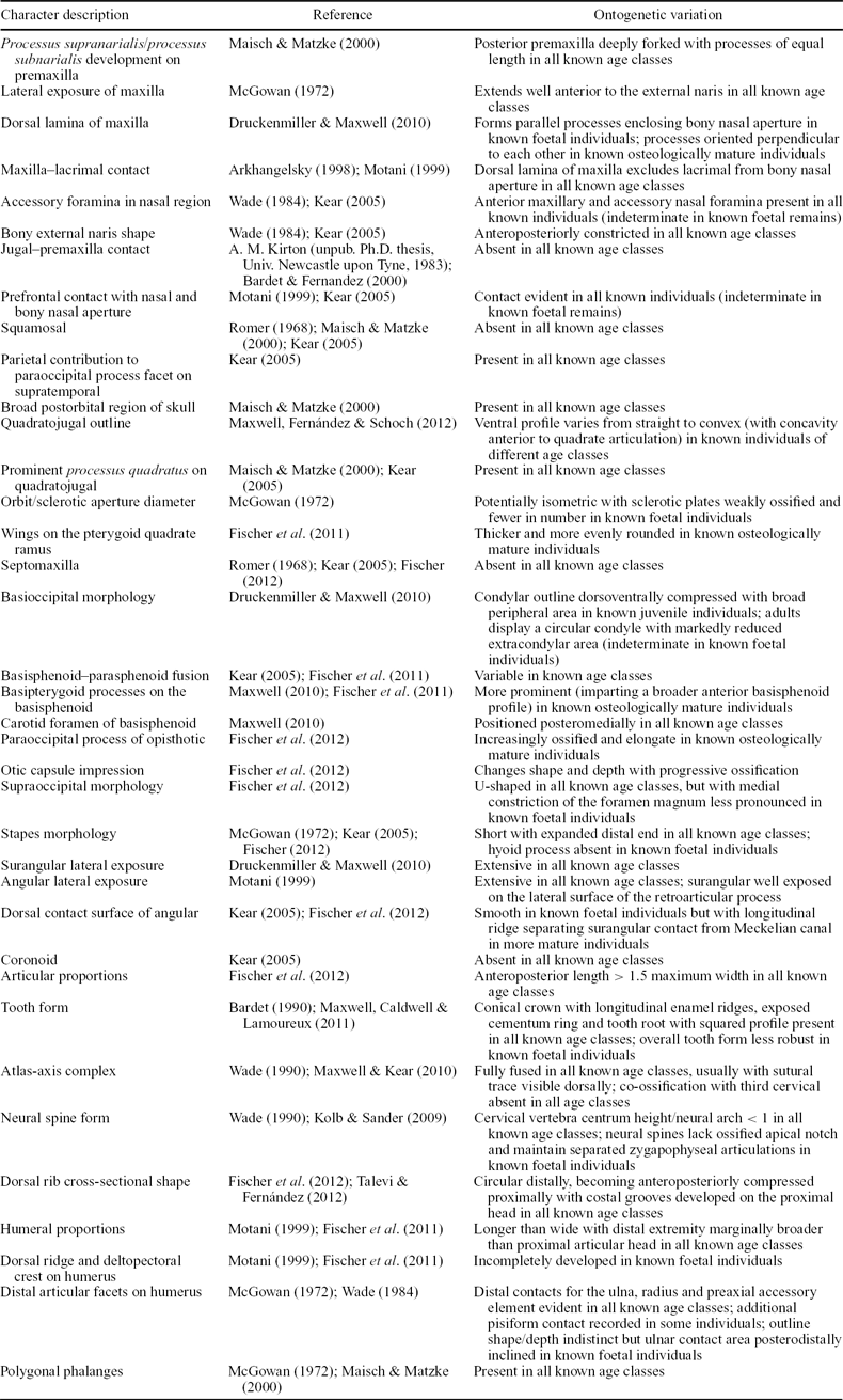
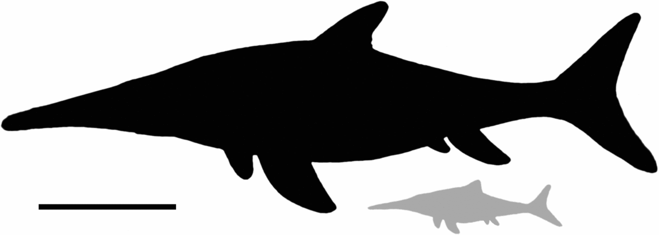
The phylogenetic implications of our foetal Platypterygius australis fossils emphasize the need for judicious selection, definition and coding of characters/state manifestations within existing ophthalmosaurian datasets/classifications (Fernández, Reference Fernández2007; Maxwell, Reference Maxwell2010; Druckenmiller & Maxwell, Reference Maxwell2010; Fischer et al. Reference Fischer, Masure, Arkhangelsky and Godefroit2011, Reference Fischer, Maisch, Naish, Kosma, Liston, Joger, Krüger, Pardo Perez, Tanish and Appleby2012; Maxwell, Fernández & Schoch, Reference Maxwell, Fernández and Schoch2012). Consideration of ontogenetic variability (e.g. Johnson, Reference Johnson1977; Wade, Reference Wade1984; A. Hungerbühler, unpub. M.Sc. thesis, Eberhard-Karls Universität, Tübingen, 1991; Maxwell, Reference Maxwell2012) is also vital, but virtually impossible to enforce without sufficient knowledge of growth trajectories in the operational taxonomic units. Nevertheless, the remarkably consistent trait expression observed between age classes in at least one well-documented species implies that many character components employed in ophthalmosaurian phylogenetics retain discriminative capacity, and thus can be used to diagnose taxa irrespective of osteological maturity. Moreover, the foetal P. australis specimens reported here provide the first detailed evidence of early ontogenetic development within a Cretaceous ichthyosaurian. The only other coeval examples include the imperfect skeletons of two very small embryos (skull length 42 mm) associated with some adult vertebrae (UALVP 45639) of Maiaspondylus (Maxwell & Caldwell, Reference Maxwell and Caldwell2003, Reference Maxwell and Caldwell2006). By comparison, the in utero P. australis are much larger: the minimum skull length of ~ 500 mm in QM F16812 yielding a total body length estimate of > 1 m (Fig. 3) based on the regression equation calculated for Stenopterygius (Deeming et al. Reference Deeming, Halstead, Manabe and Unwin1993). Deeming et al. (Reference Deeming, Halstead, Manabe and Unwin1993) also proposed an orbit [OL]/skull length [SL] proportional relationship derived from morphometric observations of intra-ovum crocodilians (Deeming & Ferguson, Reference Deeming and Ferguson1990) and used this to determine developmental stages within Ichthyosaurus and Stenopterygius. Applying the same methodology suggests that QM F16812 was probably close to parturition because of its low OL/SL ratio (0.17) and well-formed teeth and bones. In addition, orientation of QM F16812 with its tail towards the mother's pelvic region is consistent with the positioning of most larger-bodied Stenopterygius embryos (Böttcher, Reference Böttcher1990; A. Hungerbühler, unpub. M.Sc. thesis, Eberhard-Karls Universität, Tübingen, 1991; Deeming et al. Reference Deeming, Halstead, Manabe and Unwin1993) and conforms to the cetacean-like ‘tail-first’ birthing scenario postulated for all ichthyopterygians (Böttcher, Reference Böttcher1990). Furthermore, the seemingly isolated occurrences of QM F16812 and AM F90830 within the body-cavities of their parents are compatible with the typically low numbers of in utero young (usually between one and three but sometimes up to as many as 11) found in other gravid ichthyosaurians (Böttcher, Reference Böttcher1990; A. Hungerbühler, unpub. M.Sc. thesis, Eberhard-Karls Universität, Tübingen, 1991; Deeming et al. Reference Deeming, Halstead, Manabe and Unwin1993; Brinkmann, Reference Brinkmann1996; Lomax & Massare, Reference Lomax and Massare2012), and might suggest a K-selected reproductive strategy similar to that of some other advanced marine amniotes (e.g. cetaceans and plesiosaurs: O'Keefe & Chiappe, Reference O'Keefe and Chiappe2011). Taken in conjunction with the robust teeth and skull of QM F16812, this could advocate precocial habits (Deeming et al. Reference Deeming, Halstead, Manabe and Unwin1993), perhaps incorporating independent capture and dismemberment of prey by neonates. The structural requirements of active predation at such an early life stage might have influenced the differential ossification patterns observed in P. australis. For example, the ‘adult’ traits expressed by QM F16812 all relate to formation and suturing of the dermocranium (Table 1), and especially the anterior orbital region, which would have experienced high stress loads during feeding (see Kear, Reference Kear2005 for discussion). In contrast, noticeable ontogenetic variation (detailed in Table 1) affected the proportions of the orbit/sclerotic aperture (Fernández et al. Reference Fernández, Archuby, Talevi and Ebner2005) and relative ossification of some basicranial components (wing shape on the pterygoid quadrate rami, hyoid process development on the stapes, basioccipital condyle dimensions relative to the extracondylar area, formation of the basipterygoid processes on the basisphenoid, length of the paraoccipital process, excavation of the otic capsule impression, completion of the exoccipital facets on the supraoccipital), and to a lesser extent the lower jaw (angular–surangular contact reinforced by ridges). Developmental changes in the axial (formation of the neural arch processes) and appendicular skeleton (ossification of the humeral articular surfaces and muscle attachment areas) are similar to those of other ichthyosaurians (see Johnson, Reference Johnson1977; A. Hungerbühler, unpub. M.Sc. thesis, Eberhard-Karls Universität, Tübingen, 1991) and parallel bone mineralization sequences observed in extant aquatic diapsids (e.g. Rieppel, Reference Rieppel1993).
Summary of phylogenetically pertinent character trait conditions expressed by in utero versus osteologically more mature specimens of Platypterygius australis

Proportionately scaled silhouettes of an osteologically mature (black) Platypterygius australis individual (based on reconstructions in Kear & Hamilton-Bruce, Reference Kear and Hamilton-Bruce2011) of 4.5–5 m snout–tail length, versus the in utero foetus QM F16812 (grey) estimated at 1–1.5 m (based on the growth series of Stenopterygius: McGowan, Reference McGowan1973). Scale bar equals 1 m.

Acknowledgements
Robert Jones and Yong Yi Zhen (Australian Museum), Oliver Rauhut (Bayerische Staatssammlung für Paläontologie und historische Geologie), Gabrielle Baglione, Cédric Cremiere and Arnaud Duchemin (Muséum d'Histoire Naturelle du Havre), Andrew Rozefelds and Kristen Spring (Queensland Museum) and Michael Cassiliano (Geological Museum, University of Wyoming) generously assisted with access to specimens and information. Thanks to Erin Maxwell (Universität Zürich) for images and discussion on the type remains of Platypterygius hercynicus. We are also indebted to Marta Fernández (Museo de La Plata) and Patrick Druckenmiller (University of Alaska, Museum of the North) for their constructive reviews. The Australian Research Council and Uppsala University provided financial support for BPK.