Introduction
Studies conducted over the past decade have provided evidence of pituitary gland volume abnormalities among individuals with schizophrenia, with a variable pattern of aberration apparently demarcating illness phase. Typically, cross-sectional studies have observed larger pituitary volumes among patients with first-episode psychosis relative to healthy controls (Pariante et al. Reference Pariante, Vassilopoulou, Velakoulis, Phillips, Soulsby, Wood, Brewer, Smith, Dazzan, Yung, Zervas, Christodoulou, Murray, McGorry and Pantelis2004, Reference Pariante, Dazzan, Danese, Morgan, Brudaglio, Morgan, Fearon, Orr, Hutchinson, Pantelis, Velakoulis, Jones, Leff and Murray2005; Büschlen et al. Reference Büschlen, Berger, Borgwardt, Aston, Gschwandtner, Pflueger, Kuster, Radu, Stieglitz and Riecher-Rossler2011; Takahashi et al. Reference Takahashi, Zhou, Nakamura, Tanino, Furuichi, Kido, Kawasaki, Noguchi, Seto, Kurachi and Suzuki2011) while smaller volumes relative to controls have been reported among those with established schizophrenia (Pariante et al. Reference Pariante, Vassilopoulou, Velakoulis, Phillips, Soulsby, Wood, Brewer, Smith, Dazzan, Yung, Zervas, Christodoulou, Murray, McGorry and Pantelis2004; Upadhyaya et al. Reference Upadhyaya, El-Sheikh, MacMaster, Diwadkar and Keshavan2007). Not all studies, however, have shown differences in pituitary volume between psychosis patients (either those with first-episode or chronic illness) and healthy controls (Nicolo et al. Reference Nicolo, Berger, Garner, Velakoulis, Markulev, Kerr, McGorry, Proffitt, McConchie, Pantelis and Wood2010; Klomp et al. Reference Klomp, Koolschijn, Hulshoff Pol, Kahn and Haren2012). These abnormalities may reflect changes in hypothalamic–pituitary–adrenal (HPA) axis activity, the primary system involved in coordinating the physiological response to stressors. Specifically, it has been proposed that the pituitary volume enlargements characterizing first-episode psychosis patients may reflect an increase in the size and number of corticotroph cells within the anterior pituitary that produce HPA axis hormones, thus resulting in an increase in these hormones (Pariante, Reference Pariante2008). Consistent with this hypothesis, abnormal cortisol levels (including elevated diurnal cortisol, a blunted cortisol awakening response, and a decreased cortisol response during psychosocial stressor tasks) have been observed among first-episode psychosis patients (Borges et al. Reference Borges, Gayer-Anderson and Mondelli2013; Shah & Malla, Reference Shah and Malla2015); although results are heterogeneous across studies (Bradley & Dinan, Reference Bradley and Dinan2010).
The neural diathesis–stress model of schizophrenia (Walker & Diforio, Reference Walker and Diforio1997; Walker et al. Reference Walker, Mittal and Tessner2008) proposes that, among those with an underlying vulnerability for the disorder, psychosocial stress may act via the HPA axis to trigger psychosis onset and that persistent cortisol elevations (e.g. due to stress or immunological factors) may increase psychobiological risk for psychosis. In support of this model, our group (Cullen et al. Reference Cullen, Zunszain, Dickson, Roberts, Fisher, Pariante and Laurens2014b ) and others (Walker et al. Reference Walker, Walder and Reynolds2001, Reference Walter, Studerus, Smieskova, Tamagni, Rapp, Borgwardt and Riecher-Rossler2013; Mittal et al. Reference Mittal, Dhruv, Tessner, Walder and Walker2007; Collip et al. Reference Collip, Nicolson, Lardinois, Lataster, van Os and Myin-Germeys2011; Sugranyes et al. Reference Sugranyes, Thompson and Corcoran2012) have observed cortisol abnormalities among individuals at risk for psychosis. However, the extent to which pituitary volume abnormalities precede psychosis onset, and the relationship between these abnormalities and experiences of psychosocial stress, is currently unclear.
Studies examining pituitary volume among individuals at increased risk for schizophrenia (due to a family history of illness and/or clinical presentation) have yielded inconsistent findings. Enlarged pituitary volumes have been observed among adult first-degree relatives (predominately parents) of patients with schizophrenia (Mondelli et al. Reference Mondelli, Dazzan, Gabilondo, Tournikioti, Walshe, Marshall, Schulze, Murray, McDonald and Pariante2008), yet a subsequent study found no difference between adult siblings and controls (Habets et al. Reference Habets, Collip, Myin-Germeys, Gronenschild, van Bronswijk, Hofman, Lataster, Lardinois, Nicolson, van Os and Marcelis2012). However, the extent to which these adult relatives (mean age 49.7 and 28.3 years, respectively) remain ‘at risk’ for psychosis is unclear. More recently, no pituitary volume abnormalities were observed among adolescent/young adult relatives (mean age 16.6 years), although pituitary volume was positively associated with schizotypy symptoms (Shah et al. Reference Shah, Tandon, Howard, Mermon, Miewald, Montrose and Keshavan2015). Both larger (Takahashi et al. Reference Takahashi, Suzuki, Velakoulis, Lorenzetti, Soulsby, Zhou, Nakamura, Seto, Kurachi and Pantelis2009) and smaller pituitary volumes (Romo-Nava et al. Reference Romo-Nava, Hoogenboom, Pelavin, Alvarado, Bobrow, Macmaster, Keshavan, McCarley and Shenton2013) relative to healthy controls have been reported among at-risk individuals with schizotypal personality disorder (SPD); although these contrasting findings may relate to differences in illness severity and antipsychotic use across studies. Studies of help-seeking youth at ultra high risk (UHR) for psychosis due to their clinical presentation (typically, attenuated psychotic symptoms; Fusar-Poli et al. Reference Fusar-Poli, Borgwardt, Bechdolf, Addington, Riecher-Rossler, Schultze-Lutter, Keshavan, Wood, Ruhrmann, Seidman, Valmaggia, Cannon, Velthorst, De Haan, Cornblatt, Bonoldi, Birchwood, McGlashan, Carpenter, McGorry, Klosterkotter, McGuire and Yung2013) have also yielded conflicting findings. Whilst two early studies indicated that UHR youth who later developed illness were characterized by larger pituitary volumes relative to those who did not (Garner et al. Reference Garner, Pariante, Wood, Velakoulis, Phillips, Soulsby, Brewer, Smith, Dazzan, Berger, Yung, van den Buuse, Murray, McGorry and Pantelis2005; Büschlen et al. Reference Büschlen, Berger, Borgwardt, Aston, Gschwandtner, Pflueger, Kuster, Radu, Stieglitz and Riecher-Rossler2011), subsequent studies have observed no association between transition status and pituitary volume (Takahashi et al. Reference Takahashi, Nakamura, Nishiyama, Furuichi, Ikeda, Kido, Nakamura, Kawasaki, Noguchi, Seto and Suzuki2013; Walter et al. Reference Walter, Studerus, Smieskova, Tamagni, Rapp, Borgwardt and Riecher-Rossler2015). Takahashi et al. (Reference Takahashi, Nakamura, Nishiyama, Furuichi, Ikeda, Kido, Nakamura, Kawasaki, Noguchi, Seto and Suzuki2013) did, however, observe larger volumes in the total sample of UHR youth (i.e. regardless of transition status) relative to controls.
The extent to which pituitary volume abnormalities relate to experiences of psychosocial stress has scarcely been examined among individuals at risk for schizophrenia, or, indeed, in any population. The only study of at-risk individuals to examine this relationship observed only a weak positive association between pituitary volume and emotional reactivity to daily stressors among adult siblings of psychosis patients (Habets et al. Reference Habets, Collip, Myin-Germeys, Gronenschild, van Bronswijk, Hofman, Lataster, Lardinois, Nicolson, van Os and Marcelis2012). However, studies of children with post-traumatic stress disorder (Thomas & De Bellis, Reference Thomas and De Bellis2004) and adolescents with borderline personality disorder (Garner et al. Reference Garner, Chanen, Phillips, Velakoulis, Wood, Jackson, Pantelis and McGorry2007) have reported associations between pituitary volume and childhood maltreatment, albeit in opposite directions, thus suggesting that pituitary volume may be influenced by external stressors.
In line with the neural diathesis–stress model, we aimed to determine whether antipsychotic-naive children at putatively elevated risk for schizophrenia because they present multiple antecedents of schizophrenia (ASz) or a family history of illness (FHx) are characterized by pituitary volume enlargements relative to typically developing (TD) children. Our previous work indicates that ASz and FHx children are more likely to be exposed to daily hassles and negative life events, respectively, than their TD peers, and that they are more distressed by these exposures (Cullen et al. Reference Cullen, Fisher, Roberts, Pariante and Laurens2014a ). Thus, a secondary aim was to examine associations between these psychosocial stressors and pituitary volume. In light of previous studies (Thomas & De Bellis, Reference Thomas and De Bellis2004; Garner et al. Reference Garner, Chanen, Phillips, Velakoulis, Wood, Jackson, Pantelis and McGorry2007), it was also of interest to explore the relationship between pituitary volume and physical punishment, thought to lie on a continuum with childhood maltreatment (Afifi et al. Reference Afifi, Mota, Dasiewicz, MacMillan and Sareen2012).
Method
Participants
ASz and TD children were recruited using a novel community-screening procedure described previously (Laurens et al. Reference Laurens, Hodgins, Maughan, Murray, Rutter and Taylor2007, Reference Laurens, Hodgins, Taylor, Murray, David, McGuffin and Kapur2011). Briefly, children aged 9–12 years and their caregivers independently completed questionnaires, at school and at home, respectively, assessing a triad of antecedents of schizophrenia, including: (i) a speech and/or motor delay or abnormality; (ii) a social, emotional and/or behavioural problem; and (iii) a psychotic-like experience. Delays or abnormalities in speech and/or motor development were assessed via nine items included in the caregiver questionnaire (Laurens et al. Reference Laurens, Hodgins, Maughan, Murray, Rutter and Taylor2007). Social, emotional and behavioural problems were defined as a score in the clinical range on at least one of the four psychopathology scales of the Strengths and Difficulties Questionnaire (SDQ; Goodman, Reference Goodman2001): emotional symptoms (child-reported); conduct problems; hyperactivity–inattention; and peer relationship problems (caregiver-reported). Psychotic-like experiences were assessed via nine items in the child questionnaire (Laurens et al. Reference Laurens, Hobbs, Sutherland, Green and Mould2012); each item was rated on a three-point scale (0 = not true, 1 = somewhat true, 2 = certainly true) with a score of 2 on any item indicating a positive rating. Children presenting abnormalities in all three antecedent domains (10% of the screening sample; Laurens et al. Reference Laurens, Hodgins, Taylor, Murray, David, McGuffin and Kapur2011) were eligible for the ASz group; TD children presented none of the antecedents and had no first-, second- or third-degree relatives with a schizophrenia spectrum disorder, as confirmed subsequently using the Family Interview for Genetic Studies (FIGS; Maxwell, Reference Maxwell1992). Whilst only longitudinal follow-up can determine the proportion of ASz children who will later develop psychosis/schizophrenia, transition rates of 8% at age 18 years (Zammit et al. Reference Zammit, Kounali, Cannon, David, Gunnell, Heron, Jones, Lewis, Sullivan, Wolke and Lewis2013) and 23% at age 38 years (Fisher et al. Reference Fisher, Caspi, Poulton, Meier, Houts, Harrington, Arseneault and Moffitt2013), respectively, have been observed among individuals reporting psychotic-like experiences at age 11–12 years. We therefore anticipate higher transition rates among ASz children who present psychotic-like experiences in combination with other well-replicated antecedents of schizophrenia.
FHx children were identified via the caregiver screening questionnaire, which included items assessing child and family mental health difficulties. Additional FHx cases were identified by reviewing medical records of mental health service users within the South London and Maudsley National Health Service (NHS) Foundation Trust to identify patients with schizophrenia or schizo-affective disorder who had a child relative aged between 9 and 12 years. All FHx children had at least one first- or second-degree relative with schizophrenia or schizo-affective disorder, as confirmed by the FIGS.
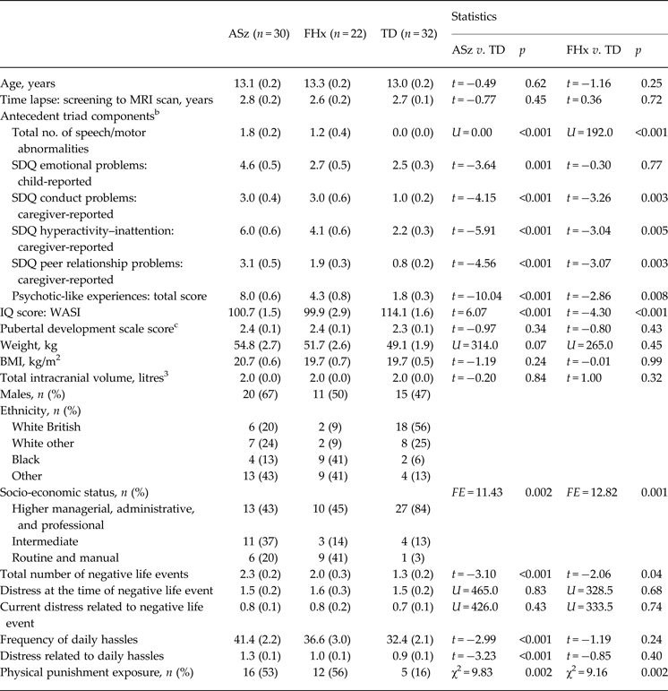
ASz and FHx groups obtained significantly higher scores on all antecedent screening variables relative to the TD group (p ⩽ 0.008), with the exception that the FHx group did not differ from the TD group on SDQ emotional problems (Table 1).
Sample characteristics by risk group a

Data are given as mean (standard error) unless otherwise indicated.
ASz, Antecedents of schizophrenia; FHx, family history of schizophrenia; TD, typically developing; MRI, magnetic resonance imaging; SDQ, Strengths and Difficulties Questionnaire (Goodman, Reference Goodman2001); IQ, intelligence quotient; WASI, Wechsler Abbreviated Scale of Intelligence (Wechsler, Reference Wechsler1999); BMI, body mass index; FE, Fisher's exact.
a Groups are not mutually exclusive (five children meeting both FHx and ASz criteria are retained in both risk groups).
b Antecedents of schizophrenia assessed at screening.
c Higher scores indicate more advanced pubertal development and mean scores in all groups equate to mid-pubertal stage. Missing data: weight (n = 4); BMI (n = 4).
Procedure
Caregivers and children provided written informed consent and assent, respectively, for participation. Ethical permission for the study was granted by the Joint South London and Maudsley and the Institute of Psychiatry NHS Research Ethics Committee. The current study uses cross-sectional data drawn from the overall longitudinal investigation involving biennial assessment of participants; the data presented are derived predominantly from the second assessment, completed when participants were aged between 11 and 14 years. Child participants and their caregivers attended an assessment session at the Institute of Psychiatry, Psychology & Neuroscience which included a structural magnetic resonance imaging (MRI) scan (described below). Caregivers provided information on ethnicity (determined during the FIGS interview) and parental occupation status (coded according to the UK National Statistics Socio-economic Classification; Office for National Statistics, 2010). Participants completed the self-report Pubertal Developmental Scale (Carskadon & Acebo, Reference Carskadon and Acebo1993) to assess pubertal status; the two-subtest version of the Wechsler Abbreviated Scale of Intelligence (Wechsler, Reference Wechsler1999) was used to provide age-adjusted intelligence quotient (IQ) scores.
MRI procedures
Scans were conducted on a 3T GE Medical Systems MRI scanner (General Electric, USA) based at the Centre for Neuroimaging Sciences, Institute of Psychiatry, Psychology & Neuroscience. Structural images were acquired using a 5 min, three-dimensional spoiled gradient recalled echo sequence yielding 196 slices each of 1.1 mm thickness (repetition time = 6.0 ms, echo time = 2.8 ms, flip angle = 20°, field of view = 28 × 21 cm2, and acquisition matrix = 256 × 256).
Pituitary measurements
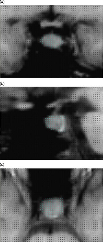
Anonymized scans were imported into Measure (version 0.8, John Hopkins University, USA) and the pituitary was traced manually in the coronal view using the stereological Cavalieri method employing a point counting procedure. To compute total pituitary volumes (mm3), pixel grids were superimposed on each scan slice and pixels falling within the pituitary gland were selected on each slice on which the pituitary could be visualized (Fig. 1). Anatomical boundaries were determined using a protocol implemented in previous studies (Pariante et al. Reference Pariante, Vassilopoulou, Velakoulis, Phillips, Soulsby, Wood, Brewer, Smith, Dazzan, Yung, Zervas, Christodoulou, Murray, McGorry and Pantelis2004, Reference Pariante, Dazzan, Danese, Morgan, Brudaglio, Morgan, Fearon, Orr, Hutchinson, Pantelis, Velakoulis, Jones, Leff and Murray2005; Mondelli et al. Reference Mondelli, Dazzan, Gabilondo, Tournikioti, Walshe, Marshall, Schulze, Murray, McDonald and Pariante2008). Measurements excluded the pituitary stalk but included the posterior bright spot, thought to correspond to the posterior pituitary. The superior and inferior boundaries of the pituitary were defined by the diaphragma sellae and the sphenoid sinus, respectively, and the cavernous sinuses were used to indicate the lateral boundaries. Tracings were performed by one rater (A.E.C.) trained for the purpose of this study. Training was provided by an experienced tracer (F.L.D., who has previously trained eight individuals) with expertise in tracing the pituitary gland in UHR youth; the intraclass correlation coefficient (A.E.C. and F.L.D.) computed from ratings obtained on 10 scans was high (r = 0.97).
Coronal (a), sagittal (b) and axial (c) view of a structural magnetic resonance image in Measure (version 0.8, John Hopkins University, USA) with superimposed pixel grid. The pituitary gland (centre; grey pixels; blue pixels in the online version) was manually traced by selecting pixels falling within the boundaries of the gland. A colour figure is available in the online version of this paper.

Total intracranial volume (ICV)
Total ICV was obtained using Statistical Parametric Mapping 5 software (SPM5; http://www.fil.ion.ucl.ac.uk/spm/software/spm5). Images were first segmented in SPM5 using a study-specific template created within the Template-O-Matic (TOM) toolbox (https://irc.cchmc.org/software/tom.php). Specifically, brain images used to create the template were derived from reference data obtained from 404 healthy children (Wilke et al. Reference Wilke, Holland, Altaye and Gaser2008), who were selected according to the age and sex characteristics of the total sample of participants in the current study. Total grey matter, white matter and cerebrospinal fluid volumes were obtained from segmented images and summed to derive a total ICV for each participant.
Psychosocial stress
As detailed previously (Cullen et al. Reference Cullen, Fisher, Roberts, Pariante and Laurens2014a ), an adaptation of the questionnaire by Heubeck & O'Sullivan (Reference Heubeck and O'Sullivan1998) was used to elicit self-reported exposure and distress relating to child-appropriate negative life events (eight items) and school-related daily hassles (37 items) (Heubeck & O'Sullivan, Reference Heubeck and O'Sullivan1998). The negative life event measure provided information on the total number of events ever experienced, and the average level of distress experienced in relation to each event, both at the time of the event and currently. Previous and current levels of distress were rated for each item on a four-point scale (range: 0 ‘not at all’ to 3 ‘a lot’), scores were then summed across items and divided by the total number of events experienced to derive two average distress ratings. The daily hassles measure assessed scholastic-, peer-, teacher- and home-related hassles occurring during the past 6 months, providing a total daily hassles frequency score [sum of frequency ratings for each item (range: 0 ‘never’ to 3 ‘not at all’)] and an overall (average) distress score [sum of distress ratings for each item (range: 0 ‘not at all’ to 3 ‘a lot’), divided by the number of items endorsed].
Physical punishment exposure was assessed using the corporal punishment scale of the Alabama Parenting Questionnaire (Shelton et al. Reference Shelton, Frick and Wootton1996), completed by the child and caregiver independently at the current (second) and first (age 9–12 years) assessments. The scale includes three items: (i) spanking; (ii) slapping; and (iii) hitting with a belt, switch or other object. Children and their caregivers indicated how often each item typically occurred in their home on a five-point scale (1 ‘never’, 2 ‘almost never’, 3 ‘sometimes’, 4 ‘often’, and 5 ‘always’). Consistent with previous studies (Afifi et al. Reference Afifi, Mota, Dasiewicz, MacMillan and Sareen2012), physical punishment was defined as present if punishment was reported to have occurred ‘sometimes’ or more (child- or caregiver-report at either assessment). Thus, a single (cross-sectional) indicator of physical punishment reported within the past 24 months was derived for each participant.
Statistical analyses
Group differences on demographic variables and psychosocial stress measures were examined using independent-samples t tests, Mann–Whitney U tests, χ2 tests and Fisher's exact tests, as appropriate to the distribution of the variables. Associations between pituitary volume and demographic variables were explored using independent-samples t tests, one-way analyses of variance and correlation analyses. Linear regression analyses were used to examine the effect of risk status on pituitary volume which was approximately normally distributed across the sample. Owing to the non-mutually exclusive nature of the ASz and FHx groups (described below), the effect of each at-risk group was tested independently (i.e. ASz and FHx groups were examined relative to the TD group but were not directly compared with each other). Analyses were subsequently adjusted for demographic factors associated with either group status or pituitary volume (IQ, sex, ethnicity, socio-economic status, pubertal status and weight). Unstandardized regression coefficients (unadjusted and adjusted) were used to derive standardized mean differences (d) as indices of effect size (Lipsey & Wilson, Reference Lipsey and Wilson2001). Exploratory correlation analyses (Pearson's ‘r’ and Spearman's rho ‘ρ’ for normally and non-normally distributed variables, respectively) were then conducted in the ASz and FHx groups to examine associations between antecedents assessed at screening and pituitary volume. Within-group correlation analyses were next conducted to examine the extent to which pituitary volume was associated with exposure and distress related to negative life events and daily hassles; the relationship between physical punishment and pituitary volume was examined using within-group independent-samples t tests.
Results
Sample characteristics
Pituitary volumes were obtained for 79 participants; 25 children met ASz criteria only, 17 met FHx criteria only, five met both ASz and FHx criteria, and 32 met TD criteria. The five ASz + FHx cases were included in both at-risk groups, yielding data for 30 ASz and 22 FHx children in total. Neither the ASz nor FHx group differed from the TD group on age, lapse of time between screening and MRI scan, sex, pubertal status, weight, body mass index (BMI) or total ICV (p > 0.05; Table 1). Relative to the TD group, both the ASz and FHx groups were found to differ significantly on IQ (p ⩽ 0.001), ethnicity (p ⩽ 0.008) and socio-economic status (p ⩽ 0.001).
Sociodemographic correlates of pituitary volume
Pituitary volume was not significantly associated with age, IQ, total ICV, ethnicity or socio-economic status (p > 0.10). As is typical, females had significantly larger pituitary volumes than males [mean 504.8 (s.e. 27.4) v. 372.8 (s.e. 26.4), respectively; t = −3.47, p = 0.001]. Pituitary volume was significantly correlated with weight (ρ = 0.26, p = 0.02) and pubertal status (r = 0.38, p = 0.001), and with BMI at a trend level (ρ = 0.21, p = 0.07).
Pituitary volume by risk group
Linear regression analyses indicated no significant differences in pituitary volume when either ASz children (d = −0.12, p = 0.64) or FHx children (d = 0.04, p = 0.89) were compared with the TD group (Table 2). Results were unchanged after adjustment for factors associated with group status and/or pituitary volume (i.e. IQ, sex, ethnicity, socio-economic status, pubertal status and weight); analyses stratified by sex also demonstrated no significant effect of group status on pituitary volume (data not shown). Pituitary volume was not significantly correlated with scores on any of the antecedent screening items in either the ASz or FHx group (p > 0.05).
Linear regression analyses examining the effect of risk status on pituitary volume a

ASz, Antecedents of schizophrenia; FHx, family history of schizophrenia; TD, typically developing; d, standardized effect size; B, unstandardized regression coefficient; CI, confidence interval; s.e., standard error.
a Groups are not mutually exclusive (five children meeting both FHx and ASz criteria are retained in both risk groups).
b Linear regression analyses without adjustment for potential confounders (unadjusted) and adjusted for intelligence quotient, sex, ethnicity, socio-economic status, pubertal status and weight (adjusted).
Pituitary volume and psychosocial stress
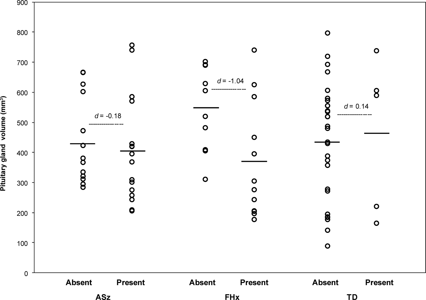
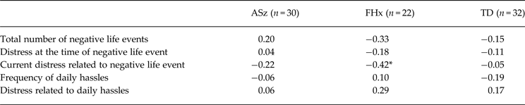
Group differences on psychosocial stress measures are presented in Table 1. Consistent with our previous analyses in a larger, overlapping sample, ASz and FHx children were exposed to a greater number of negative life events than the TD group (p < 0.05) and ASz children also reported that they more frequently experienced daily hassles and were more distressed by these hassles than TD children (p < 0.001). Additionally, ASz and FHx groups were both more likely to experience physical punishment than TD children (p = 0.002). Within-group correlation analyses (Table 3) indicated a significant negative correlation between pituitary volume and current distress relating to negative life events among FHx children only (ρ = −0.42, p = 0.05). Furthermore, as illustrated in Fig. 2, FHx children exposed to physical punishment had significantly smaller pituitary volumes compared with FHx children who had not (t = 2.43, p = 0.03), equating to a large effect size (d = −1.01). In the ASz and TD groups, however, there was no association between pituitary volume and exposure to physical punishment (p > 0.60).
Pituitary gland volume in those exposed to physical punishment (present) and those not exposed to physical punishment (absent) by risk group. Individual data are shown, with means indicated by horizontal lines. ASz, Antecedents of schizophrenia; FHx, family history of schizophrenia; TD, typically developing; d, standardized effect size.

Correlations between pituitary volume, negative life events and daily hassles

ASz, Antecedents of schizophrenia; FHx, family history of schizophrenia; TD, typically developing.
* p ⩽ 0.05.
Discussion
In the first study to examine pituitary volume among antipsychotic-naive children at putatively elevated risk for schizophrenia, in contrast to hypotheses, neither children with a family history of schizophrenia nor children presenting antecedents of schizophrenia were characterized by abnormal pituitary volume relative to their TD peers. However, among FHx children only, pituitary volume was negatively associated with current distress relating to negative life events and with exposure to physical punishment.
The finding that neither FHx nor ASz children were characterized by pituitary volume abnormalities relative to the TD group contrasts with the results of some previous studies of individuals at elevated risk for schizophrenia, but not all. Mondelli et al. (Reference Mondelli, Dazzan, Gabilondo, Tournikioti, Walshe, Marshall, Schulze, Murray, McDonald and Pariante2008) observed significantly larger pituitary volumes among adult relatives (predominately parents) of patients with schizophrenia, yet studies of younger samples of relatives have observed no differences in pituitary volume between relatives and healthy controls (Habets et al. Reference Habets, Collip, Myin-Germeys, Gronenschild, van Bronswijk, Hofman, Lataster, Lardinois, Nicolson, van Os and Marcelis2012; Shah et al. Reference Shah, Tandon, Howard, Mermon, Miewald, Montrose and Keshavan2015). Given the large differences in participant age across these investigations, age-related changes in pituitary volume may have contributed to the heterogeneous findings across studies. Inconsistent findings have also been reported by studies of young adults with SPD. Takahashi et al. (Reference Takahashi, Suzuki, Velakoulis, Lorenzetti, Soulsby, Zhou, Nakamura, Seto, Kurachi and Pantelis2009) observed significantly larger pituitary volumes in individuals with SPD relative to healthy controls, yet a further study observed smaller pituitary volumes among males, but not females, with SPD (Romo-Nava et al. Reference Romo-Nava, Hoogenboom, Pelavin, Alvarado, Bobrow, Macmaster, Keshavan, McCarley and Shenton2013). The contrast in findings is likely to reflect the fact that individuals with SPD in the former study were psychiatric patients, the majority of whom were receiving antipsychotic medication; however, individuals in the latter study were recruited from the community and were all medication-naive. Contrasting findings have also been reported among UHR youth. Two studies observed no differences in pituitary volume between UHR youth and healthy controls (Garner et al. Reference Garner, Pariante, Wood, Velakoulis, Phillips, Soulsby, Brewer, Smith, Dazzan, Berger, Yung, van den Buuse, Murray, McGorry and Pantelis2005; Büschlen et al. Reference Büschlen, Berger, Borgwardt, Aston, Gschwandtner, Pflueger, Kuster, Radu, Stieglitz and Riecher-Rossler2011), although both studies observed pituitary volume increases among UHR youth who later transitioned to psychosis compared with those who did not. However, subsequent studies have observed no association between transition status and pituitary volume (Takahashi et al. Reference Takahashi, Nakamura, Nishiyama, Furuichi, Ikeda, Kido, Nakamura, Kawasaki, Noguchi, Seto and Suzuki2013; Walter et al. Reference Walter, Studerus, Smieskova, Tamagni, Rapp, Borgwardt and Riecher-Rossler2015), although, when the total sample of UHR youth was compared with the control group, Takahashi et al. (Reference Takahashi, Nakamura, Nishiyama, Furuichi, Ikeda, Kido, Nakamura, Kawasaki, Noguchi, Seto and Suzuki2013) reported significantly larger pituitary volumes among UHR youth. Differences in age, illness severity and medication status may contribute to inconsistencies observed across studies of high-risk individuals.
Among FHx children, a significant negative correlation was observed between pituitary volume and current distress relating to negative life events. This finding contrasts with a previous study in which a weak positive association was observed between pituitary volume and emotional stress reactivity (i.e. negative affect relating to daily stressful experiences) among patients with psychosis, and, to a lesser extent, their adult siblings and healthy controls (Habets et al. Reference Habets, Collip, Myin-Germeys, Gronenschild, van Bronswijk, Hofman, Lataster, Lardinois, Nicolson, van Os and Marcelis2012). These conflicting findings may reflect differences in the age of participants and/or the type of stressor examined; whilst greater distress relating to major traumatic events appears to be associated with smaller pituitary volume (at least among children with a family history of schizophrenia), emotional reactivity to minor stressors may be related to larger pituitary volume. We also observed that FHx children who had experienced physical punishment had significantly smaller pituitary volumes than those who had not, equating to a large effect size. This finding is consistent with a previous study of adolescents with borderline personality disorder (BPD) in which there were no differences in pituitary volume between adolescents with BPD and healthy controls, yet in the BPD group, those exposed to childhood maltreatment were characterized by smaller pituitary volumes compared with unexposed youth (Garner et al. Reference Garner, Chanen, Phillips, Velakoulis, Wood, Jackson, Pantelis and McGorry2007). Our findings suggest that the relationship between pituitary volume and childhood maltreatment might extend to less severe forms of harsh physical parenting.
The above findings indicate that experiences of psychosocial stress may contribute to changes in pituitary volume. Thus, one potential explanation for the lack of consistent findings across studies examining pituitary volume among at-risk individuals is that pituitary volume abnormalities may be caused by an interaction between psychosocial stress and at-risk status. Specifically, it may be that only vulnerable individuals who subsequently experience psychosocial stress will go on to develop pituitary volume abnormalities. One alternative explanation for the finding that pituitary volume was associated with psychosocial stress among FHx children only (i.e. aside from the fact that these children have increased vulnerability for psychosis) is that other environmental stressors such as ethnic minority and low socio-economic status may have influenced HPA axis responsivity to stressors in this group (Chong et al. Reference Chong, Uhart, McCaul, Johnson and Wand2008; Marsman et al. Reference Marsman, Nederhof, Rosmalen, Oldehinkel, Ormel and Buitelaar2012). However, ASz children also differed from the TD group on these demographic factors; thus, genetic vulnerability may drive this association.
The current study provides new insights into the nature and timing of pituitary volume abnormalities in schizophrenia. A possible explanation for the lack of pre-morbid pituitary volume enlargements in ASz and FHx children (a finding observed in some at-risk samples) is that HPA axis hyperactivity is associated with more severe clinical presentation. In support of this suggestion, our previous report indicated no elevation of diurnal cortisol levels in ASz or FHx children (Cullen et al. Reference Cullen, Zunszain, Dickson, Roberts, Fisher, Pariante and Laurens2014b ). However, the fact that enlarged pituitary volumes have been observed among relatives of patients with schizophrenia who are not acutely unwell (Mondelli et al. Reference Mondelli, Dazzan, Gabilondo, Tournikioti, Walshe, Marshall, Schulze, Murray, McDonald and Pariante2008) suggests that genetic liability for psychosis may play some role in the expression of pituitary volume abnormalities. Whilst the current findings, and those of two previous studies of younger relatives (Habets et al. Reference Habets, Collip, Myin-Germeys, Gronenschild, van Bronswijk, Hofman, Lataster, Lardinois, Nicolson, van Os and Marcelis2012; Shah et al. Reference Shah, Tandon, Howard, Mermon, Miewald, Montrose and Keshavan2015), do not support this hypothesis, the larger pituitary volumes characterizing older adult relatives may nonetheless reflect genetic predisposition to HPA axis hyperactivity that emerges with increasing age following accumulated life stress.
The current study also tentatively suggests that experiences of psychosocial stress may contribute to pituitary volume abnormalities. Whilst the negative association that we observed between pituitary volume and psychosocial stress among FHx children is to some extent inconsistent with the notion that psychosocial stress may lead to HPA axis hyperactivity (as indexed by increases in pituitary volume and cortisol secretion), it has been suggested that elevated cortisol levels may have an inhibitory action on the pituitary corticotroph cells that produce HPA axis hormones, eventually leading to pituitary volume reduction (Sassi et al. Reference Sassi, Nicoletti, Brambilla, Harenski, Mallinger, Frank, Kupfer, Keshavan and Soares2001). Consistent with this notion, it has recently been proposed that chronic HPA axis hyperactivity may eventually exhaust the HPA axis such that the system then has limited capacity to mount a response to acute stressors (Shah & Malla, Reference Shah and Malla2015). Thus, whilst stressful experiences might have been positively associated with increased cortisol levels and enlarged pituitary volume at the time that they occurred, enduring distress relating to these experiences among FHx children might result in pituitary volume reduction.
Limitations
Whilst the current study is limited by the relatively small groups, this is not uncommon in high-risk studies examining biological indices of HPA axis function. The study was nonetheless sufficiently powered to detect significant associations between pituitary volume and experiences of psychosocial stress among FHx children. A further limitation relates to the fact that the transition rate among the ASz group is currently unknown, but may be lower than in FHx children (i.e. given that a positive family history remains one of the strongest known predictors of schizophrenia), and lower than among UHR youth (i.e. given that ASz children are not currently seeking treatment for their symptoms). The number of ASz children actually in the pre-morbid stages of illness may be low, thus yielding insufficient statistical power to detect pituitary volume abnormalities in this group. However, by recruiting high-risk youth from the community rather than via clinical services, we were able to obtain an antipsychotic-naive group which is particularly important for studies of pituitary volume (MacMaster et al. Reference MacMaster, El-Sheikh, Upadhyaya, Nutche, Rosenberg and Keshavan2007; Nicolo et al. Reference Nicolo, Berger, Garner, Velakoulis, Markulev, Kerr, McGorry, Proffitt, McConchie, Pantelis and Wood2010).
Regarding psychosocial stress measures; first, at the age 11–14 year follow-up we did not assess more severe forms of psychosocial stress such as childhood trauma which may have shown more robust associations with pituitary volume. However, using a well-established measure of parenting practices we were able to examine physical punishment, which lies on a continuum with childhood maltreatment. Second, whilst our decision to create a binary variable indexing physical punishment based on a rating of ‘sometimes or more’ was guided by previous research (Afifi et al. Reference Afifi, Mota, Dasiewicz, MacMillan and Sareen2012), this is an arbitrary cut-off, and reducing a complex exposure into a single indicator may lack clinical validity. Third, our ability to measure levels of distress relating to negative life events at the time which they occurred may be limited due to problems with recall. Finally, whilst attempts were made to complete psychosocial stress measures within 1 month of the structural MRI scan, this was not always possible. However, as our groups did not differ on the lapse of time between completing these measures, this cannot explain the different patterns of association observed across groups.
Conclusions
The current study indicates a lack of pituitary gland volume abnormalities among children at putatively elevated risk for schizophrenia relative to their TD peers. These findings are consistent with our previous work demonstrating that these putatively at-risk children are also not characterized by elevated cortisol levels during the day (Cullen et al. Reference Cullen, Zunszain, Dickson, Roberts, Fisher, Pariante and Laurens2014b ), and imply that these biological markers of HPA axis hyperactivity that have been observed in some older samples of high-risk individuals may emerge later, more proximally to disease onset. Alternatively, it is possible that an interactive effect of psychosocial stress and at-risk status may have obscured the examination of a main effect of at-risk status on pituitary volume. This study supports the notion that experiences of psychosocial stress are associated with pituitary volume abnormalities among those with increased vulnerability for schizophrenia, though longitudinal studies are needed to establish the temporal nature of this relationship.
Acknowledgements
This work was supported by funding to K.R.L. from a National Institute for Health Research (NIHR) Career Development Fellowship (CDF/08/01/015); a Bial Foundation Research Grant (36/06); a National Alliance for Research on Schizophrenia and Depression (NARSAD) Young Investigator Award (2005); the British Medical Association Margaret Temple Award for schizophrenia research (2006); and the Schizophrenia Research Institute, utilizing infrastructure funding from the NSW Ministry of Health. All authors are affiliated with the NIHR Specialist Biomedical Research Centre (BRC) for Mental Health at the South London and Maudsley NHS Foundation Trust and Institute of Psychiatry, King's College London, UK. The authors thank the children and caregivers who participated in the study and the researchers and students who contributed to data collection.
Declaration of Interest
None.