


The sphingolipid-storage diseases are a collection of ~40 genetically distinct disorders caused by inherited deficiencies of lysosomal hydrolytic activities or lipid transport that result in intracellular accumulations of cholesterol and lipids in the endosomal–lysosomal network. These disorders have an aggregate prevalence of approximately 1 in 8000 births worldwide (Ref. Reference Meikle1). Among this group is Niemann–Pick type C disease (NPC, MIM 257220), an autosomal recessive, progressive neurovisceral disorder characterised by abnormal cholesterol trafficking and by intracellular accumulations of unesterified cholesterol and glycosphingolipids in late endosomes and lysosomes (Refs Reference German2, Reference Patel3, Reference Vanier and Millat4, Reference Higgins5, Reference Karten6, Reference Karten7, Reference Reid, Sugii and Chang8). These lipid accumulations cause a multifaceted toxic response in the liver, spleen and brain. NPC is caused by loss-of-function mutations in either NPC1 (Ref. Reference Carstea9) or NPC2 (Ref. Reference Naureckiene10); the protein products of these genes mediate proper intracellular lipid transport through pathways that are incompletely understood.
NPC patients develop symptoms over a wide range of ages (Ref. Reference Patterson11), and there is no strict correlation between disease-causing mutations and the severity of the clinical phenotype (Refs Reference Vanier and Millat4, Reference Yamamoto12). While patients may initially exhibit systemic findings such as hepatosplenomegaly or obstructive jaundice, all eventually develop neurological and/or psychiatric symptoms, the severity of which is inversely associated with lifespan (Refs Reference Imrie13, Reference Turpin, Masson and Baumann14). Patients presenting with neurological symptoms during the first two years of life, classified as the most extreme, infantile cases of NPC, demonstrate delayed motor development and hypotonia, and typically die within their first 5 years (Ref. Reference Vanier15). The classical form of NPC, which encompasses ~60–70% of cases, presents between the ages of 3 and 15 years. Parents of these patients are often the first to notice troublesome signs of the disease, such as loss of speech or increasing clumsiness. The disorder progresses to cause a constellation of neurological symptoms that may include cerebellar ataxia, dysarthria, dysphagia, cataplexy, seizures, dystonia, vertical gaze palsy, progressive dementia and death by 8–25 years (Ref. Reference Patterson11). Adult-onset cases of NPC are well documented although much less common, and their unusual clinical presentation often initially leads to misdiagnosis. Most adults with NPC develop symptoms in their second or third decades of life, although onset can occur as late as the mid-sixth decade (Ref. Reference Sevin16). These adult patients develop symptoms similar to the classical form of juvenile NPC and usually die when in their late 30s or 40s. At the current time, there are no effective treatments available to patients with this devastating disorder.
NPC pathology and biochemical defects
While heterogeneity characterises the clinical presentation of NPC, there is less pathological heterogeneity at the biochemical level. The vast majority of cases show prominent accumulations of unesterified cholesterol, sphingolipids and complex gangliosides in late endosomes and lysosomes of most cells types of the body (Refs Reference German2, Reference Patel3, Reference Vanier and Millat4, Reference Patterson11). However, it should be noted that a subset of patients with certain mutations demonstrate much less lipid storage (Ref. Reference Millat17), and central nervous system (CNS) pathology is accompanied by particularly prominent storage of sphingolipids and complex gangliosides.
Histopathology
The progression of NPC in the periphery is characterised by an enlargement of the liver and spleen that results from the presence of lipid-laden macrophages, termed foam cells. Kupffer cells in the liver and splenic macrophages, the latter predominantly in the red pulp, exhibit marked cytoplasmic vacuolisation due to an accumulation of cholesterol, phospholipids and glycolipids.
Impairment of lipid trafficking also affects the CNS and results in neuron loss throughout the brain (Ref. Reference Walkley and Suzuki18). The occurrence of swollen neuronal cell bodies in many brain regions, including cortex, basal ganglia, thalamus, cerebellum and brainstem is also characteristic of NPC and reflects lipid accumulation within late endosomes and lysosomes. The cell body of these neurons and their axonal processes also contain granular material, which by electron microscopy appears as membrane-bound polymorphous bodies containing loosely packed lamellae or as dense osmiophilic inclusions. Additional neuronal pathology includes the formation of ectopic dendrites, swelling of proximal axons to so-called meganeurites, and the presence of axonal spheroids – changes indicative of a neuroaxonal dystrophy (Ref. Reference Walkley and Suzuki18). Like several other neurodegenerative diseases, NPC is classified as a tauopathy due to the presence of intracellular aggregates of the microtubule-binding protein tau, which are biochemically identical to aggregates in Alzheimer disease (Ref. Reference Auer19). These changes in the NPC brain are accompanied by gliosis in grey and white matter, microglial activation and cerebral atrophy.
Biochemistry
Lipid-trafficking defects within the NPC brain reflect defects in the pathway by which cholesterol and other lipids arrive in neurons and are sorted intracellularly. Studies in normal mice have demonstrated that cholesterol accumulation in the CNS markedly increases during the first three weeks of life, a period of late development and myelination (Refs Reference Dietschy and Turley20, Reference Quan21). Lipoprotein cholesterol from the periphery does not cross the blood–brain barrier to enter the CNS, even during this early phase of brain development, and therefore de novo synthesis is required to meet the needs of both the developing and mature nervous system (Refs Reference Herz and Bock22, Reference Turley23, Reference Dietschy and Turley24). Neurons and other CNS cell types obtain the cholesterol they require though endogenous synthesis or by uptake of lipoprotein cholesterol particles produced and released within the nervous system, possibly by astrocytes (Ref. Reference Mauch25). Once target cells internalise these particles, unesterified cholesterol and companion lipids are trafficked from the endosomal–lysosomal system to compartments such as the Golgi complex and endoplasmic reticulum for processing and to serve as substrates for further reactions (Ref. Reference Pagano26). Derivatives of cholesterol include oxysterols, which signal a decrease in the de novo synthesis of cholesterol and therefore provide negative feedback (Refs Reference Fu27, Reference Janowski28).
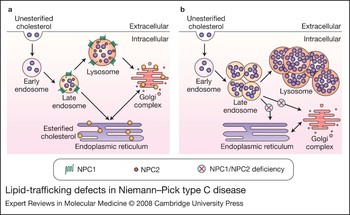
In NPC fibroblasts, lipoprotein cholesterol particles are internalised without disruption but become entrapped within trafficking vesicles, particularly late endosomes and lysosomes (Fig. 1) (Refs Reference Ory29, Reference Liscum, Ruggiero and Faust30, Reference Sokol31). The insufficient efflux of cholesterol and lipids out of these structures causes a twofold problem: unesterified cholesterol, sphingolipids and complex gangliosides accumulate within vesicles in the cytoplasm of the cell, while there is a simultaneous paucity of cholesterol and other essential lipids in organelles to which these compounds were originally destined. Decreased cholesterol in the endoplasmic reticulum, Golgi complex and other internal organelles causes deleterious effects on a multitude of processes dependent on proper membrane composition (Ref. Reference Wojtanik and Liscum32). Disrupted trafficking also results in a paucity of substrate for further synthetic reactions. For example, decreased availability of unesterified cholesterol impairs production of oxysterols, which may lead to increased cholesterol synthesis and an exacerbation of cholesterol accumulation in NPC cells (Ref. Reference Vance33).
Lipid-trafficking defects in Niemann–Pick type C disease. (a) Under normal conditions, lipoprotein cholesterol particles enter the endosomal network after binding to cell-surface receptors. Unesterified cholesterol and companion lipids are trafficked from the endosomal–lysosomal system to the Golgi complex, endoplasmic reticulum and other intracellular compartments. (b) Loss of function of NPC1 or NPC2 inhibits the egress of unesterified cholesterol and sphingolipids from late endosomes and lysosomes, resulting in their accumulation at these sites and a paucity of these lipids at the intracellular compartments to which they were destined. Abbreviations: NPC, Niemann–Pick type C.
Genetics of NPC
Almost a century has passed since the original reports by Albert Niemann and Ludwick Pick described patients with hepatosplenomegaly and rapidly progressive brain damage. Since then, biochemical and genetic studies have helped define the classification of the Niemann–Pick disease group. Niemann–Pick types A and B disease (MIM 257200 and 607616), distinct from NPC, are caused by loss of function mutations in the acid sphingomyelinase gene, encoding an enzyme that metabolises sphingomyelin to ceramide and phosphocholine. Although fibroblasts from NPC patients may show reduced sphingomyelinase activity in culture, this reduction is due to an imbalanced lipid and cholesterol environment, and removal of cholesterol from the media recovers enzymatic activity (Ref. Reference Thomas34). By contrast, NPC fibroblasts exhibit an accumulation of unesterified cholesterol indicative of a defect in cholesterol trafficking. The major complementation group for NPC (Ref. Reference Patterson11) was mapped to the proximal long arm of chromosome 18, and mutations in the NPC1 gene were found in ~95% of patients with this disease (Ref. Reference Carstea9). The remaining 5% of NPC patients were subsequently found to have loss-of-function mutations in a separate gene, NPC2 (Ref. Reference Naureckiene10).
The NPC1 gene encodes a multipass transmembrane protein, NPC1, which is localised to late endosomes and lysosomes (Refs Reference Higgins35, Reference Neufeld36). The encoded protein is predicted to contain a sterol-sensing domain, similar to that found in regulators of cholesterol metabolism and in Patched (PTCH1), a receptor for the secreted signalling protein Hedgehog (SHH) (Refs Reference Carstea9, Reference Watari37). Experimental evidence indicates that NPC1 is involved in the efflux of low-density lipoprotein (LDL)-derived unesterified cholesterol from late endosomes and lysosomes to other intracellular compartments (Refs Reference Liscum, Ruggiero and Faust30, Reference Sokol31). Multiple disease-causing mutations within the NPC1 coding region have been identified that lead to its loss of function (Ref. Reference Runz38). The NPC1 protein is conserved within the animal kingdom, and loss-of-function mutations in orthologous genes in yeast (Ref. Reference Malathi39), worms (Ref. Reference Sym, Basson and Johnson40), flies (Ref. Reference Huang41), mice (Ref. Reference Pentchev42), cats (Ref. Reference Brown43) and dogs (Ref. Reference Kuwamura44) disrupt lipid trafficking. Introduction of the Saccharomyces cerevisiae NPC1 orthologue, NCRP1, into Chinese hamster ovary cells deficient in NPC1 is sufficient to reverse lipid-trafficking defects associated with NPC1 deficiency, demonstrating that this protein is functionally conserved from yeast to mammals (Ref. Reference Malathi39).
The NPC2 gene encodes a small, soluble protein that is localised to lysosomes, but is also secreted (Refs Reference Naureckiene10, Reference Mutka45). Loss-of-function mutations in this gene result in a disease phenotype that is clinically and biochemically indistinguishable from the disease caused by NPC1 mutations. The NPC2 protein binds cholesterol, and structural analysis has identified a hydrophobic pocket that serves as a putative cholesterol-binding domain (Ref. Reference Friedland46). Disease-causing mutations in NPC2 that affect amino acid residues surrounding this pocket impair cholesterol binding (Ref. Reference Ko47). Analysis of Npc1–Npc2 double mutant mice demonstrates that the disease phenotype is similar to that occurring in Npc1 or Npc2 single mutants, providing genetic evidence that the NPC1 and NPC2 proteins function in the same pathway to regulate intracellular lipid transport (Ref. Reference Sleat48).
NPC1-deficient mice
Phenotype
The most commonly used mammalian model of NPC, BALB/cNctr-Npc1m1 N/J mice (Jackson Laboratory #003092), contains a transposon insertion into Npc1 exon 9 (Ref. Reference Loftus49). Animals homozygous for this insertional mutation fail to express functional NPC1 protein and exhibit a degenerative phenotype that begins around 7 weeks (Refs Reference Loftus49, Reference Morris50). The severity of the phenotype progresses with age and includes weight loss, tremors, ataxia, and death by 10–12 weeks. The liver and spleen contain an abundance of foam cells, reminiscent of the histopathological changes seen in humans with this disorder (Fig. 2). Recently, in vivo antisense knockdown of Npc1 expression only in the liver of normal mice has been shown to recapitulate the hepatic pathology associated with NPC, generating a novel model to examine liver dysfunction associated with disease independent of CNS involvement (Ref. Reference Rimkunas51).
Niemann–Pick type C disease pathology. Foamy macrophages are abundant in the liver and spleen of NPC1-deficient mice (examples at arrowheads). In the brain, loss of cerebellar Purkinje cells is a prominent pathological feature; these large, pyramidal-shaped neurons normally reside at the interface of the molecular layer and granular cell layer of the cerebellar folia (examples at arrowheads). These cells die in NPC1-deficient mice and are replaced by reactive astrocytes. The histopathology of NPC1-deficient mice has been previously described (examples include Refs Reference Loftus49, Reference Morris50, Reference Takikita55, Reference Higashi56). Abbreviation: NPC, Niemann–Pick type C.

Global loss of Npc1 expression leads to CNS pathology including vacuolated neurons that accumulate glucosylceramide, lactosylceramide, complex gangliosides and cholesterol (Refs Reference Walkley52, Reference Xie53, Reference Zervas, Dobrenis and Walkley54), abnormally swollen axons, and decreased myelination in the cerebral white matter (Ref. Reference Takikita55). A prominent feature of CNS pathology is loss of Purkinje cells, the major neuronal cell type transmitting output from the cerebellum (Ref. Reference Higashi56). These highly specialized neurons are responsible for timing and smoothness of movements, and dysfunction of or lesions within the cerebellum can lead to ataxia. Loss of Purkinje cells is initially apparent in the cerebellar vermis by 7 weeks. Progressive Purkinje cell loss proceeds from the anterior to posterior vermis, yet largely spares lobules IX and X, even in mice with end-stage disease (Ref. Reference Higashi56). Interestingly, 2-year-old Npc1 +/− mice also exhibit mild Purkinje cell loss, suggesting that haploinsufficiency for NPC1 may have adverse consequences in the ageing brain (Ref. Reference Yu57).
Lipid-trafficking defects
NPC1-deficient mice have been used to explore how lipid-trafficking defects lead to progressive neurodegeneration. Attention has focused on the role of glycosphingolipids, including complex gangliosides such as GM2, which accumulate in NPC neurons (Ref. Reference Zervas, Dobrenis and Walkley54); although involved in normal brain development, these are not commonly found in the adult brain. Their presence in the NPC brain has been implicated in pathogenesis, including the formation of ectopic dendrites that branch from swollen axons (Ref. Reference Walkley and Suzuki18). These abnormally long and inappropriately located processes may contribute to neuronal dysfunction. Based on this notion, on indications that some glycosphingolipids are toxic, and on findings that cholesterol accumulation in the NPC brain is ganglioside-dependent (Ref. Reference Gondre-Lewis, McGlynn and Walkley58), glycosphingolipid synthesis has emerged as a therapeutic target in NPC and related sphingolipid-storage disorders (Refs Reference Cox59, Reference Zervas60, Reference Patterson61).
Trafficking defects also lead to diminished production of cholesterol derivatives including neurosteroids, which normally help mediate brain development, growth and differentiation (Ref. Reference Mellon62). A single injection of the neurosteroid allopregnanolone, whose production is significantly decreased in NPC mice, is sufficient to promote Purkinje cell survival and delay disease phenotype (Ref. Reference Griffin63). Although initial evidence suggested that this beneficial effect was mediated via signalling from the GABAA receptor (class A receptor for γ-aminobutyric acid), more recent studies indicate that it occurs by activation of the pregnane X receptor (PXR) or liver X receptor (LXR) (Ref. Reference Langmade64). These steroid receptors are ligand-activated transcription factors that bind the oxysterols 25-hydroxycholesterol and 27-hydroxycholesterol (Refs Reference Fu27, Reference Janowski28). Pathways activated by the receptors promote efflux of cholesterol from late endosomes and lysosomes, and suppress endogenous cholesterol synthesis. Exogenous application of either 25-hydroxycholesterol or 27-hydroxycholesterol is sufficient to correct sterol defects in NPC1-deficient human fibroblasts and to mobilise cholesterol from the late endosomal–lysosomal compartment (Ref. Reference Frolov65). LXR agonists also promote cholesterol efflux from the NPC mouse brain, slow neurodegeneration and prolong life (Ref. Reference Repa66), suggesting that activation of these pathways is a promising therapeutic strategy.
Cell-autonomous cell death
Defects in lipid trafficking that occur in NPC lead to cell-autonomous neuronal death. This was established by an analysis of chimaeric mice that express functional NPC1 protein in only a subset of cells (Ref. Reference Ko67). In this model, NPC1-deficient Purkinje cells died, even when neighbouring cells harboured normal levels of NPC1 protein. Conversely, Purkinje cells with functional NPC1 were spared, even in an NPC1-deficient environment. Examination of NPC1-deficient Purkinje cells in mutant mice revealed evidence of autophagy, suggesting that this pathway is activated in the NPC brain and may play a role in pathogenesis (Ref. Reference Ko67).
Autophagy
Autophagy is a conserved cellular pathway that mediates the turnover of damaged or aged macromolecules and organelles. Three types of autophagy have been described: microautophagy, chaperone-mediated autophagy and macroautophagy (Ref. Reference Mizushima68). For the purposes of this review, we discuss only macroautophagy and refer to it simply as ‘autophagy’.
At basal levels, autophagy is a house-keeping mechanism for the constitutive degradation of long-lived proteins and other macromolecules inaccessible to the ubiquitin–proteasome pathway. Regulated degradation is governed by activation of autophagy-related genes (ATGs) (Ref. Reference Klionsky69). Autophagy begins with the generation of an isolation membrane – a crescent-shaped double membrane originating from either smooth endoplasmic reticulum or another subcellular location that has yet to be defined (Ref. Reference Xie and Klionsky70). The isolation membrane surrounds cargo targeted for degradation (Fig. 3). During the engulfment process, two ubiquitin-like conjugation reactions are required for progression through autophagy: the formation of an ATG12–ATG5–ATG16 complex; and the conjugation of ATG8 (also known as microtubule-associated protein 1 light chain 3 protein; LC3) to phosphatidylethanolamine (Ref. Reference Ohsumi and Mizushima71). This latter step is commonly used as a biochemical marker for the induction of autophagy (Refs Reference Kabeya72, Reference Tanida, Ueno and Kominami73). Following engulfment, mature autophagosomes fuse with lysosomes to enable degradation of contents and recycling of their components, a process indicative of flux through the autophagic pathway (Ref. Reference Tanida74).
The autophagic pathway. Autophagy occurs at low, basal levels and can be induced by multiple stimuli, including amino acid starvation or mTOR inhibition. It begins with the formation of an isolation membrane – a crescent-shaped sequestering membrane also termed a phagophore – and the induction of a ubiquitin-like conjugation reaction that couples soluble LC3-I with phosphatidylethanolamine. Lipidated LC3 (LC3-II) studs the phagophore membrane as it engulfs macromolecules, organelles and other cargo destined for degradation. Mature autophagosomes fuse with lysosomes to form autolysosomes, thereby degrading cargo and enabling components to be recycled. Not shown is the fusion of some autophagosomes with endosomes to form so-called amphisomes, another vesicle that fuses with lysosomes to facilitate cargo degradation. Abbreviations: mTOR, mammalian target of rapamycin; LC3, microtubule-associated protein 1 light chain 3 protein (also known as ATG8).

Autophagy can be induced above basal levels by several stimuli, including amino acid starvation or cellular stress. This induction is mediated by signalling through the mammalian target of rapamycin (mTOR) or through a complex of the class III phosphoinositide 3-kinase (PI3K) and Beclin-1 (BECN1) (Ref. Reference Klionsky and Emr75). mTOR is a negative regulator of autophagy and its chemical inhibition by rapamycin relieves this blockade (Refs Reference Klionsky and Emr75, Reference Noda and Ohsumi76). The PI3K–Beclin-1 complex is necessary for the induction of autophagy and can activate autophagy independently of mTOR inhibition (Ref. Reference Yamamoto, Cremona and Rothman77).
Autophagy is necessary for normal CNS development and function, and has been implicated in the pathogenesis of a diverse array of neurodegenerative diseases, including protein-aggregation disorders such as Alzheimer (Ref. Reference Nixon78), Parkinson (Ref. Reference Pan79) and Huntington disease (Ref. Reference Ravikumar80), as well as the sphingolipid-storage disorders (Ref. Reference Kiselyov81). For age-related diseases associated with protein misfolding, inducing autophagy in model systems enhances the clearance of misfolded or mutant proteins that are not efficiently degraded by the proteasome. This enhancement of protein clearance abrogates disease phenotypes (Ref. Reference Williams82), indicating that autophagy may have beneficial effects in some disease processes. Conversely, genetic deletion in mouse CNS of either of two autophagy-related genes – Atg5 (Ref. Reference Kuma83) or Atg7 (Ref. Reference Komatsu84) – leads to neurodegeneration and the accumulation of ubiquitinated protein aggregates, demonstrating that basal autophagy contributes significantly to protein quality control and cell survival in the nervous system.
Although the induction of autophagy may alleviate disease in some instances, in other situations autophagy can have deleterious effects. In mouse embryonic fibroblasts deficient in apoptosis – the classic programmed cell death pathway – because they lack the apoptotic effectors BAX and BAK, application of death stimuli such as staurosporin or etoposide triggers an apoptosis-independent cell death pathway (Ref. Reference Shimizu85). Further genetic manipulation of these cells through RNA interference knockdown of two autophagy regulators – Atg5 and Becn1 – inhibited cell death, demonstrating the occurrence of a death program mediated by autophagy. Contributions from autophagic cell death have been implicated in several processes, including normal development and disease (Refs Reference Mizushima68, Reference Shintani and Klionsky86).
Autophagy in NPC and other lysosomal storage disorders
Induction of autophagy in NPC1-deficient mice was first reported by Ko et al., who observed frequent autophagic vacuoles in Purkinje cells of mutant mice accompanied by increased accumulation of lipidated LC3 in cerebellar lysates (Ref. Reference Ko67). Subsequent independent findings (Refs Reference Liao87, Reference Pacheco, Kunkel and Lieberman88) have confirmed autophagy induction in the NPC brain, and extended this observation to additional pathologically affected organs, such as liver, and to primary human fibroblasts from NPC patients. Flux through the autophagic pathway is also increased in NPC1-deficient cells, as confirmed by monitoring the degradation of long-lived proteins and by establishing that inhibition of lysosomal proteases increases lipidated LC3 levels in mutant cells (Ref. Reference Pacheco, Kunkel and Lieberman88); in contrast, inhibition of lysosome function does not alter LC3 levels in pathological situations characterised by impaired autophagosome–lysosome fusion (Ref. Reference Tanida74). The induction of autophagy in NPC1 deficiency is not mediated by signalling through the AKT–mTOR–S6K1 pathway, but rather by the complex of Beclin-1 and class III PI3K (Ref. Reference Pacheco, Kunkel and Lieberman88). Beclin-1 expression is mildly elevated in NPC1-deficient mouse tissue and human fibroblasts, and Beclin-1 knockdown by small interfering RNA decreases degradation of long-lived proteins (Ref. Reference Pacheco, Kunkel and Lieberman88). Induction of autophagy and increased Beclin-1 expression are similarly observed in NPC2-deficient primary human fibroblasts and in control cells treated with U18666A (Refs Reference Pacheco, Kunkel and Lieberman88, Reference Cheng89, Reference Lajoie90), a compound known to induce an accumulation of unesterified cholesterol. By contrast, Gaucher disease fibroblasts, which traffic sphingolipids normally (Refs Reference Pagano26), show wild-type levels of Beclin-1 and lipidated LC3, suggesting that autophagy induction is sensitive to alterations in these lipids (Ref. Reference Pacheco, Kunkel and Lieberman88). We envision that the complex of Beclin-1 and class III PI3K senses changes due to the dysregulation of lipid trafficking in NPC and other disorders and activates autophagy.
Although NPC1 deficiency is characterised by elevated induction and flux through the autophagic pathway, ubiquitinated proteins accumulate in the mutant mouse brain. These ubiquitinated proteins can be visualised in cerebellar tissue of NPC1-deficient mice by immunohistochemistry (Ref. Reference Higashi56). Fractionation studies indicate that much of this material is in the endosomal–lysosomal compartment (Ref. Reference Liao87). The paradoxical accumulation of ubiquitinated proteins in the setting of increased autophagic degradation suggests that flux though this pathway is not sufficient to handle the quantity of proteins targeted for degradation (Fig. 4). Situations where induction and flux are disproportionate to each other may lead to autophagic stress, a possible mediator of neuronal dysfunction and a precursor to cell death (Ref. Reference Levine and Kroemer91).
Autophagic induction and flux in Niemann–Pick type C and other lysosomal storage disorders. In healthy cells, macromolecules are efficiently targeted for degradation by autophagy in a process characterised by proportionate induction and flux through the pathway. This steady-state equilibrium is disrupted in Niemann–Pick type C disease (NPC) and other lysosomal storage diseases. In NPC, our working model holds that sphingolipid-trafficking defects alter membrane lipid composition and induce autophagy through the activity of the complex of class III phosphoinositide 3-kinase and Beclin-1. This induction can be measured experimentally by an accumulation of lipidated LC3 in NPC1- or NPC2-deficient protein lysates. The enhanced induction of autophagy occurs co-ordinately with a more modest increase in autophagic flux, resulting in the accumulation of ubiquitinated proteins in tissues from NPC patients and animal models. Disproportionate induction and flux through the autophagic pathway can lead to cell stress (so-called ‘autophagic stress’) and may trigger cell death, with dire consequences for NPC patients. Abbreviations: LC3, microtubule-associated protein 1 light chain 3 protein (also known as ATG8).

Impaired flux through the autophagic pathway has also been observed in other lysosomal storage disorders, including Pompe disease (Ref. Reference Raben, Roberts and Plotz92), mucolipidosis type IV (Ref. Reference Vergarajauregui93), multiple sulphatase deficiency and mucopolysaccharidosis type IIIA (Ref. Reference Settembre94). Mouse models of many of these disorders exhibit increased lipidated LC3, an accumulation of enlarged autophagosomes, and impaired autophagosome–lysosome fusion resulting in decreased substrate degradation. A similar block of autophagosome–lysosome fusion has been documented in a mouse model of juvenile neuronal ceroid lipofucinosis, a degenerative disorder caused by mutations in a novel, multipass transmembrane protein localised to late endosomes and lysosomes (Ref. Reference Cao95). These data suggest that impaired flux through the autophagic pathway is common to many lysosomal storage diseases, and that an imbalance between induction and flux may contribute to the pathology of this diverse array of disorders by causing autophagic stress or triggering autophagic cell death.
Clinical implications and outstanding research questions
Current therapeutic options for patients with the sphingolipid-storage disorders are limited (see Table 1 for a summary of NPC therapeutic strategies). For diseases caused by deficiencies of lysosomal hydrolases, ideal therapies would replace the deficient enzymes; however, administration presents a significant challenge. Intravenous infusion of active enzymes has yielded some success in Gaucher disease, Fabry disease, mucopolysaccharidosis I and glycogen-storage disease type II (Ref. Reference Beck96). Yet, this approach treats only the systemic and not CNS disease manifestations because of limited blood–brain barrier permeability. Similarly, bone-marrow transplantation is effective at reducing systemic but not neurological symptoms in NPC (Ref. Reference Hsu97), as is the case for most, but not all, sphingolipid-storage disorders (Ref. Reference Escolar98). An alternative treatment strategy is substrate-reduction therapy, which aims to reduce accumulating material in endosomes and lysosomes based on the notion that excess substrate contributes to pathogenesis. In the case of NPC, reduction of intracellular cholesterol by a combination of cholestyramine, lovastatin and nicotinic acid was able to lower liver and blood levels of cholesterol, but had little effect on disease progression (Refs Reference Patterson99, Reference Sylvain100). For disorders characterised by neurodegeneration and the accumulation of glycosphingolipids in the CNS, small-molecule inhibitors of glycosphingolipid synthesis have attracted considerable attention. Iminosugars such as miglustat (N-butyl-deoxynojirimycin) are reported to act through this mechanism, and delay symptom onset and prolong survival in mouse models of NPC (Ref. Reference Zervas60) and Sandhoff disease (Ref. Reference Jeyakumar101). A recently completed clinical trial of miglustat in a small number of NPC patients similarly yielded a mild, encouraging effect after one year of treatment (Ref. Reference Patterson61).
Niemann–Pick type C disease therapeutic targets and strategies

Abbreviations: LXR, liver X receptor; NPC, Niemann–Pick type C; PXR, pregnane X receptor.
Despite these various treatment strategies, only modest clinical improvements have been achieved and new therapeutic approaches are needed. The recently described relationship between autophagy and NPC may lead to novel therapeutic targets. A critical, unanswered question is whether activation of this pathway in NPC patients is detrimental and promotes cell death, or is compensatory and promotes cell survival. To resolve this issue, autophagy will need to be modulated in NPC model systems and effects on disease phenotype established. If it is determined that increasing autophagy is beneficial, then administration of compounds like rapamycin would be an interesting therapeutic strategy. Rapamycin, in the form of Rapamune® (also termed sirolimus), has been tested in humans as an immune suppressant for organ-transplant patients (Ref. Reference Gaumann, Schlitt and Geissler102). Alternatively, if autophagy is found to enhance the NPC phenotype, small-molecule inhibitors of this pathway may provide therapeutic benefit. A search for compounds that specifically modulate the activity of the autophagic pathway in the CNS will likely yield important reagents for investigational studies of disease mechanisms, and may provide lead compounds for the development of new treatments.
Acknowledgements and funding
We thank the reviewers for their helpful comments on our manuscript. Work in our laboratory on NPC is supported by grants from the National Institutes of Health (F31 NS51143 to C.D.P.; R03 NS057150 to A.P.L.).



