Implications
Finishing pigs kept in pens with concrete floors are often affected by claw and limb lesions. While straw bedding is not suitable for many production systems, specifically designed rubber mats could constitute a functionally adequate alternative. In this study, we compared rubber mats with concrete and varied the amount of floor perforation in the lying area. Our results show that rubber mats have beneficial effects on some aspects of claw and leg health, whereas the effects of floor perforation are minor.
Introduction
In modern pig production systems the majority of finishing pigs are kept on concrete floors with slats. These floors differ markedly from the woodland habitat of the wild boar, which is the principal ancestor of modern European domestic pigs (Larson et al., Reference Larson, Dobney, Albarella, Fang, Matisoo-Smith, Robins, Lowden, Finlayson, Brand, Willerslev, Rowley-Conwy, Andersson and Cooper2005) and have raised welfare concerns in respect to claw health. In particular, finishing pigs are often affected by claw and limb lesions. For example, Mouttotou et al. (Reference Mouttotou, Hatchell and Green1999a) investigated 4038 finishing pigs reared on 17 farms in the United Kingdom at slaughter and found an overall prevalence of foot lesions of 93.8%. In Switzerland, Lippuner (Reference Lippuner2012) examined 692 finishing pigs of different breeds kept in pens with partly slatted concrete floors and found wall bleeding in 66.8%, longitudinal wall cracks in 39.7% and heel erosions in 29.3% of the claws. Furthermore, lesions of the integument of the limbs, in particular wounds and pathologic remodelling processes such as hyperkeratosis and the formation of auxiliary bursae, are often seen in finishing pigs. For example, Quinn (Reference Quinn2014) reported a prevalence of 25.4% for wounds, 99.5% for hyperkeratosis and 29.6% for bursae in 1289 finishing pigs housed in pens with fully or partly slatted floors without bedding on 68 farms in Ireland. Several studies have shown that the prevalence of limb lesions differs between housing systems with fully and partly slatted flooring. Gillman et al. (Reference Gillman, KilBride, Ossent and Green2008) observed a lower prevalence of bursae in pigs housed in pens with a partly slatted concrete floor (46.6%) compared with those reared in pens with a fully slatted concrete floor (61.5%). In line with this, the risk of bursae and wounds was higher in fully slatted pens than in pens with a partly slatted floor in a cross-sectional study by Quinn (Reference Quinn2014). With regard to claw lesions, studies comparing pens with fully and partly slatted flooring have been contradictory. Some studies found no effect on claw lesions of perforated compared with solid concrete floors (Newton et al., Reference Newton, Booram, Hale and Mullinix1980; Rähse, 2006), whereas others did (Geyer, Reference Geyer1979; Mouttotou et al., Reference Mouttotou, Hatchell and Green1999a; Gillman et al., Reference Gillman, KilBride, Ossent and Green2009). In summary, these data indicate that the pigs’ claws and limbs are overly strained by slatted concrete floors.
Several factors may be responsible for the prevalence of claw and limb lesions observed in finishing pigs kept on slatted concrete floors. Newton et al. (Reference Newton, Booram, Hale and Mullinix1980) addressed the degree of traction, roughness and abrasiveness of the flooring as main sources of claw damage, whereas Savary et al. (Reference Savary, Gygax, Wechsler and Hauser2009) attributed the occurrence of hyperkeratosis to the flooring material alone. In several studies, the prevalence or severity of claw and limb lesions in finishing pigs was found to be reduced on softer floorings, that is on straw-bedded floors (Jorgensen, Reference Jorgensen2003; Scott et al., Reference Scott, Chennells, Campbell, Hunt, Armstrong, Taylor, Gill and Edwards2006). Rähse (2006) reported an overall prevalence of 28.4% for moderate to severe claw lesions (deformities, pressure sores and contusions, indentations, heel lesions, cracks) in finishing pigs kept in pens with deep-straw bedding v. 58.8% in pens without bedding and 65.9% in pens with sparse straw bedding. In a cross-sectional study by Mouttotou et al. (Reference Mouttotou, Hatchell and Green1999b), bedding in the lying area and in the whole pen was associated respectively with a 0.29- and 0.11-fold decreased risk of bursae.
Despite its predominantly positive effects on claw and limb health, straw is not used as a bedding material in modern pig production, as it is incompatible with the widely used liquid manure systems. To improve floor quality without use of straw, rubber mats were developed recently for pig housing systems and could constitute an adequate alternative in regard to claw and limb health. They provide a softer, more flexible surface and offer more traction (Von Wachenfeldt et al., Reference Von Wachenfeldt, Nilsson and Pinzke2010) compared with concrete flooring. Several studies have investigated the effects of rubber mats on claw and limb health in gilts and pregnant sows. Calderon Diaz et al. (Reference Calderon Diaz, Fahey, KilBride, Green and Boyle2013) observed less severe swellings, but also a greater risk of higher scores for hyperkeratosis on the legs and claw lesions, in gilts kept in pens with a slatted concrete floor covered with rubber slat mats compared with pens without such a covering. In a study by Bos et al. (Reference Bos, Van Riet, Maes, Millet, Ampe, Janssens and Tuyttens2016), sows kept in pens covered with rubber mats in the lying area and a slatted area during gestation scored better for several claw lesions (heel overgrowth and erosion, heel-sole crack, white line), but also worse for the lesion type vertical crack in the wall horn, compared with sows housed in pens with partially slatted concrete floor. It is therefore of interest to know whether rubber flooring also has effects on claw and limb health in finishing pigs.
In the present study, we compared the development of claw lesions and lesions of the integument of the limbs as well as claw measures of finishing pigs housed in pens with either rubber mats or concrete flooring in the lying area. At the same time, the effects of different amounts of perforation (unperforated, 5%, 10%) were investigated for both types of floors. We expected claw and limb lesions to rise in prevalence over the fattening period. With regard to floor quality, pigs kept in pens with rubber mats in the lying area were expected to show fewer and less severe claw and limb lesions compared with pigs reared in pens with no mats. We also expected claw angles to be smaller and claw width to be wider in pigs kept in pens with mats. Finally, we hypothesised that pigs kept on floors with perforation of 5% or 10% in the lying area would have more and more severe claw and limb lesions compared with animals housed in pens with an unperforated lying area.
Material and methods
Experimental design and housing conditions
The study was conducted on the experimental farm of Agroscope in Tänikon, Switzerland, from September 2013 to November 2014. A total of 240 sex balanced Swiss Large White fattening pigs were included in the study. After weaning, all animals were kept in pens where the solid floor was covered completely with deep-straw litter and thus had no access to concrete floor before being introduced into the experimental pens. The experiment consisted of four batches in six groups of 10 animals each. The experimental period was 12 weeks per batch, covering the growth interval from 25 to 100 kg BW.
The groups were kept in six rectangular pens which initially measured 1.85×3.55 m, providing a total space allowance of 0.6 m2 per pig. After 6 weeks, the pens were extended to 1.85×5.25 m to provide 0.9 m2 per pig. This was done by removing a partition in the rear and relocating the front wall of the pen. By doing so, the pen measurements met the minimum standards, defined in the Swiss animal welfare legislation, over the whole experimental phase. Each pen contained a lying area (initially 0.4 m2 per pig, extended to 0.6 m2 per pig after 6 weeks) and a defecating area (initially 0.2 m2 per pig, extended to 0.3 m2 per pig after 6 weeks).
The quality of the lying area differed between the six pens in respect to material and the percentage of floor perforation. In three pens, it was covered with rubber mats for pigs with a shore value of 60 (‘Porca Relax’, Kraiburg Elastik, Tittmoning, Germany). One pen each was equipped with an unperforated mat, a mat with 5% perforation and a mat with 10% perforation, respectively. The perforation was in the form of longitudinal gaps with a width of 15 mm, a gap length of 180 mm and a slat width of 153 mm (5% perforation) or a gap length of 190 mm and slat width of 110 mm (10% perforation). The rubber mats were attached to the underlying concrete slats as recommended by the manufacturer, using specifically designed nail plugs. In the remaining three pens the lying area consisted of concrete elements which were either solid or had 5% or 10% perforation and gap widths of 18 mm with the same gap length and slat width as the rubber mats. To help the drainage of fluids, the floor of the lying area in the two unperforated variants was sloped (3%). The six different variants of lying areas were distributed randomly in one room with no positional change between the four batches due to the difficulty of re-arranging the different floor types in respect to construction.
The manure area consisted of a slatted floor (15% perforation), with a gap width of 18 mm, a gap length of 285 mm and a slat width of 830 mm in all six pens. All groups were fed ad libitum on one two-space wet mix feeder per pen with a commercial feed (UFA 330-4: 13.5 MJ DE, 9.7 g/kg lysine, 3.3 g/kg methionine, 50 g/kg crude fibre, 150 g/kg CP). Water was freely available from one nipple drinker in each pen. For enrichment, every pen was equipped with a compressed straw block in a dispenser and a free-hanging hay ball. The enrichment material was available ad libitum. The pigs directed only little attention to the rubber mats. Two mats were slightly damaged on the surface at a few places. The pen was cleaned daily if necessary. The pigs did not leave the pens during the 12-week experimental phase except for the two inspections of the claws. A total of 10 pigs died or were removed from the stable during the experimental period, of which four animals were from a pen with rubber mats and six from a pen with concrete elements in the lying area. In all pens, at least one animal had to be excluded. The maximum number of excluded animals was three in the pen with 5% perforated concrete. In five cases, the animal died due to a peracute process (necropsy of one body by researcher: intestinal torsion), one animal was euthanized because of severe lameness (necropsy by Institute of Veterinary Pathology, Vetsuisse Faculty Zurich: abscess in the pelvic region) and four animals were removed and transferred into a separate pen because of lameness or, in one case, inflammation of the tail.
Claw and limb lesions
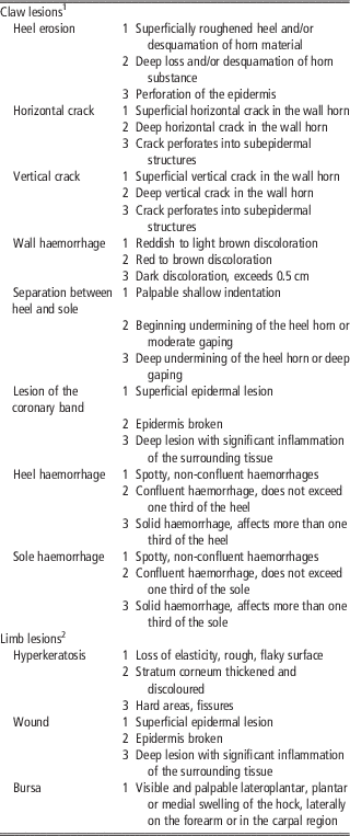
Each individual was assessed clinically for claw lesions as well as lesions of the limbs at the beginning (average weight: 24.9±3.8 kg), in the middle (average weight: 59.3±8.2 kg) and at the end (average weight: 102.3±12.2 kg) of the 12-week fattening period. This was done by restraining and lifting the animals in a self-designed metal chute. The findings were recorded separately for each claw and limb. The side (right v. left), the position on the animal (front or hind) and the position on the foot (lateral or medial) was noted. The assessment was done using the scheme presented in Table 1. Scores between 0 (no lesion) and 3 (severe lesion) were given for each lesion type.
Description of the lesion scores for the assessment of claw and limb lesions in fattening pigs

1 Adapted from Lippuner (Reference Lippuner2012).
2 Adapted from Savary et al. (Reference Savary, Gygax, Wechsler and Hauser2009).
For bursae, we did not follow the previous definitions. Rather, we used the term ‘bursa’ for all visible and palpable swellings in regions that have been described as being predisposed to the development of auxiliary bursae (Mouttotou et al, Reference Mouttotou, Hatchell and Green1998). In a cross-sectional study, KilBride (Reference KilBride2008) found no correlation between the histopathological degree of severity and the previously given scores in a clinical examination of what they assumed to be auxiliary bursae. As this is in accordance with our own observations, we did not score this lesion type, but only recorded its presence or absence.
Claw measures
To keep the number of measurements small, only claw width and claw angle were measured on both lateral and medial claws of the right fore and hind limb only. Claw width was measured using a caliper rule on the widest part of the heel. For the measurement of the claw angle a goniometer was used. Measurements were carried out without inflicting pressure on the tissue. All assessments of claw health and all measurements of claw dimensions were done by the same trained researcher.
Statistical analyses
All statistical analyses were carried out in R (version 3.4.0, R Development Core Team, Reference Rähse2017). It is likely that the occurrence of each lesion type on the different claws (front, hind, lateral, medial) as well as the different lesion types are correlated. Therefore, we condensed the single claw assessments. In a first approach, we subjected the lesion scores of the four claws and the five types of lesions ‘heel erosion’, ‘wall haemorrhage’, ‘crack horizontal’, ‘crack vertical’, ‘sole haemorrhage’ (4 claws×5 types totalling in 20 variables) to a principle component analysis. We used a standard principal component approach (function princomp in R) but based this approach on a correlation matrix of polychoric correlations (function hector in package polycor; Fox, Reference Fox2016) between the lesion scores because they are represented as categorical variables. In this evaluation, the first principle component only explained very little of the variance and the cumulative variance explained increased only incrementally from one principle component to the next. Accordingly, the pattern of factor loadings was inconsistent and virtually impossible to interpret.
In a second approach, we first summarised the four lesion scores of the different claws for a given pig at a given time point. We did so by choosing the most severe score that was observed on any of the four legs. This maximum score was then subjected to a principle component analysis based on polychoric correlations including the five types of lesions ‘heel erosion’, ‘wall haemorrhage’, ‘crack horizontal’, ‘crack vertical’, ‘sole haemorrhage’. All maximum scores loaded on the first principle component (with values between 0.36 and 0.52) explaining 48% of the overall variance. All further components were contrasts between the lesion types and therefore not easily interpreted. Due to the estimation based on the polychoric correlations, it was not possible to estimate directly the values of the first principle component for each of our observations. Because the loadings were quite similar, we decided to use the sum of the scores of these five lesion types as our first outcome variable for further statistical evaluation (total claw lesion score).
We proceeded in an analogous way for the leg lesions and the continuous measurements on the legs. We proceeded in an analogous way for the leg lesions and the continuous measurements on the legs (claw width und claw angle). We found that ‘hyperkeratosis’ and ‘bursa’ loaded on a first principle component explaining 53% of the variance with values of 0.70 each, whereas the much rarer wounds loaded on the second component (loading 0.98, cumulative variance explained: 0.86). Therefore, we used the summed score of hyperkeratosis and bursa as our second outcome measure (total leg lesion score). We also found that claw width and claw angle were highly correlated in that they both loaded with a value of 0.71 on a first principle component explaining 84% of the variance. We used ‘average claw angle’ across the four claws as our third outcome measurement.
The total claw lesion score, total leg lesion score and average claw angle were each used as an outcome variable. Model assumptions were examined using graphical analysis of residuals in respect to the normality of errors and random effects as well as the homoscedasticity of the errors. To satisfy assumptions, the total scores plus 0.5 were log-transformed whereas the claw angle was left untransformed. The experimental design was reflected in the random effect of the model: we used animal ID nested in group nested in batch as our hierarchically nested random effect. In respect to the fixed effects, we selected a final statistical model for each outcome based on the model probabilities (model weights) derived from the Bayesian information criterion within a set of models. The model probability reflects the probability of each model in the set being optimal in respect to high predictive power but few predictor variables given the data. The set comprised models that differed in how main effects and their possible interactions were combined. Starting with a maximum model, we performed an all subset analysis using the function ‘dredge’ in the package ‘MuMin’ (Barton, Reference Barton2012) because we had little prior knowledge of the expected effects.
The maximum model included the fixed effects material (factor with two levels: rubber mat or concrete elements), grade of perforation (factor with three levels: 0%, 5%, 10%), assessment time (factor with three levels: B, M, E), and all their potential interactions. These were included because they all make biologically sense and we had little prior knowledge about which interactions to expect. In addition, sex (factor with two levels: females, castrated males) was added as a main effect to account for this nuisance variable.
Results
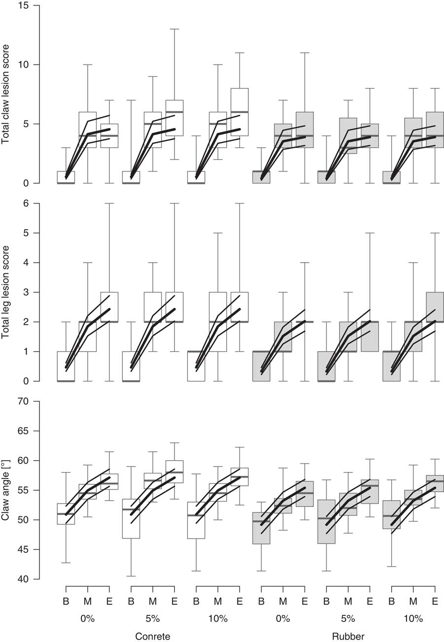
The total claw lesion score increased from the beginning to the end of the fattening period with a smaller increase in the second half of the period (model with assessment time only: w i =0.48, ER0>956; Figure 1). The values were slightly lower for animals kept on rubber mats than on concrete (second most likely model with main effects of material and assessment time: w i =0.40, ER0>806; all other models had w i <0.06).
Distribution of total claw lesion score, total leg lesion score and claw angle (from top to bottom) in fattening pigs kept on concrete and rubber floors at three assessments (B=begin of fattening period; M=middle of fattening period; E=end of fattening period) in different perforation percentages (0%, 5%, 10%). For the prevalence data: white=in pens with concrete lying area; grey=in pens with rubber mats. Lines represent model estimates and 95% confidence intervals.

The total leg lesion score showed a more monotonous increase throughout the fattening period and also had lower values in animals kept on rubber mats than on concrete (model with main effects of material and assessment time: w i =0.59, ER0>1174; Figure 1). The effect of the material was more weakly supported because the second best model included assessment time only (w i =0.29, ER0>574; all other models had w i <0.08).
Claw angle also increased almost continuously throughout the fattening period and were lower in animals kept on rubber mats than on concrete (model with main effects of material and assessment time: w i =0.61, ER0>1222). There was a weak indication that castrated males had larger claw angles than females (second best model additional including the sex of the animals w i =0.21, ER0>414; all other models had w i <0.10).
We found no indication that the grade of perforation had an effect on any of these outcome variables (the fixed effect was never chosen in a model with high w i ).
Discussion
Effect of time
Claw lesions
The total claw lesion score rose from the beginning to the end of the fattening period on all floor variants. The total claw lesion score in this study was composed of the lesion types heel erosion, wall haemorrhage, horizontal crack, vertical crack and sole haemorrhage.
Generally, heel erosions were highly prevalent in all claw locations. This is in accordance with findings of Lippuner (Reference Lippuner2012) and Gillman et al. (Reference Gillman, KilBride, Ossent and Green2009) who reported a prevalence of heel lesions of 28.4% and 10.8%, respectively. The heel region is characterised by horn material with fewer horn tubuli (Geyer, Reference Geyer1979) and a difference in keratin composition (Webb, Reference Webb1984) compared with other parts of the horny box. It is therefore relatively soft. Besides abrasive wear of the volar surface (McKee and Dumelow, Reference McKee and Dumelow1995), it may be assumed that prior impairment of the tissue provokes heel lesions (Newton et al., Reference Newton, Booram, Hale and Mullinix1980). Gillman et al. (Reference Gillman, KilBride, Ossent and Green2009) found a relationship to bleeding of the heel. Trauma to the heel region may have occurred in all different floor conditions in the experiment presented here, possibly either because the rubber mats provided were harder than the soft heel horn or because all animals had access to a perforated concrete floor in the defecation area. Newton et al. (Reference Newton, Booram, Hale and Mullinix1980) and Penny et al. (Reference Penny, Osborne and Wright1963) assumed that the weakened and roughened heel horn is susceptible to secondary bacterial infection which possibly leads to continuous degradation of the regrowing horn tissue. Mouttotou et al. (Reference Mouttotou, Hatchell and Green1999a) related lesions on the volar face of the claw to enhanced pressure on the weight-bearing surface. Accordingly, it could be hypothesised that the pressure strains may lead to or worsen heel erosions as they grow with the animals’ weight over time.
Wall haemorrhages have been described as occurring when a claw sinks into a gap hitting its wall against the slat rim (Geyer, Reference Geyer1979). They are visible in the horn substance while they grow down (Geyer, Reference Geyer1979) until they are worn off with the horn. Therefore, an accumulation of older and fresher lesions over the fattening period may have occurred.
Horizontal cracks are considered to be outgrowing remains of earlier wounds of the coronary band (Newton et al., Reference Newton, Booram, Hale and Mullinix1980), which also occur when the claw sinks into a gap. Therefore, the explanations we gave for the prevalence of wall haemorrhages may also apply for horizontal cracks. Vertical cracks typically occurred at the junction between the abaxial horn wall and the softer heel horn in our study. This region of the claw has been described as being vulnerable and therefore predisposed to the formation of cracks (Geyer, Reference Geyer1979). As the strains rise with the rising BW of the animals, this lesion type may have become more prevalent towards the end of the fattening period. Vertical cracks can suddenly progress and become painful when they reach into deeper layers or become infected (Geyer, Reference Geyer1979).
Limb lesions
The total leg lesion score also increased in prevalence over the fattening period. Limb lesion types examined in this study were auxiliary bursae, hyperkeratosis and wounds. Auxiliary bursae can be seen as pathologic remodelling processes and typically form over bony prominences with little soft tissue cover. Persistent or recurring friction and pressure lead to the formation of a fluid-filled cavity in the subcutaneous connective tissue (Adams, Reference Adams1974; Jensen et al., Reference Jensen, Nielsen, Vinther and D’Eath2012, cited in Mouttotou et al., Reference Mouttotou, Hatchell and Green1999b). Oberländer (Reference Oberländer2016) found signs of inflammation in more than 85% of bursal tissue samples in histopathologic examination. This emphasises the meaning of this lesion type for the well-being of pigs, as inflammation is likely to be painful in its acute state. Areas predisposed to the formation of bursae on the hind legs are mainly located on the lateroplantar, plantar and medial aspect of the hock and the point of the hock (Mouttotou et al., Reference Mouttotou, Hatchell and Green1999b; Gillman et al., Reference Gillman, KilBride, Ossent and Green2008). A high prevalence of bursae in finishing pigs was reported in previous studies. Oberländer (Reference Oberländer2016) observed a prevalence of 91.8% in pigs kept on fully or partly slatted concrete floors and examined at slaughter. Quinn (Reference Quinn2014) found a prevalence of 29.6% in weaner and finishing pigs kept on floors made of different materials, without bedding and with different perforation percentages. The increase of bursae over time is in accordance with other studies investigating bursae in finishing pigs. Ekkel et al. (Reference Ekkel, Spoolder, Hulsegge and Hopster2003) found that the increase in the prevalence of bursae was related to weight gain and the greater amount of time older pigs spent lying. KilBride (Reference KilBride2008) suggested that the rising prevalence may indicate that some bursae become chronic and therefore accumulate over time. Hyperkeratosis of the skin is provoked by chronic excessive pressure or friction (Freeman, Reference Freeman2002). As pressure strains on the joint areas increase with the animals’ weight gain during the fattening period, hyperkeratosis progressed in affected animals over time, similar to observations made by Savary et al. (Reference Savary, Gygax, Wechsler and Hauser2009). Unlike all other lesion types, hyperkeratosis was already highly prevalent at the first assessment, but only on the fore legs, where they possibly remain from the suckling period. However, hyperkeratosis on the hind legs developed in the experimental phase of our study. The results on limb lesions show that all tested variants of floor in the experimental pens did strain the skin.
Effect of the surface material in the lying area
Claw lesions
The total claw lesion score was lower in pigs kept on rubber mats compared with pigs kept on concrete. The greater softness and therefore flexibility of rubber absorbs pressure forces (De Carvalho et al., Reference De Carvalho, de Alencar Nääs, Neto and Lucas de Souza2009), may reduce slipping by providing more traction (Von Wachenfeldt et al., Reference Von Wachenfeldt, Nilsson and Pinzke2010), and may lead to more even weight distribution between the lateral and medial claws.
Several authors describe a decrease in prevalence of claw lesions in pigs kept in bedded pens, which is also likely to be an effect of the greater softness compared with concrete floors without bedding. For example, Rähse (2006) found an overall prevalence of 28.4% for moderate to severe claw lesions (deformities, pressure sores and contusions, indentations, heel lesions, cracks) in finishing pigs kept in pens with deep-straw bedding compared with 58.8% in pens without bedding and 65.9% in pens with sparse straw bedding. Scott et al. (Reference Scott, Chennells, Campbell, Hunt, Armstrong, Taylor, Gill and Edwards2006) also found reduced severity scores for some (sole and heel erosions), but not all (toe erosions, white line lesions) claw lesions in post-slaughter examinations of pigs kept in pens with straw bedding compared with pigs kept on fully slatted concrete floors. They explain the even higher incidence of toe erosions in straw-bedded pens, which is also in line with findings of Mouttotou et al. (Reference Mouttotou, Hatchell and Green1999a), with dampness and poor hygiene in the straw bedding which weakens the claw horn substance. This disadvantage of straw bedding probably does not apply for rubber mats. Still, first studies on the effect on claw lesions in breeding sows are contradictory, as Calderon Diaz et al. (Reference Calderon Diaz, Fahey, KilBride, Green and Boyle2013) found a greater risk for toe erosion, heel-sole crack, crack in the wall and white line lesion in breeding sows kept in pens equipped with rubber slat mats during two parities. Baumann et al. (Reference Baumann, Pflanz, Gallmann and Schrader2012) reported a stagnation in incidence of breeding sows with abrasion of the wall and haemorrhage from the time they were stalled in pens with rubber mats in the lying area, whereas sows in pens with concrete in the lying area showed a rising incidence in these characteristics. These results, of course, relate to heavier adult animals, which also probably already acquired predispositions (Newton et al., Reference Newton, Booram, Hale and Mullinix1980) and deformities before entering the studies. Therefore they might not be directly comparable with the present study, in which the animals showed almost no claw lesions at the initial examination and for which we assume that almost all lesions newly developed under the experimental floor conditions.
Limb lesions
Pigs kept in pens with rubber mats showed a lower total leg lesion score. Gillman et al. (Reference Gillman, KilBride, Ossent and Green2008) suggested that the formation of auxiliary bursae is a response to lying rather than walking on hard surfaces, as the risk of this lesion type decreased when bedding material was provided, as also shown by Mouttotou et al. (Reference Mouttotou, Hatchell and Green1999b) and Oberländer (Reference Oberländer2016). Studies by KilBride (Reference KilBride2008) showed that roughness of the floor can add to the higher risk of bursae in pigs lying on hard floor. The rubber mats in the present study seemed to be flexible and smooth enough to reduce this risk. KilBride (Reference KilBride2008) also identified slipperiness as a risk factor for bursae. The greater traction of rubber compared with concrete (Von Wachenfeldt et al., Reference Von Wachenfeldt, Nilsson and Pinzke2010) may lead to a decrease in slipping events. This may also explain a reduced prevalence of wounds, which, among other reasons, may occur as injuries when the animals slip. As the occurrence of hyperkeratosis, as explained before, seems to be related to pressure strains, a reduction of this lesion type is also thinkable.
Claw angles
Claw angles of all claws were smaller in pigs kept in pens with rubber mats. As rubber is softer (Von Wachenfeldt et al., Reference Von Wachenfeldt, Nilsson and Pinzke2010) than concrete, we assume that there was less mechanical wear-off in the claws of pigs kept on the mats. The resulting greater total length-growth of the claw horn resulted in smaller claw angles.
Effect of floor perforation
There was no effect of floor perforation on neither the total claw lesion score nor the total leg lesion score in this study. Some other studies which investigated the effect of floor perforation show an increased risk for different limb and claw lesions (Geyer, Reference Geyer1979; Scott et al., Reference Scott, Chennells, Campbell, Hunt, Armstrong, Taylor, Gill and Edwards2006; KilBride, Reference KilBride2008), but several authors also report a lack of an effect (Mouttotou et al., Reference Mouttotou, Hatchell and Green1999b; Rähse, 2006). Newton et al. (Reference Newton, Booram, Hale and Mullinix1980) hypothesize that perforation of the floor has no overriding effect on claw and leg health and suggests that traction, roughness and abrasiveness are the main influence factors on the development of lesions. In the present study, it also has to be taken into account, that all animals had access to a slatted concrete manure area.
Conclusions
The present data show that rubber mats in the lying area bring about improvements in some aspects of claw and leg health in fattening pigs, whereas there was no effect of floor perforation. The reduced incidence of lesions may contribute to the animals’ welfare, as some of the investigated lesion types are likely to be painful.
Acknowledgements
The authors would like to thank the staff of the experimental stables, Andreas Hagenbüchli and Barbara Ammann, for their support and excellent care of the animals. Special thanks goes to Esther Bürgi for her generous professional advice. We would also like to thank Agroscope, Tänikon, for the use of their research facilities and the Swiss Federal Food Safety and Veterinary Office for funding this study (Project No. 2.13.05).