Double-barrelled aorta is a rare anomaly characterized by presence of a second systemic-to-systemic channel connecting the ascending and descending components of the aorta. In the past, this malformation has been interpreted as persistence of the fifth aortic arch,1–3 despite the fact that, thus far, there is no embryological evidence to support the existence of this enigmatic structure during normal development. We report the first case, as far as we are aware, of double-barrelled aorta associated with tetralogy of Fallot and pulmonary atresia.
Case report
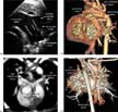
A two-day-old female presented with cyanosis and a continuous murmur being audible over the left precordium. The liver was not enlarged. The chest radiograph demonstrated an enlarged heart, and increased pulmonary vascularity. Cross-sectional echocardiography demonstrated normal segmental connections and tetralogy of Fallot with pulmonary atresia. The pulmonary arterial supply was derived from major systemic-to-pulmonary collateral arteries. The supraster-nal sagittal view also revealed dual lumens within the aortic arch, which was on the left side of the trachea (Fig. 1a). The superior arch gave rise to the brachiocephalic, left common carotid and left subclavian arteries in sequence, but became atretic immediately distal to the origin of the left subclavian artery. The inferior arch connected the ascending aorta to the descending aorta, with mild narrowing of its distal part. Contrast-enhanced computed tomography confirmed the anatomy of the arch as seen at echocardiography (Fig. 1b). It also revealed that the small pulmonary trunk bifurcated into the right and left pulmonary arteries (Fig. 1c). The right pulmonary artery gave rise to branches to the right middle lobe and basal part of the right lower lobe. The left pulmonary artery gave rise to the branches to a part of the left lower lobe. There were two major systemic-to-pulmonary collateral arteries (Fig. 1d). One arose from the left common carotid artery, and supplied the right upper lobe and superior segment of the right lower lobe. The second one arose from the descending aorta, and supplied the left upper lobe and posterior part of the left lower lobe. A branch of this collateral artery anastomosed to a branch of the left pulmonary artery in the left lower lobe, being the sole source of arterial supply to the confluent pulmonary arteries. Subsequent cardiac catheterization at 6 weeks of age confirmed the echocardiographic and computed tomographic findings. The child underwent successful surgical correction at 2 months of age. The narrowed segment of the inferior arch was repaired by augmentation with a pericardial patch. The child was soon discharged, with no pressure gradient between the upper and lower limbs.

Figure 1. The echocardiogram of the aortic arch obtained through the suprasternal approach (a) shows the double-barrelled aorta. The superior arch, at colour Doppler interrogation, was shown to be atretic distal to the origin of the left subclavian artery. (b) Shows the volume rendered contrast-enhanced computed tomogram. The aortic arch seen from the left anterior aspect shows the double-barrelled aorta. The superior arch is atretic at its connection to the descending aorta. The distal part of the inferior arch shows mild narrowing (asterisk). A contrast-enhanced computed tomogram reformatted in oblique axial plane (c) shows the vestigial pulmonary trunk that gives rise to the small right and left pulmonary arteries. The volume rendered computerised tomographic image of the thorax seen from behind (d) shows two collateral arteries; one (C1) arising from the proximal part of the left common carotid artery and the other (C2) arises from the descending aorta.
Discussion
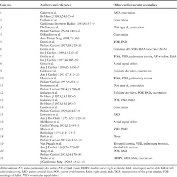
Double-barrelled aorta is a rare condition, in which an aortic arch with dual lumens is present on the same side of the trachea. It is in contrast to the double aortic arch, in which each arch is on opposite side of the trachea. The first documentation of this anomaly was presented by Van Praagh and Van Praagh4 in 1969. In their report, they described dual lumens of the left aortic arch, the inferior channel extending from the ascending aorta proximal to the origin of the brachiocephalic artery to the descending aorta opposite to the origin of the left subclavian artery.4 Since their description, 21 additional cases have been published. (Table 1). Van Praagh and Van Praagh,4 in their initial description, explained the malformation on the basis of persistence of the fifth aortic arch. Since their report, any inferior aortic channel arising from the distal ascending aorta has also been interpreted as persistence of this purported structure. Subsequently, the hypothesis of the persistence of the embryologic fifth aortic arch was further extended to explain cases in which an abnormal branch arises from the distal ascending aorta and connects to the pulmonary arteries. This hypothetical interpretation of anatomy that is already complex, however, has induced unnecessary grouping of heterogeneous abnormalities under the title of “persistent fifth aortic arch”, with subcategorization into those with a systemic-to-systemic connection as opposed to those with a systemic-to-pulmonary connection.5, 6 It is doubtful whether this approach facilitates our understanding of the pathology, and questions must be raised as to whether evidence exists relating to cardiac development to substantiate its usage. In fact, the existence of a fifth aortic arch in human and mammalian embryos is controversial.1–3 In a recent elegant study of the mouse embryo using scanning electron microscopy of casts, Hiruma and colleagues found no evidence of the purported fifth arch.7 In the light of the absence of convincing evidence supporting the existence of this structure during normal development, therefore, we prefer to use the term “double-barrelled aorta” as an appropriate and simple term to describe the malformation in which two channels exist between the ascending and descending components of the aorta on the same side of the trachea. As far as we know, our case found in the setting of tetralogy of Fallot with pulmonary atresia is the first description in the English literature of this combination.
Table 1. Double-barrelled aorta.