Introduction
Migraine in general affects approximately 6 per cent of men and 18 per cent of women.Reference Lipton, Bigal, Diamond, Freitag, Reed and Stewart1, Reference Lempert and Neuhauser2 It is estimated that 38 per cent of the migraine population also have vertigo.Reference Troost3 People with migraine and vertigo can be divided into two categories: those with migrainous vertigo (vertigo is directly related to the migraine and there is no other cause of vertigo present)Reference Lempert and Neuhauser4 and those with migraine-associated vertigo (vertigo exists separately but in association with the migraine).Reference Cass, Ankerstjerne, Yetiser, Furman, Balaban and Aydogan5
Lempert and Neuhauser have developed criteria which aid in the diagnosis of migrainous vertigo.Reference Lempert and Neuhauser4 Such criteria are necessary because migrainous vertigo is often underdiagnosed, despite being a relatively prevalent problem,Reference Troost3–Reference Furman, Sparto, Soso and Marcus9 and because this condition is currently not recognised as a specific category of migraine by the International Headache Society's classification of migraines.10 Using Lempert and Neuhauer's categorisation,Reference Lempert and Neuhauser4 the current study examined the vestibular system of those with migrainous vertigo by comparing findings of this sample with a migraine without vertigo group and a non-migraine group.
Vestibular rehabilitation for migrainous vertigo was suggested by a small study to be effective in treating abnormalities in vestibular ocular reflexes, vestibular spinal reflexes (and balance) and function.Reference Gottshall, Moore and Hoffer11 Two larger studies showed the effectiveness of vestibular rehabilitation for migraine-associated vertigo.Reference Whitney, Wrisley, Brown and Furman12, Reference Wrisley, Whitney and Furman13 However, assessments of vestibular function, such as the gaze stabilisation test, the dynamic visual acuity test for vestibular ocular reflexes, the head shake sensory organisation test for vestibular spinal reflexes (specifically for balance) and the functional gait assessment for overall function, have not been studied in a migrainous vertigo population. Therefore, it is not known whether there is an underlying functional abnormality in the vestibular system of individuals with migrainous vertigo during a non-migrainous period, and if so, whether the problem exists in both the vestibular ocular reflexes and the vestibular spinal reflexes (and balance).
The vestibular ocular reflexes and vestibular spinal reflexes (and balance) are part of a complex vestibular system composed of peripheral apparatus and a collection of central nuclei.Reference Herdman14–Reference Crutchfield and Barnes16 The peripheral apparatus sends messages to the central nuclei regarding angular and linear acceleration of the head.Reference Herdman14–Reference Crutchfield and Barnes16 The central nuclei process these messages and relay them up and down the spinal cord.Reference Herdman14–Reference Crutchfield and Barnes16 The superior and medial vestibular nuclei relay messages up to the eyes and control the vestibular ocular reflexes.Reference Kandel, Schwartz and Jessel15, Reference Crutchfield and Barnes16 The lateral vestibular nuclei relay messages down to the skeletal muscles and control the vestibular spinal reflexes.Reference Kandel, Schwartz and Jessel15, Reference Crutchfield and Barnes16 Communication between the vestibular nuclei and the cerebellum is controlled by the medial vestibular nuclei.Reference Kandel, Schwartz and Jessel15, Reference Crutchfield and Barnes16 Exploration of this complex vestibular system can be conducted in several ways using otoneurological tests and functional clinical tests.
Researchers utilising otoneurological tests have generally found that the working of the vestibular system is normal during the non-migrainous period in people with migrainous vertigo.Reference Lempert, Neuhauser and Daroff7, Reference Gottshall, Moore and Hoffer11, Reference Casani, Sellari-Franceschini, Napolitano, Muscatello and Dallan17–Reference Cutrer and Baloh22 However, 20 per cent of people with migrainous vertigo have hypoexcitability to caloric stimulation and 10 per cent have directional preponderance of caloric-induced nystagmus.Reference Celebisoy, Gokcay, Sirin and Bicak21, Reference Cutrer and Baloh22 These findings are not unique to the migrainous vertigo population and may also be seen in the migraine without vertigo population.Reference Celebisoy, Gokcay, Sirin and Bicak21, Reference Cutrer and Baloh22 There are uncertainties as to why vestibular symptoms are different between those who suffer from migraine without vertigo and those who suffer from migrainous vertigo, and why vestibular rehabilitation performed during a non-migrainous period may be effective for migrainous vertigo. In order to determine if all components of the vestibular system are functioning normally in migraine sufferers (both those without vertigo and those with migrainous vertigo) compared with a normal population, studies need to examine vestibular ocular reflexes, vestibular spinal reflexes (and balance) and function. The identification of specific underlying abnormalities in the vestibular system during a non-migrainous period could benefit vestibular rehabilitation in those with migrainous vertigo.
Vestibular ocular reflexes can be measured functionally in two ways, using the gaze stabilisation test and the dynamic visual acuity test.Reference Ward, Mohammad, Whitney, Marchetti and Furman23 The gaze stabilisation test measures how fast the head can move whilst maintaining a visual image.Reference Ward, Mohammad, Whitney, Marchetti and Furman23, Reference Pritcher, Whitney, Marchetti and Furman24 Pritcher et al. found that the gaze stabilisation test results were abnormal in people with vestibular dysfunction, especially during pitch plane movements.Reference Pritcher, Whitney, Marchetti and Furman24 The findings of Whitney et al. also showed that people with vestibular disorders had difficulty with gaze stabilisation during yaw movements, but the effect was greater during pitch movements.Reference Whitney, Marchetti, Pritcher and Furman25 The dynamic visual acuity test compares visual acuity with the head still and the head moving.Reference Schubert, Migliaccio, Clendaniel, Allak and Carey26 This is also a test of vestibular ocular reflex function. Again, studies have been conducted exploring dynamic visual acuity in people with vestibular disorders,Reference Ward, Mohammad, Whitney, Marchetti and Furman23–Reference Schubert, Migliaccio, Clendaniel, Allak and Carey26 but not specifically in those who suffer from migraine without vertigo or migrainous vertigo.
Vestibular spinal reflexes and balance, which can be assessed using the head shake sensory organisation test, has not been studied in the migrainous vertigo population. The head shake sensory organisation test was developed to provide additional information on those with mild vestibular disorders who may be well compensated (an abnormal vestibular system but normal sensory organisation test result).Reference Shepard, Cole, Bradshaw, Hyder and Parent27–Reference Honaker, Converse and Shepard30 Three studies found that the test results were worse in those with vestibular disorders than those without vestibular disorders.Reference Roma28–Reference Honaker, Converse and Shepard30 However, this test has not been employed to study migraine without vertigo or migrainous vertigo.
Functional testing, specifically gait testing, has been used extensively to assess those with vestibular disorders.Reference Wrisley, Marchetti, Kuharsky and Whitney31–Reference Wrisley and Kumar33 Several gait tests have been developed to test gait function in those with vestibular disorders.Reference Wrisley, Marchetti, Kuharsky and Whitney31–Reference Wrisley and Kumar33 The most recently developed test is the functional gait assessment, which was developed to evaluate postural stability during gait.Reference Wrisley, Marchetti, Kuharsky and Whitney31–Reference Wrisley and Kumar33 In one study that used the functional gait assessment, it was found that slow, pitch plane movements of the head were predictive of gait abnormalities in people with vestibular disorders (BJ Baker, unpublished data). In a pilot study examining gait in those affected by migraine, higher functional gait assessment scores were demonstrated for the migraine without vertigo group compared with the migrainous vertigo group during a non-migrainous period.Reference Ishikawa, Cao, Wang, Wong, Tanaka and Miyazaki34
Differences in the test results of vestibular ocular reflexes, vestibular spinal reflexes (and balance) and function have been found between those with normal and abnormal vestibular systems.Reference Ward, Mohammad, Whitney, Marchetti and Furman23–Reference Ishikawa, Cao, Wang, Wong, Tanaka and Miyazaki34 However, no studies have attempted to define vestibular function, vestibular ocular reflexes, vestibular spinal reflexes (and balance) and function in a migraine without vertigo population or a migrainous vertigo population during a non-migrainous period, and examined the findings in relation to a non-migraine population.
This study aimed to determine whether there is underlying vestibular dysfunction in the migrainous vertigo population compared with the migraine without vertigo population and the non-migraine population, as measured by the gaze stabilisation test, dynamic visual acuity test, head shake sensory organisation test and functional gait assessment. It was hypothesised that there would be a difference in the scores of these tests for the migrainous vertigo population when compared with the other two groups. Evaluation of vestibular functioning in those with migrainous vertigo during a non-migrainous period should aid the efficacy of vestibular rehabilitation for this group, and may even improve treatment techniques.
Materials and methods
Study design and sample
Sixty-six participants aged between 18 and 65 years were recruited from a university campus (via an online listing service) and from local neurological clinics. The study was a descriptive, case–control study. Twenty-two participants were recruited for each of three subject groups: a migraine without vertigo group, a migrainous vertigo group and a control group.
For the 44 participants with a history of migraine, the diagnosis was confirmed by a physician where possible. Attempts were made to verify diagnosis by faxing the physicians for confirmation. Response from physicians was poor; therefore, self-report was used when physician confirmation was not available. Credibility of self-reported diagnosis was increased with the use of a survey (developed by Furman et al.), which determined possible and probable migrainous vertigo.Reference Furman, Marcus and Balaban35
Exclusion criteria included the use of an assistive device, reports of a neurological condition or vestibular condition other than migrainous vertigo, a history of head trauma, or an orthopaedic disorder which affected balance or head movement.
This study was approved by the Western Michigan University Human Subjects Institutional Review Board and the Grand Valley State University Human Research and Review Committee. Each subject was given an informed consent to read, query and then sign. Two different demographic forms were used: one for the participants without migraine and another for those with migraine. The participants with migraine were then asked to sign several disclosure forms and to include the name of their physician (used for contacting the physicians).
Assessments and procedures
Participants were tested during a non-migrainous period and during a time when they did not have vertigo, as confirmed by a visual analogue scale for pain and vertigo. Each participant in the migraine groups responded to the questions in the Furman et al. survey,Reference Furman, Marcus and Balaban35 which were based on Lempert and Neuhauser criteria for migrainous vertigo.Reference Lempert and Neuhauser4
All participants were tested with the same battery of tests, which included the gaze stabilisation test (yaw and pitch), the dynamic visual acuity test (yaw and pitch), the head shake sensory organisation test (yaw and pitch) and the functional gait assessment. The order of the tests were randomised using a random number table; the order of yaw and pitch movements were also randomised.
Performance in the gaze stabilisation test and dynamic visual acuity test (employed to examine vestibular ocular reflexes) was quantified using the inVision System™, which is part of the NeuroCom EquiTest system (NeuroCom International, Clackamas, Oregon, USA). Static visual acuity and perception time were measured, with the participant seated approximately 3 metres from the computer screen. A head piece was placed on the participant's head to detect the speed and direction of head movement. The participant performed both yaw and pitch head movements (in a random order). The participant moved his or her head, stopping when a visual cue appeared on the screen.
The gaze stabilisation test has been determined to be reliable (intraclass correlation coefficient = 0.75) and valid.Reference Ward, Mohammad, Whitney, Marchetti and Furman23 The results of this test are recorded in degrees per second and range from 0–200, with 200 being the optimal gaze stabilisation score. The dynamic visual acuity test compares static visual acuity to dynamic visual acuity, and the results are calculated by the computer. Dynamic visual acuity loss was recorded in terms of the logarithm of the minimum angle of resolution, with possible scores ranging from 0 to 1, with 1 indicating a total loss of dynamic visual acuity.Reference Schubert, Migliaccio, Clendaniel, Allak and Carey26
Vestibular spinal reflexes and balance were examined using the head shake sensory organisation test, which is an instrumental test of the vestibular spinal reflexes.Reference Roma28–Reference Honaker, Converse and Shepard30, Reference Pang, Lam, Wong, Au and Chow36 This test was performed on the Smart EquiTest system (NeuroCom International), which is a computerised dynamic posturography instrument that has been shown to be reliable and valid.Reference Pang, Lam, Wong, Au and Chow36 A harness was placed on the participant for safety, and the participant's feet were positioned as per the Smart EquiTest system protocol. Each participant wore flat, non-slip shoes.
The head shake sensory organisation test required performance in two conditions which physiologically stressed the vestibular system.Reference Roma28–Reference Honaker, Converse and Shepard30, Reference Pang, Lam, Wong, Au and Chow36 In the first condition, referred to as the fixed head shake sensory organisation test, participants' eyes closed and they were stood on a firm surface. The second condition, the sway head shake sensory organisation test, was also performed with eyes closed, but took place on a moveable surface. In each condition, the participant performed yaw and pitch head movements. Equilibrium scores for both conditions of the head shake sensory organisation test were determined by the computer (which entailed a comparison of the head-still condition with the head-moving condition, for both yaw and pitch movements). Total possible scores for the head shake sensory organisation test ranged from 0 to 100 per cent, with 100 per cent demonstrating optimal postural control.Reference Pang, Lam, Wong, Au and Chow36
This was followed by the functional gait assessment, a test which required the use of vestibular ocular reflexes and vestibular spinal reflexes (and balance). This 10-item walking test battery has demonstrated reliability (intraclass correlation coefficient = 0.93).Reference Wrisley, Marchetti, Kuharsky and Whitney31–Reference Wrisley and Kumar33 During this assessment, the subject was guarded to prevent falls. Each item was scored according to the criteria, based on a 4-point scale with a score of 3 being normal.Reference Wrisley, Marchetti, Kuharsky and Whitney31 The total score for the 10 tasks was recorded. The total possible score ranges from 0 to 30, with 30 being a perfect score.Reference Wrisley, Marchetti, Kuharsky and Whitney31
Results and analysis
Analyses were performed using the Statistical Package for the Social Sciences version 18.0 (SPSS, Chicago, Illinois, USA). The variables were not normally distributed. Analysis of variance was conducted using Helmert planned comparisons when variables were statistically significant at the alpha = 0.05 level. For the planned comparisons, the control group were first compared with both migraine groups combined and then the two migraine groups were compared.
There were 66 participants in total: 22 in the non-migraine group, 22 in the migraine without vertigo group and 22 in the migrainous vertigo group. Of the 66 participants, 59 were female. There were four males in the non-migraine group, no males in the migraine without vertigo group and three males in the migrainous vertigo group. Participant ages ranged from 18 to 61 years of age. The mean age was 26 years for the non-migraine group (range of 23–57 years), 27 years for the migraine without vertigo group (range of 18–61 years) and 35 years for the migrainous vertigo group (range of 20–57 years). No history of neurological disorders or vestibular disorders other than migrainous vertigo were reported by participants in any of the three groups, and none of the participants suffered from any orthopaedic disorders that would limit participation.
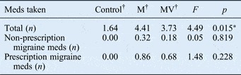
As shown in Table I, there was a significant difference (p < 0.015) between the total number of medications used by the non-migraine group (mean = 1.64), the migraine without vertigo group (mean = 3.73) and the migrainous vertigo group (mean = 4.41). However, the difference between the migraine without vertigo group and migrainous vertigo group was not significant when examined using planned comparisons. There were no significant differences between the migraine without vertigo group and the migrainous vertigo group in terms of either the number of non-prescription migraine medications (p < 0.819) or that of prescription migraine medications (p < 0.228). It should be noted that many of the medications used for vertigo in the migrainous vertigo group were also used for nausea in the migraine without vertigo group.
Group medication data

*Bonferroni correction post-hoc analyses revealed that: the control group versus the migraine without vertigo group comparison was not significant (p = 0.102), the control group versus the migrainous vertigo group comparison was significant (p = 0.015) and the migraine without vertigo group versus the migrainous vertigo group comparison was not significant (p = 1.00). †n = 22. M = migraine without vertigo; MV = migrainous vertigo; meds = medications
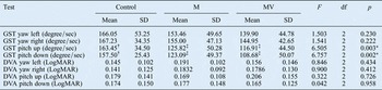
Vestibular ocular reflexes testing (shown in Table II) indicated no statistically significant differences between the three groups for left and right gaze stabilisation test performance in the yaw plane (p < 0.230 and p < 0.222, respectively). The mean left gaze stabilisation test scores for the yaw plane were: 166.05 degrees per second (standard deviation (SD) = 53.25) for the non-migraine group, 153.46 degrees per second (SD = 49.65) for the migraine without vertigo group and 139.90 degrees per second (SD = 44.78) for the migrainous vertigo group. The mean right gaze stabilisation test scores for the yaw plane were: 167.23 degrees per second (SD = 34.35) for the non-migraine group, 155.00 degrees per second (SD = 47.13) for the migraine without vertigo group and 144.95 degrees per second (SD = 42.65) for the migrainous vertigo group.
Vestibular ocular reflex data

*p < 0.05. †Helmert planned comparison revealed a significant difference between the control group and the two migraine groups (p < 0.0001). ‡Helmert planned comparisons between the two migraine groups were not statistically significant for up and down movements (p = 0.500 and 0.273, respectively). M = migraine without vertigo; MV = migrainous vertigo; df = degrees of freedom; SD = standard deviation; GST = gaze stability test; degree/sec = degrees per second; DVA = dynamic visual acuity loss; LogMAR = logarithm of the minimum angle of resolution
There was a statistically significant difference between the three groups for up and down gaze stabilisation test performance in the pitch plane (p < 0.003 and p < 0.002, respectively) (see Table II). The mean up gaze stabilisation test scores for the pitch plane were: 163.45 degrees per second for the non-migraine group, 125.82 degrees per second for the migraine without vertigo group and 116.91 degrees per second for the migrainous vertigo group. The mean down gaze stabilisation test scores for the pitch plane were: 157.50 degrees per second for the non-migraine group, 123.09 degrees per second for the migraine without vertigo group and 108.68 degrees per second for the migrainous vertigo group. Planned comparisons showed that the difference between the control group and both migraine groups was statistically significant (p < 0.0001), but there was no significant difference between the migraine without vertigo group and the migrainous vertigo group in up and down pitch movements (p = 0.500 and p = 0.273, respectively).
Vestibular ocular reflex testing using the dynamic visual acuity test indicated no statistically significant differences between the three groups for left and right dynamic visual acuity in the yaw plane (p < 0.434 and p < 0.412, respectively), or up and down in the pitch plane (p < 0.726 and p < 0.958, respectively).
The vestibular spinal reflex and balance testing results are shown in Table III. There were no statistically significant differences in the head shake sensory organisation test results for the yaw plane, in either the fixed and sway conditions (p < 0.197 and p < 0.068, respectively), between the non-migraine group (fixed mean = 96 per cent (SD = 4) and sway mean = 74 per cent (SD = 23)), the migraine without vertigo group (fixed mean = 89 per cent (SD = 24) and sway mean = 65 per cent (SD = 27)) and the migrainous vertigo group (fixed mean = 96 per cent (SD = 13) and sway mean = 81 per cent (SD = 16)). There were also no differences in the head shake sensory organisation test results for the pitch plane, in either the fixed or sway conditions (p < 0.606, p < 0.419, respectively), between the non-migraine group (fixed mean = 93 per cent (SD = 5) and sway mean = 71 per cent (SD = 17)), the migraine without vertigo group (fixed mean = 90 per cent (SD = 21) and sway mean = 64 per cent (SD = 20)) and the migrainous vertigo group (fixed mean = 94 per cent (SD = 7) and sway mean = 71 per cent (SD = 25)).
Vestibular spinal reflex, balance and gait data

*p < 0.05. †Helmert planned comparison revealed a significant difference between the control group and the two migraine groups (p < 0.0001). ‡Helmert planned comparison revealed a significant difference between the between two migraine groups (p = 0.002). M = migraine without vertigo; MV = migrainous vertigo; df = degrees of freedom; SD = standard deviation; HS-SOT = head shake sensory organisation test; FGA = functional gait assessment
The functional gait assessment results (Table III) indicated statistically significant differences between the control group (mean = 29.5, SD = 0.67), the migraine without vertigo group (mean = 27.8, SD = 0.19) and the migrainous vertigo group (mean = 26.0, SD = 0.23) (p < 0.0001 for the control group vs both migraine groups, p < 0.0001 for the migraine without vertigo group vs the migrainous vertigo group). Planned comparisons showed that there were differences between the control group and the two migraine groups (p < 0.0001), and between the migraine without vertigo group and the migrainous vertigo group (p = 0.002).
Discussion
Vestibular ocular reflexes, vestibular spinal reflexes (and balance) and function were examined in a control group, a migraine without vertigo group and a migrainous vertigo group during a non-migrainous period. Vestibular ocular reflexes, as measured by the gaze stabilisation test (pitch movements), were faster in the control group than in the two migraine groups. Scores for function, as measured by the functional gait assessment, were highest in the control group and lowest the migrainous vertigo group.
Vestibular spinal reflexes and balance (measured using the head shake sensory organisation test) were not statistically significantly different between the three groups. As vestibular spinal reflexes are controlled by the lateral vestibular nucleus,Reference Kandel, Schwartz and Jessel15, Reference Crutchfield and Barnes16 it is possible that the latter is not involved in migraine without vertigo and migrainous vertigo; hence, vestibular spinal reflexes and balance responses are normal in these populations. This finding suggests that vestibular rehabilitation for migrainous vertigo should not concentrate on balance or other vestibular spinal reflex functions.
Vestibular ocular reflexes were measured (in pitch and yaw movements) by both the dynamic visual acuity test and the gaze stabilisation test. The dynamic visual acuity test results showed no statistically significant differences between the three groups. Because dynamic visual acuity is not as reliable between subjects as it is within subjects, it may not have been the most appropriate test for this study.Reference Schubert, Migliaccio, Clendaniel, Allak and Carey26 The gaze stabilisation test results revealed significantly faster up and down pitch movements for the control group compared with the two migraine groups (during a non-migrainous period), but there were no differences between the groups for yaw plane movements. Other researchers' results indicate that movements in a pitch plane are more difficult to perform than movements in a yaw plane, and this may be why significant differences were seen in the pitch plane but not in the yaw plane.Reference Pritcher, Whitney, Marchetti and Furman24, Reference Whitney, Marchetti, Pritcher and Furman25
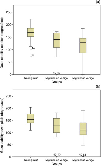
Pitch velocity means are clinically important. A gaze stabilisation speed of 120 degrees per second is considered normal for pitch movements, and encompasses everyday functional activities.Reference Pritcher, Whitney, Marchetti and Furman24 The control group mean and the migraine without vertigo group mean were both above 120 degrees per second for pitch movements (150 and 125 degrees per second, respectively), but the migrainous vertigo group mean was below 120 degrees per second (108 degrees up and 116 degrees down per second), which is slower than normal. However, there was no statistically significant difference between the two migraine groups in terms of the participants who scored below 120 degrees per second. There was wide variability in the gaze stabilisation test scores in both the migraine without vertigo group and the migrainous vertigo group (Figure 1), which helps to explain this finding.
Gaze stability test pitch results for (a) up and (b) down movements. Degree/sec = degrees per second

The differences found for vestibular ocular reflexes could be explained by the lateral and inferior nuclei, which control the vestibular ocular reflexes;Reference Kandel, Schwartz and Jessel15, Reference Crutchfield and Barnes16 these may function differently in those who suffer from migraine compared with those who do not. The large variability, especially that observed in the migrainous vertigo group, needs to be studied further. Perhaps the migrainous vertigo population is heterogeneous in regards to vestibular function and cannot be categorised.
Normal vestibular function requires vestibular spinal reflexes, specifically balance, and vestibular ocular reflexes. Functional testing with the functional gait assessment can distinguish between those with vestibular dysfunction and those with normal vestibular functioning.Reference Wrisley, Marchetti, Kuharsky and Whitney31–Reference Wrisley and Kumar33 A small pilot study showed that functional gait assessment scores were higher in a migraine without vertigo group compared with a migrainous vertigo group.Reference Ishikawa, Cao, Wang, Wong, Tanaka and Miyazaki34 This supports our finding of a difference between the three groups, with the migrainous vertigo group scoring the lowest and the control group scoring the highest. This also indicates that the vestibular systems of individuals with migrainous vertigo function differently to those in the other populations. Furthermore, the difference between the control group and the migraine without vertigo group suggests that there might even be some structural vestibular differences affecting function in those suffering from migraine without vertigo.
The functional gait assessment scores for each of the experimental groups can be compared with age-based norms found in another study.Reference Walker, Austin, Banke, Foxx, Gaetano and Gardner32 Walker et al. determined the normative means of community dwelling adults between the ages of 40 and 89 years on the functional gait assessment.Reference Walker, Austin, Banke, Foxx, Gaetano and Gardner32 They found an inverse relationship between age and functional gait assessment mean score, wherein the mean score decreased every 10 years. For the 40–49 year age group, the average functional gait assessment score was 29. Normative values were not determined for the 20–29 year age group or the 30–39 year age group, and the values for these groups may be higher than in the 40–49 year age group.Reference Walker, Austin, Banke, Foxx, Gaetano and Gardner32 In the current study, all three groups had a mean age of less than 40 years. The mean age of the control group was 26 years; this group had a mean functional gait assessment score of 29.5. The mean age of the migraine without vertigo group was 27 years, with a mean score of 27.8. The mean age of the migrainous vertigo group was 35 years, with a mean score of 26.0. Both the migraine without vertigo group and the migrainous vertigo group had scores below those of subjects in the 40–49 year age group in the Walker et al. study.
The current study revealed a significant association between the gaze stabilisation test results for pitch movements and the functional gait assessment scores in all three groups. The results of a study by Whitney et al. who explored the relationship between gaze stabilisation test scores for pitch movements and scores on functional measures of gait in older people with vestibular dysfunction, support this finding.Reference Whitney, Marchetti, Pritcher and Furman25 Our findings demonstrate that this relationship is present even in a younger population.
Our findings suggest that there are differences in the vestibular systems of those who suffer from migraine without vertigo or migrainous vertigo (during a non-migrainous period) compared with a non-migraine population. We found that these differences were similar in both migraine groups. Although there were differences between those without migraine (control group) and those with migraine (the migrainous vertigo, and the migraine without vertigo group), these differences do not explain the presence of vertigo in migrainous vertigo only (and not in migraine without vertigo). There were no baseline differences for vestibular ocular reflexes between the migraine without vertigo group and the migrainous vertigo group. However, there may be increased susceptibility to pathophysiological changes which occur during a migraine in the migrainous vertigo population compared with migraines that occur in the migraine without vertigo population. This is a matter that should be investigated in future studies. There might be a continuum between migraine without vertigo and migrainous vertigo, causing sufferers of the former to experience ‘motion sickness’ and the latter to experience actual vertigo.
• Migrainous vertigo is common but underdiagnosed
• Otoneurological tests suggest the vestibular system is normal in migrainous vertigo during non-migrainous periods
• Gaze stabilisation, dynamic visual acuity, head shake sensory organisation and functional gait test results are abnormal in vestibular disorders
• Migrainous vertigo and migraine without vertigo have not been studied using these tests
• Gaze stabilisation (pitch) and functional gait showed differences between non-migraine, migraine and migraine with vertigo groups
• Gaze stabilisation (pitch) and functional gait performance were related
The functional gait assessment findings also indicate mild structural vestibular differences in those who suffer from migraine without vertigo and migrainous vertigo, even during a non-migrainous period. The migrainous vertigo group performed significantly worse in the functional gait assessment than the migraine without vertigo group, and both groups performed worse than the control group. This is an important finding and supports the use of vestibular rehabilitation in the treatment of patients with migrainous vertigo during a non-migrainous period. Our research findings can explain why vestibular rehabilitation for migrainous vertigo might be effective. There may be mild underlying vestibular dysfunction that specifically affects vestibular ocular reflexes during non-migrainous periods in both migraine without vertigo and migrainous vertigo. This underlying vestibular abnormality may be accentuated during the migrainous period, resulting in the vertigo reported by the migrainous vertigo population.
Limitations and future directions
The sample size was large enough to provide an appropriate power (80 per cent) to determine differences in the tests examined. However, the sample was non-random. In addition, although we attempted to attain physician confirmation of the participant-reported diagnosis, we were unsuccessful in this attempt for most of the participants. Nevertheless, patient diagnoses were supported by the number and type of migraine medications, and the current findings. Another factor which improved the current method of identifying the correct diagnosis was the use of the Furman et al. interview form.Reference Furman, Marcus and Balaban35 This survey has been shown to be reliable and valid.Reference Furman, Marcus and Balaban35 In the current study, the results of the form agreed in all cases with the participant-reported diagnosis of migraine without vertigo or migrainous vertigo. Future studies on the migrainous vertigo population should include assessments of gaze stabilisation pre- and post-vestibular rehabilitation, and there should be further investigation of the relationship between gaze stabilisation and gait in different populations.
Conclusion
There was a statistically significant difference in the gaze stabilisation test results for pitch movements (a measure of vestibular ocular reflexes) between the non-migraine group which was faster, and the two migraine groups (which were slower) during a non-migrainous period. The functional gait assessment also revealed significant differences between all three groups, with the control group scoring the highest and the migrainous vertigo group scoring the lowest during a non-migrainous period. Tests of vestibular spinal reflex components, specifically balance, did not differ between the groups. These findings support the suggestion that there are underlying differences in the vestibular ocular reflexes and function of both the migraine without vertigo population and the migrainous vertigo population, but that these differences are most significant for those with migrainous vertigo.
Acknowledgement
We would like to acknowledge the Blue Cross Blue Shield of Michigan (granting agency).